Gersten J.I., Smith F.W. The Physics and Chemistry of Materials
Подождите немного. Документ загружается.

454 CHARACTERIZATION OF MATERIALS
point D on the image plane, the net amplitude is zero. It will therefore show up as a
dark line. This line is the phase-contrast image of the step on the surface.
Although there are other means of creating the phase contrast, such as defocusing
by a small amount, the foregoing scheme illustrates the basic method of how a surface
may be imaged using low-energy electrons in a microscopy arrangement. In practice a
small area of the sample is illuminated with the incident beam. Information from the
diffraction pattern is then processed. The beam is rastered over the sample and data
are stored for presentation. The spatial resolution is a function of the electron energy
used, varying from 60 nm at 250 eV to 2 nm at 30 keV.
An illustration of a LEEM micrograph is given in Fig. W22.26. The dark-field
micrographs show various stages of the nucleation of vacancy islands formed during
the etching of a 10
µm terrace on the Si(100) surface in an oxygen atmosphere.
ELECTRON SPECTROSCOPY AND ION SCATTERING
In the following sections we describe methods for obtaining the energy distribu-
tion of charged particles. These distributions provide important information about the
elementary excitations of the solid. In photoemission experiments a beam of elec-
tromagnetic radiation is used to produce energetic electrons that are emitted from
the surface and are analyzed and detected. Both ultraviolet radiation and x-rays are
used. Low-energy electron beams are scattered from solids to provide information
concerning the surface and adsorbates on the surface. Extended x-ray absorption fine
structure may be used to obtain accurate information about short-range order in solids.
Auger emission spectroscopy is an important tool for obtaining quantitative informa-
tion concerning the chemical composition on or near surfaces. Secondary-ion mass
spectrometry and Rutherford backscattering provide additional information regarding
the chemical composition and defect structure.
W22.16 Photoemission
Photoemission involves the absorption of a photon by a material and the immediate
emission of an electron into vacuum. It has been studied in some detail in Section 19.9.
The energy spectrum and photoelectron yield are measured, often as a function of
photon energy. Photoemission may be carried out with ultraviolet radiation, in which
case it is called ultraviolet photoemission spectroscopy (UPS), or with x-rays, in which
case it is called x-ray photoelectron spectroscopy (XPS) or electron-spectroscopy for
chemical analysis (ESCA). Since the mean free path of electrons is limited in mate-
rials, photoemission provides information concerning the surface region of the solid,
especially in the case of UPS. Photoemission may be used to study either crystalline
or amorphous solids. It is not useful for liquids because of the need to have a good
vacuum present, so that electrons may reach the detector without making collisions
with gas molecules.
Ultraviolet Photoemission Spectroscopy (UPS). In UPS electrons are promoted
from occupied states below the Fermi level to states above the vacuum level. The
photon’s energy must exceed the work function e of the material being studied. The
maximum energy the electron may have is given by a famous formula of Einstein:
E D ¯hω e, W22.98
CHARACTERIZATION OF MATERIALS 455
hw
Vacuum
Solid
EKE
r
s
(
E
) r
v
(
E
)
–e
Φ
I
(
E
)
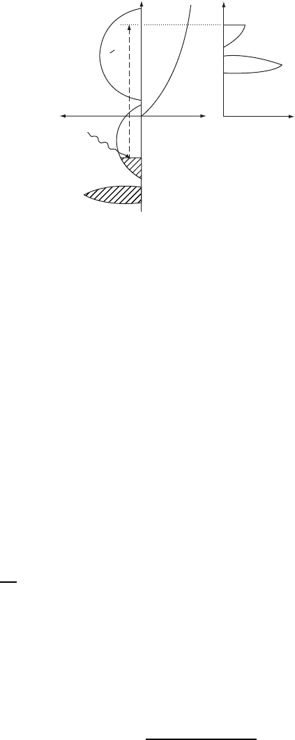
Figure W22.27. Photoemission from a metal with an occupied valence band and a partially
occupied conduction band. The density of electron states in the solid and vacuum, and the
energy-distribution curve IE are shown.
where ¯hω is the energy of the incident photon. Since the energy of the ultraviolet photon
is relatively small, electrons are extracted from the conduction band and the upper
valence bands. A schematic of the photoemission process is given in Fig. W22.27.
Three quantities are sketched in this figure. The left-hand side shows the density of
states in the solid, ;
s
E. The vacuum level is taken to be the zero of energy. The
Fermi level lies at energy e. Those states below the Fermi level are occupied
and are shaded on the diagram. The density of states in the vacuum ;
v
E is also
sketched in the figure. It corresponds to that of a free electron. On the right-hand side
of the figure the energy distribution curve of the emitted electrons, IE, is sketched.
Ideally, this curve is (aside from possibly smoothly varying distortions due to the
energy dependence of the dipole matrix elements) a replica of the density of states of
the solid below the Fermi energy. More realistically, there are significant contributions
due to secondary electrons.
A formula for the energy-distribution curves may be derived from Fermi’s golden
rule. The rate of absorption of photons is
ω D
2
¯h
i,f
s
jMj
2
υE
f
E
i
¯hωfE
i
,T[1 fE
f
,T],W22.99
where M is the dipole matrix element of the interaction of the photon with the electron,
and i and f refer to the initial and final states of the electron, respectively. There is
a sum over the two spin states, s, of the electron. The radiation interaction preserves
spin projection. There are also Fermi–Dirac distribution function factors introduced in
Chapter 7 and Appendix WB,
fE, T D
1
exp[ˇE ] C1
.W22.100
Here is the chemical potential (approximately equal to the Fermi energy, E
F
,atlow
temperatures). The first Fermi factor guarantees that there is an electron in state i,the

456 CHARACTERIZATION OF MATERIALS
second factor guarantees that state f is empty, so a transition can occur. Introduce the
electron density of states ;E as in Eq. (7.67). The absorption rate may be expressed as
D
E
0
dE
0
,W22.101
where E
0
dE
0
is the rate of absorption of photons leading to excited electrons within
the energy band E
0
to E
0
C dE
0
.Thisrateisgivenby
E
0
D
¯h
jMj
2
;
v
E
0
;
s
E
0
¯hωfE
0
¯hω, T[1 fE
0
,T],W22.102
where an average squared matrix element is used as an approximation. The rate of
producing photoemitted electrons is
IE
0
D E
0
PE
0
, W22.103
where PE
0
is the probability that if a photoelectron is produced, it will emerge from
the surface.
The graph of IE
0
versus E
0
is called the energy-distribution curve (EDC). The
previous formulas show that IE
0
is proportional to the product of the density of states
for the initial and final states. If the photon energy is sufficiently high, the final density
of states may be approximated by a free-electron density of states ;
v
E
0
/ E
0
1/2
.
The energy-distribution curve may then be used to determine the density of states
;
s
E
0
¯hω below the Fermi surface.
The total photoelectric current divided by the incident current of photons is called
the photoelectric yield. It is seen to be proportional to the joint density of states,
Iω ¾
¯h
jMj
2
P
;
s
E
0
;
v
E
0
¯hωfE
0
¯hω, T[1 fE
0
,T] dE
0
,W22.104
where an average escape probability factor P has been extracted from the integral.
As the electron leaves the solid, it can undergo inelastic-scattering processes with
other electrons. Some of these other electrons emerge as secondary electrons. One
therefore finds a large number of low-energy secondary electrons emerging from the
solid as well as the photoemitted electron. The energy-distribution curve therefore rises
at low energies.
In some experiments the angular distribution of the emitted electrons is analyzed
as well as the energy distribution. The study is called angular-resolved photoemission
spectroscopy (ARPES). This is particularly useful for obtaining information about the
surface layer of the solid or atoms or molecules adsorbed on the surface. Different
orbitals in these atoms or molecules point in different directions, and this influences
the emission pattern. For example, those orbitals pointing perpendicular to the surface
are more likely to photoemit electrons in a direction perpendicular to the surface. This
can reveal interesting information regarding the nature of the chemical bonds or the
particular bonding sites of adsorbed species.
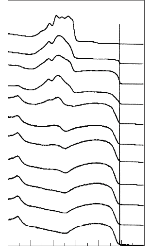
An example of a UPS spectrum is given in Fig. W22.28 for sputter-deposited
Ge
100x
Ag
x
with 0 x 39.6 at room temperature. The spectra were taken with 21.2-
eV photons from a He I ultraviolet light source. The Ge 4p valence band extends from
CHARACTERIZATION OF MATERIALS 457
10 8 6 4
Binding energy (eV)
20−2
Intensity (arb. units)
100.0
39.6
15.3
10.2
5.6
3.9
2.9
2.0
1.5
1.3
0.0
Ge4p
Ge4
s
Ag4d E
F
x
Figure W22.28. Ultraviolet photoemission spectrum of sputter-deposited Ge
100x
Ag
x
for
0 x 39.6 [From A. Suzuki and K. Tanaka, Jpn. J. Appl Phys., 37, 4872 (1998). Copyright
1998 by the Japanese Journal of Applied Physics.]
a binding energy of 4.5 to 0 eV, and the 4s band is at a binding energy of 9 eV. The
peak that develops at 5.5 eV is due to the Ag–4d band. For 0 x 5.6thespectra
show that Ag is dissolved in a Ge matrix, since a single Ag–4d peak appears. For
x ½ 5.6, phase separation occurs as silver clusters begin to form and the UPS spectrum
evolves toward that of bulk Ag, shown at the top of the figure.
X-ray Photoemission Spectroscopy (XPS or ESCA). Often, x-rays rather than UV
light are used in a photoemission experiment. The high energy of the x-ray permits
the observation of photoemitted core electrons of the solid. The bandwidths of the
core electrons are very narrow and the levels may be approximated as having a single
energy, E
core
. The energy of the emitted electron is
E
0
D ¯hω E
core
.W22.105
For a given x-ray photon energy ¯hω there will be a sharp peak in the EDC.
The precise value of the core energy is sensitive to the distribution of valence
electrons surrounding the core. To photoionize the core electron, the electron must
exit the atom by passing through the valence shells. There is a difference of potential
between the core and the outside world determined by the charge distribution of the
valence electrons. To get a qualitative feeling for this, consider a simple example.

458 CHARACTERIZATION OF MATERIALS
Suppose that a distribution of valence electrons is described by a charge distribution
;r, which will be taken to be spherically symmetric, for the sake of simplicity.
Poisson’s equation gives the potential
r
2
Vr D
1
r
2
∂
∂r
r
2
∂
∂r
Vr
D
;r
0
.W22.106
Here Vr is the contribution to the potential from the valence electrons. The contri-
bution to the potential due to the nucleus is fixed, and will be ignored. Taking the
position of the core to be approximately at r D 0, this gives a difference of potential
V1 V0 D
1
0
1
0
1
r
2
r
0
;r
0
r
02
dr
0
dr. W22.107
For example, suppose that the valence-electron charge distribution is given by
;r DQ
3
8
expr, W22.108
so that the total valence charge is Q. The parameter in this model represents the
inverse of the length over which the valence charge distribution decays outside the
atom in question. Then the difference of potential will be
V1 V0 D Q
8
0
.W22.109
The energy of an electron residing in the core may be written as the sum of a constant
plus the difference in potential energy between the electron at the core position and
the electron at infinity:
E
core
D constant e[V0 V1].W22.110
For the model above, therefore,
E
core
D constant C
eQ
8
0
.W22.111
For more compact charge distributions will be larger and the core level will be
shifted upward (i.e., less tightly bound). Correspondingly, for more spread-out valence
charge distributions, the core level will be lowered. In forming chemical bonds, the
electron distribution around atoms is distorted. This gives rise to core-level shifts
characteristic of the particular bonds that are formed. By measuring the difference
between the energy of the incident photon and the emitted electron, the energy of the
core level may be found.
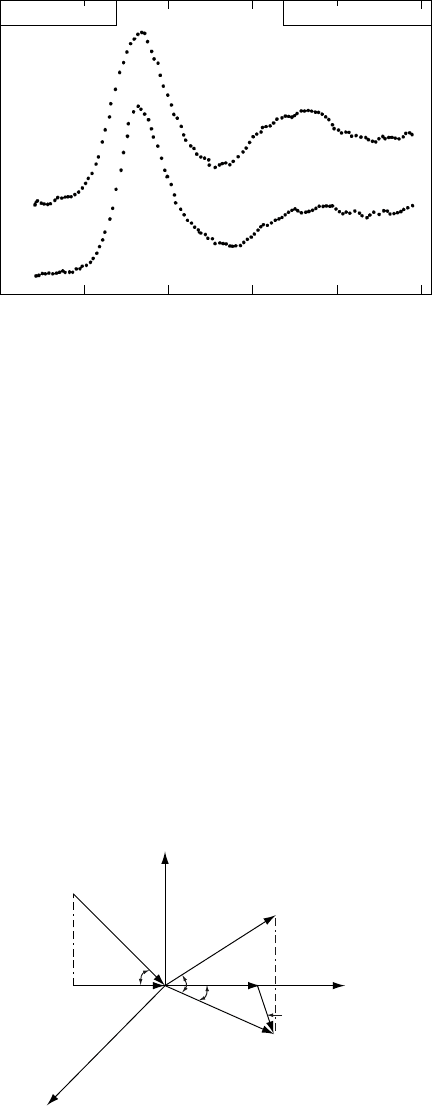
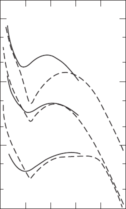
Examples of x-ray core-level spectra are given in Fig. W22.29. Data for
La
1.85
Sr
0.15
CuO
4
are taken at T D 300 K, where it is semiconducting, and T D 80 K,
where it is superconducting. The spectrum focuses on the Cu 2p
3/2
state. The data
provide evidence for a change of valence state with temperature.
CHARACTERIZATION OF MATERIALS 459
930 940
Binding energy (eV)
950
Cu 2p
3/2
XPS La
1.85
Sr
0.15
Cu O
4
Intensity (arbitrary units)
80 K
300 K
Figure W22.29. X-ray core-level spectroscopy of La
1.85
Sr
0.15
CuO
4
at T D 300 K and
T D 80 K. [From D. D. Sarma, Phys. Rev. B, 37, 7948 (1988). Copyright 1988 by the American
Physical Society.]
W22.17 Low-Energy Electron Loss Spectroscopy
As in LEED, the technique of low-energy electron loss spectroscopy (LEELS) involves
directing a beam of electrons at a surface. In LEELS, however, the energy loss of the
electron is studied rather than the elastic scattering. Electrons of energy E impinge
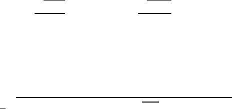
on a solid, making an angle with respect to the surface and come off at a variety
of angles. A detector is positioned so it accepts electrons that emerge at an angle
0
and an azimuthal angle (Fig. W22.30). The current of the scattered beam, I,isthen
analyzed as a function of the energy of the electron, E
0
. LEELS data generally can
consist of a table of IE
0
,
0
, as a function of E and , but more often are presented
as an angular-integrated function IE
0
, showing loss peaks. As with LEED, LEELS
provides information primarily about what is occurring on or near the surface.
When the electron scatters from the surface, it may emit (or absorb) an elemen-
tary excitation from the solid. This excitation is usually a phonon, but other types of
z
x
k
y
KK
E'
E
k'
K'
θ
θ'
Q, ω(Q)
φ
Figure W22.30. Scattering geometry for a LEELS experiment.
460 CHARACTERIZATION OF MATERIALS
excitations, such as two-dimensional plasmons associated with charged layers on the
surface, are also possible. The excitation carries with it both energy and momentum.
In general, the LEELS spectrum consists of energy-loss peaks from three origins: bulk
excitations of the substrate, surface excitations of the substrate, and excitations of
adsorbed species on the surface. Because of the limited penetration of electrons into
the solid, LEELS is particularly useful for studying the latter two surface excitations.
Surface excitations of the substrate are characterized by having a wave vector
parallel to the surface, Q, and a frequency ωQ. For the case of a periodic lattice there
is conservation of wave vector in the plane of the surface, modulus a reciprocal-net
vector (i.e., surface reciprocal-lattice vector):
K
0
D K CQ C G,W22.112
where K and K
0
are the surface components of k and k
0
. In the case of surface
adsorbates, unless the adsorbates form an ordered net, there will be no wave-vector
conservation.
In the following, attention will be restricted to the case where there is energy loss.
Energy gain, however, is possible if the temperature of the surface is high enough for
a thermal excitation to be present and absorbed by the electron. The basic equation of
LEELS is the energy conservation condition:
E
0
D E ¯hω. W22.113
For example, in the case of the excitation during inelastic scattering from an adsorbed
molecule, the energy of the electron will be reduced by the difference in energy between
two vibrational levels of the adsorbed molecule. It is also possible to study the vibra-
tional spectrum of the adsorbate bonded to the surface. As an analytical tool one may
make a quantitative analysis of the adsorbates, since the vibrational frequencies of each
molecule are a unique fingerprint for that molecule. The strength of the LEELS signal
is proportional to the number of adsorbed molecules.
Suppose that a substrate surface excitation is excited. It is possible to obtain the
dispersion curve of the excitation [i.e., to find ωQ]. The procedure follows from the
energy conservation law:
E
0
D E ¯hωQ. W22.114
Attention will be restricted to the case of near-specular scattering (i.e., let G D 0).
Using the following expressions for the wave-vector components (see Fig. W22.30),
K D
p
2ME
¯h
cos , K
0
D
p
2ME
0
¯h
cos
0
,W22.115
and the law of cosines
Q
2
D K
2
C K
2
0
2KK
0
cos , W22.116
the following expression for the wave-vector transfer is found:
Q D
1
¯h
2mE
0
cos
2
0
C E cos
2
2
p
EE
0
cos cos
0
cos . W22.117
CHARACTERIZATION OF MATERIALS 461
0 100 200 300 400 500
Energy loss (meV)
10°
5°
0°
Relative plasmon loss intensity (arbitrary units)
∆N = 2 × 10
13
cm
−2
Figure W22.31. LEELS spectra for ZnO for several scattering angles. [Reprinted From
Y. Goldstein et al., Surf. Sci., 98, 599 (1980), Copyright 1980, with permission from Elsevier
Science.]
Since E
0
is measured and E is known, the value of ωQ may be determined. Equation
(W22.117) gives Q in terms of E, E
0
, ,
0
and . Thus the dispersion relation for the
excitation can be measured.
An example of a LEELS spectrum is presented in Fig. 19.17 for n-type GaAs. The
spectrum shows phonon loss and gain peaks as well as a surface-plasmon loss peak.
In Fig. W22.31 data for angular-resolved LEELS are presented for electrons scattering
from a ZnO surface with an accumulation layer. The data are interpreted in terms
of the excitation of two-dimensional plasmons in the accumulation layer. From this
data, using Eq. (W22.117), it is possible to obtain information about ωQ for the
two-dimensional plasmon. The breadth of the peaks is due to the large dispersion of
the two-dimensional plasmon.
W22.18 Extended X-ray Absorption Fine Structure
An accurate determination of interatomic distances in a crystal may be obtained by
carefully studying the x-ray absorption spectrum. The absorption spectrum exhibits
oscillatory structure that comes about due to an interference effect involving the
electrons. The method is called extended x-ray absorption fine-structure (EXAFS)
spectroscopy.
When x-rays pass through a sample of thickness d the intensity of the emerging
beam, I, is related to the intensity of the incident beam, I
0
, through Beer’s law:
Id D I
0
exp˛d, W22.118
462 CHARACTERIZATION OF MATERIALS
where the very small surface reflection of the x-rays is neglected. The attenuation
constant, ˛, has contributions arising from both the absorption of x-rays and the Bragg
scattering of x-rays out of the incident beam (extinction). In this section attention
centers on the absorption contribution.
Absorption comes about when an electron is photoionized from an atom. The elec-
tron is promoted from some low-lying state to a state in the conduction band. In the case
of deep-core levels the bandwidths are very narrow and there is a threshold absorption
energy from a given band equal to the difference in energy between the Fermi energy,
E
F
, and the core-level energy, E
core
. For simplicity’s sake, restrict the discussion to
the case of a parabolic conduction band. When the excited electron travels through the
crystal it has a wave vector
k D
1
¯h
2m[¯hω E
c
E
core
],W22.119
where E
c
is the energy of the bottom of the conduction band. Thus the wave vector is
a function of the x-ray frequency.
The rate at which photon absorption takes place depends on how probable it is
to find the excited electron at the position of the nucleus. Technically, this comes
about because the rate depends on a matrix element of the radiation operator between
wavefunctions governing the initial and final states of the electron. In particular, it is
sensitive to the magnitude of the final-state wavefunction at the position of the atom.
If this magnitude were somehow to increase, the absorption would increase, whereas
if it were to decrease, the absorption would decrease.
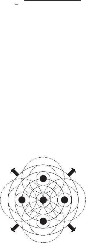
Upon absorption of the photon a spherically outgoing electron wave is created
with the wave vector above. This wave may scatter off neighboring atoms in the
crystal a distance a
j
away. The waves reflected interfere with the wave emitted as in
Fig. W22.32. What is of primary interest is the situation at the location of the ionized
atom. If there is constructive interference, the amplitude of the final-state electron
wavefunction will be maximum. If there is destructive interference, the amplitude will
be minimum. The condition for constructive interference is
2ka
j
C υ
j
D 2N. W22.120
Figure W22.32. Spherically outgoing excited electron waves scatter off neighboring atoms and
these reflected waves interfere with the emitted wave.
CHARACTERIZATION OF MATERIALS 463
34
−0.4
−0.2
0.0
0.4
0.2
5 6 7 8 9 10 11 12
χ (k)
k (Å
−1
)
6% Co [O]
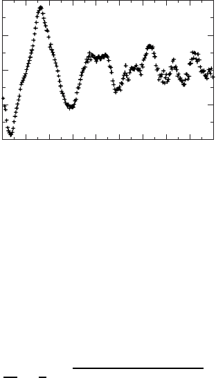
Figure W22.33. EXAFS oscillations for YBa
2
(Cu
1y
Co
y
)
3
O
6Cx
. [From H. Renevier et al., Phys.
Rev. B, 47, 11398 (1993). Copyright 1993 by the American Physical Society.]
Here 2a
j
is the round-trip distance to atom j and υ
j
is a phase shift characteristic of
the scattering of the electrons from the atoms. One expects the phase shift to be a
slowly varying function of electron energy. Thus interference oscillations in the x-ray
absorption spectrum are expected. The separation between neighboring interference
maxima (Fig. W22.33) provides a measurement of the various distances to shells of
nearby atoms. Thus
k D
a
j
D
1
¯h
2m[¯hω E
c
E
core
].W22.121
In practice, the absorption spectrum is Fourier analyzed as a function of k and the
peak positions in r space appear directly. Separate peaks may be identified with NNs,
next-NNs, and so on.
An example of EXAFS oscillations appears in Fig. W22.33 for excitation of a
Co core level. The data are for the compound YBa
2
(Cu
1y
Co
y
)
3
O
6Cx
. The quan-
tity Ek is the modulated part of the absorption constant. It is defined by Ek D
[˛k ˛
0
k]/˛
0
k,where˛k is the absorption coefficient, including its oscilla-
tions, and ˛
0
k is obtained by averaging ˛k (a smoothly varying function of k) over
the oscillations. By using the oscillations to determine the NN distance, it is possible
to determine that the Co ion has a valence state of C3. It is also possible to determine
the coordination number (5) of the Co ions to the oxygen ions.
In addition to EXAFS there is a technique called SEXAFS, which is surface EXAFS.
Grazing-incidence x-rays are used so that the radiation does not penetrate the solid
deeply and the surface region of the solid is probed. A technique closely related to
EXAFS is XANES (x-ray absorption near-edge structure).
W22.19 Auger Emission Spectroscopy
A useful tool for characterizing the chemical composition of a solid in the vicinity of
the surface is Auger emission spectroscopy (AES). A monoenergetic beam of high-
energy (1 to 10 keV) primary electrons impinges on the surface of the solid and causes
collisional ionization events to occur. Some of these events result in deep core-level
electrons being knocked out. In light elements (Be to Si), typically a K-shell electron