Gersten J.I., Smith F.W. The Physics and Chemistry of Materials
Подождите немного. Документ загружается.


434 CHARACTERIZATION OF MATERIALS
the “half-silvered” mirror, are directed through sample S, and are finally detected at
detector D (usually, a bolometer). A recording is made of the intensity as a function
of time, which is then Fourier analyzed.
Let d be the distance from m to mirror M and d
0
C vt be the distance from m to
mirror M
0
. The amplitude of the recombined wave is the superposition of the amplitudes
of the two beams
Et D
E
0
ω
2
exp2ikd C
E
0
ω
2
exp[2ikd
0
C vt].W22.62
The intensity incident on the sample is proportional to the absolute square of E.The
detected intensity is
It D 2
1
0
dωI
0
ωTω
1 C cos
2
ω
C
d
0
d C vt
.W22.63
where I
0
ω DjE
0
ωj
2
, Tω being the transmission coefficient for the sample. Now
take the Fourier transform of this to obtain
I D
1
1
dt
2
It expit
D
1
0
dωI
0
ωTω
2υ C υ
2
v
c
ω
expi?
Cυ
C 2
v
c
ω
expi?
,W22.64
where ? D 2ωd d
0
/c. Focusing attention on the resonant (second) term and
computing its amplitude gives
jIj'
c
2v
I
0
c
2v
T
c
2v
.W22.65
In the ideal case, since the blackbody spectrum is known, the functional dependence
of I
0
ω is known. Therefore, a measurement of I permits the determination of
jTc/2
vj.Sincev will typically be on the order of 1 mm/s, the ratio c/2v will
be 1.5 ð10
11
. Thus a measurement in the frequency range of ³ 1 kHz is used to
determine the spectrum in the range of 10
14
Hz! A replica of the infrared spectrum
has been produced at low frequencies.
In reality, the situation is more complicated, since the source is not a blackbody.
Usually, a baseline spectrum is taken without a sample. In this way the output can be
normalized to the response of the system, including the source spectrum and detector
sensitivity.
FTIR permits one to obtain data simultaneously over a large frequency range and
over a large collection angle. Multiple scans are used to improve the signal-to-noise
ratio. The technique is readily extended to other forms of spectroscopy, such as Raman
spectroscopy.
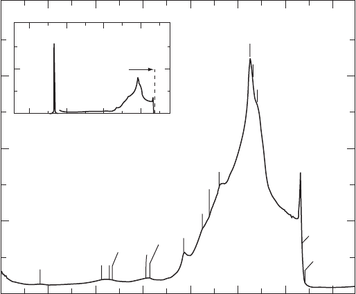
The FTIR spectrum for diamond is presented in Fig. W22.12. The spectrum clearly
shows various critical points and combinations of critical points in the phonon spectrum.

CHARACTERIZATION OF MATERIALS 435
Three-phonon
1400 1600
0
5
10
15
20
1800 2000 2200 2400 2600 2800
Absorption coefficient (cm
−1
)
Wave number (cm
−1
)
2
3
7
8
9
10
12
13
15
14
17
19
24
0
10
1000 2000 3000 4000
w
0
Two-phonon
T
= 300 K
Figure W22.12. FTIR spectrum for diamond at T D 300 K. [From R. Vogelgesang et al., Phys.
Rev. B, 58, 5408 (1998). Copyright 1998 by the American Physical Society.]
This spectrum may also be contrasted with the Raman spectrum given in the following
section. The spectrum should be compared with the phonon density of states presented
in Fig. 5.9.
W22.9 Raman Spectroscopy
The Raman effect was originally discovered in molecular physics. Monochromatic light
of frequency ω was directed at a gas sample, and the scattered light was passed through
a monochromator and onto a photodetector. The scattered light consisted mainly of
radiation at frequency ω (Rayleigh scattering), but also possessed sidebands at lower
(Stokes shifted) and higher (anti-Stokes shifted) frequencies. The displacement of the
sidebands is characteristic of the type of molecule under study and is related to the
vibrational frequencies associated with nuclear motion. The angular momentum selec-
tion rules J D 0, š2 are obeyed, where J is the total angular momentum, consistent
with what is expected for scattering of a spin 1 particle, the photon. This differs from
the absorption case where the selection rules are J D 0, š1.
A simple classical theory provides a heuristic explanation of the effect, although a
quantum-mechanical treatment is required to understand the effect quantitatively. Let
the molecule be described by a polarizability tensor
$
˛, defined in Chapter 8. Incident
light provides an electric field with amplitude E
0
, which induces an oscillating electric
dipole
m D
$
aω · E
0
expiωt. W22.66
This dipole will radiate in accordance with the Larmor radiation formula. The energy
emitted per frequency interval dω is
Uω D
ω
4
12
0
c
3
j
$
˛ω Ð
E
E
0
j
2
.W22.67

436 CHARACTERIZATION OF MATERIALS
This is elastically scattered light and is called Rayleigh scattering. Now suppose that
the molecule is allowed to vibrate in a particular normal mode with a vibrational
frequency (which is much less than ω). The polarizability tensor will also fluctuate
at this frequency. Let Q be the normal-mode coordinate displacement associated with
. Then, to a first approximation,
aω, t D a
0
ω C
∂
aω
∂Q
Q cos t. W22.68
The oscillating dipole now produces sidebands at frequencies ω C and ω ,in
addition to the oscillation at ω. The emission at these frequencies constitutes the
Raman anti-Stokes and Stokes radiation, respectively. Rayleigh scattering occurs at
frequency ω.
In the quantum-mechanical description the molecule is originally in the ground-
electronic state in some vibrational state, and the light causes it to make a virtual
transition to an excited-electronic state. This is followed by the molecule radiating a
photon and falling into any vibrational state associated with the ground-electronic state.
If the state happens to be the original one, it produces Rayleigh scattering. If it is to
a higher-energy state, it is Stokes Raman scattering, whereas if it is to a lower-energy
state, it is anti-Stokes Raman scattering. In Raman scattering the outgoing photon is
either lowered in energy or raised in energy by the vibrational quantum ¯h.Inorder
for anti-Stokes scattering to occur, there must be population in the excited vibrational
state to begin with, which arises from thermal excitation. Stokes scattering can always
occur. The ratio of the anti-Stokes to the Stokes scattering is given by the Boltzmann
factor:
I
antiStokes
I
Stokes
D exp
¯h
k
B
T
.W22.69
Raman scattering is useful in condensed matter physics and chemistry in several
instances. In solids the vibrational motions of the molecules are coupled and the exci-
tations spread out in energy. In crystals they assume the character of phonons and
are delocalized over the entire crystal. In highly disordered materials they may remain
as localized oscillations extending over many nearby neighbors. The phonons may be
categorized as being optical or acoustic. Raman scattering from the acoustic phonons
is called Brillouin scattering.
For example, consider the scattering by conduction electrons in a lightly n-doped
semiconductor. An electron may be virtually excited to some higher energy band and
then reemit a different photon in returning to the original band. However, the wave
vector of the photon is small compared with the size of the Brillouin zone. Therefore,
there cannot be much of a change in the wave vector of the electron. It could emit an
optical phonon with k D 0, selection rules permitting. It could also produce Brillouin
scattering. If anharmonic effects are taken into account, however, terms involving the
simultaneous excitation of two phonons are also present. In terms of the simple classical
model introduced earlier,
at D a
0
ω C
2
iD1
∂aω
∂Q
i
Q
i
cos
i
t C
1
2
ij
∂
2
aω
∂Q
i
∂Q
j
Q
i
cos
i
tQ
j
cos
j
t.
W22.70

CHARACTERIZATION OF MATERIALS 437
Sidebands now include terms with frequencies ω
1
2
, among others. Extending
this concept to solids implies that two-phonon production is possible. The net wave
vector carried off by a pair of optical phonons may be small (i.e. k
1
C k
2
D 0). Thus
light is able to create such a state with little momentum transfer.
Surface-enhanced Raman scattering (SERS) has emerged as a powerful tool for
studying adsorbed species on the surfaces of solids. The Raman cross section for
adsorbed species is found to be enhanced by as much as six orders of magnitude over
the gaseous cross sections. Much of this enhancement is due to the increase in the
strength of the local electromagnetic field at the surface over its value in free space.
The amplification occurs because of local surface roughness, which creates miniature
“lightning rods,” and also because of particular electronic resonances of the solid, such
as surface plasmons. At frequencies approaching these resonances the surface acts
as a high-Q resonator and has high-frequency (ac) electric fields due to the incident
and outgoing radiation. There is also considerable evidence that the formation of the
chemical bond between the adsorbed molecule and the substrate enhances the value of
the Raman tensor, ∂˛/∂Q.
An example of a Raman spectrum is given in Fig. W22.13. The intensity of the
Raman scattering for diamond is plotted as a function of the frequency shift (in wave
numbers). The Raman spectrum may be contrasted with the infrared absorption spec-
trum given in Fig. W22.12. The Raman spectrum is due to both a single-phonon
process at ω
0
(as shown in the inset to Fig. W22.13) and to much weaker two-phonon
processes. The one-phonon Raman peak at ω
0
D 1332.4cm
1
corresponds to the zone-
center optic mode at ³ 2.5 ð10
14
rad/s of Fig. W22.12. Note that Raman scattering
1400 1600
0
10000
20000
30000
40000
1800 2000 2200 2400 2600 2800
Intensity (arb. units)
Raman shift (cm
−1
)
4
5
6
11
1
12
15/16
18
19
20
23
22
21
T
= 300K
l
L
= 4762 Å
25
26
27
ω
0
1000 1500 2000 2500
X 100
Two-phonon
Figure W22.13. Raman spectrum for diamond at T D 300 K. The incident light is polarized in
the (111) plane. The backscattered light is in the [111] direction. [From R. Vogelgesang et al.,
Phys. Rev. B, 58, 5408 (1998). Copyright 1998 by the American Physical Society.]

438 CHARACTERIZATION OF MATERIALS
provides a much higher precision measurement of mode frequencies than does neutron
scattering. See also Fig. 11.21, which gives a Raman spectrum for a Si–Ge alloy.
W22.10 Luminescence
Light is absorbed by materials and a fraction of the light is reemitted, usually with
photons of lower frequencies. The process is called luminescence. The light may come
out promptly, on a time scale of the order of a nanosecond, in which case the process
is called fluorescence. It may come out on a much longer time scale, in which case it is
called phosphorescence. Just how much light comes out depends on the nature of the
competing channels for nonradiative decay. The quantum efficiency for luminescence
may be defined as the ratio of the number of output photons per unit time to the number
of input photons per unit time:
? D
P
N
output
P
N
input
ð 100%.W22.71
In metals, where the excitation of electrons–hole pairs requires no activation energy,
the nonradiative decay mechanism is probable and the quantum efficiency is very small.
In semiconductors, where there is a substantial energy gap, the quantum efficiency may
be quite large.
In Fig. W22.14 a typical luminescence process for a semiconductor is illustrated.
An incident photon is absorbed by the solid, promoting an electron from a filled
valence-band state (v) to a vacant conduction-band state (c). The photon must, in most
instances, have an energy that exceeds the energy gap, E
g
. The notable exception is the
case where excitons exist just below the bottom of the conduction band. The processes
above, in which an electron jumps from one band to the other band, is called an
interband process. A hole is left behind in the valence band. The electron is generally
produced in an excited state of the conduction band. By a sequence of phonon-emission
processes the electron relaxes to the bottom of the band. Similarly, the hole migrates
to the top of the valence band by such intraband processes. The time scale for these
transitions is typically picoseconds or less. Luminescence takes place when the electron
makes a radiative-decay transition from the bottom of the conduction band to the top
of the valence band. The radiative lifetime is longer than a nanosecond.
CB
E
g
VB
hw'
hw
Figure W22.14. Luminescence in a direct-gap semiconductor.

CHARACTERIZATION OF MATERIALS 439
0.29 0.30 0.31 0.32
Photon energy (eV)
0.33 0.34 0.35 0.36
−1
0
1
2
3
4
5
6
7
4.2 4 3.8
Wavelength (µm)
PI. intensity (a.u.)
3.6
InAs
0.911
Sb
0.089
T
= 4 K
1 µm diode laser power
320 mW
160 mW
80 mW
40 mW
20 mW
10 mW
5 mW
x 10
28.2 meV
(27-29 meV typical)
4.3 meV
(4-8 meV
typical)
9.4 meV
(7-13 meV typical)
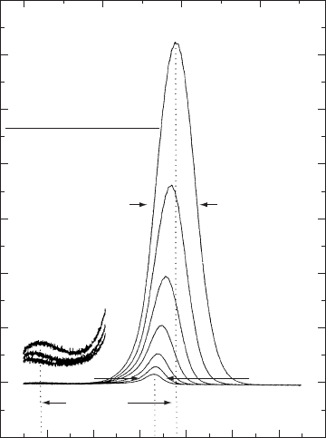
Figure W22.15. Photoluminescence spectra for MBE-grown InAs
0.911
Sb
0.089
on a GaSb
substrate at T D 4 K. [From M. A. Marciniak et al., J. Appl. Phys., 84, 480 (1998). Copyright
1998 by the American Institute of Physics.]
Hot luminescence occurs when the radiative recombination occurs not from the
bottom of the band but from some excited state in the conduction band. If the relaxation
occurs primarily with optical-phonon emission, a series of bumps will be seen in the
emission spectrum, corresponding to the photon energy less some number of optical-
phonon energies.
It is possible to study luminescence either in the frequency domain or in the time
domain. In the latter case the procedure is called a time-resolved luminescence study.
Luminescence may also be used to study defects. Cathodoluminescence is produced
by an electron beam striking the surface of a solid.
An example of a photoluminescence spectrum is given in Fig. W22.15 for a film
of InAs
0.911
Sb
0.089
lattice-matched to a GaSb substrate. In addition to the main lumi-
nescence peak, there is a sideband lowered by the energy of a single LO phonon
(³ 28.2 meV). The narrow line width (³ 5 meV) indicates that the material is of high
quality.
W22.11 Nonlinear Optical Spectroscopy
With the advent of the laser it has become very easy to generate high-intensity elec-
tromagnetic fields. Materials no longer necessarily respond in a linear manner to

440 CHARACTERIZATION OF MATERIALS
these fields, and it is important to understand their nonlinear properties. A number of
phenomena are associated with nonlinear optics, such as second- and third-harmonic
generation, three- and four-wave mixing, parametric excitation, self-focusing, self-
phase modulation, and self-induced transparency, etc. Closely related to the pure
nonlinear optical properties are the electro-optical and acousto-optical properties of
materials. One is often interested in knowing how the optical properties of a material
can be altered by applying electric fields or sound waves.
Attention will be focused on the polarization vector induced when an electric field
exists in a medium. For a linear isotropic medium,
Pω D
0
EωEω W22.72
where Eω is the electric susceptibility. For an anisotropic linear material the corre-
sponding formula is [see Eq. (8.44)]
Pω D
0
$
Eω · Eω W22.73
where
$
Eω is the electric susceptibility tensor. The anisotropy of this tensor is respon-
sible for birefringence (i.e., the variation of the speed of light in a material with the
polarization direction).
In nonlinear optics there are also nonlinear susceptibilities that may be defined. For
example, there is the second-order susceptibility defined by [see Eq. (8.46)]
P
˛
ω D
0
ˇ8
d
2
˛ˇ8
ω
1
,ω
2
; ωE
ˇ
ω
1
E
8
ω
2
dω
1
dω
2
.W22.74
This process describes the interaction of two photons of frequencies ω
1
and ω
2
in a
material to create a photon of frequency ω. Energy conservation requires that
ω
1
C ω
2
D ω. W22.75
For this process to proceed it is also necessary to guarantee wave vector conservation,
that is,
k
1
C k
2
D k.W22.76
This is called phase matching. The concept appears in Section W8.1, where the index
ellipsoid is introduced. Methods for achieving phase matching in inhomogeneous
media were discussed in Sections W20.6 and W20.8. Using the dispersion formula
it implies that
ω
1
nω
1
c
C ω ω
1
nω ω
1
c
D ω
nω
c
.W22.77
This will, in general, not be valid for arbitrary frequencies. By rotating the crystal and
making use of the different indices of refraction for the ordinary and extraordinary
waves, however, it is possible to achieve phase matching.

CHARACTERIZATION OF MATERIALS 441
A particular application of the second-order nonlinearity is in the process of second-
harmonic generation. In that case [see Eq. (8.46)]
P
˛
2ω D
0
ˇ8
d
2
˛ˇ8
ω, ω;2ωE
ˇ
ωE
8
ω. W22.78
Depending on the symmetry of the crystal, there will only be a small number of inde-
pendent components of d
2
˛ˇ8
. The various components of the second-order polarization
may be measured by focusing lasers of various polarizations onto a crystal volume and
measuring the amount of second-harmonic light that is generated. Values for the d
2
˛ˇ8
components for various materials are given in Table 8.4.
The second-order polarizability exists only in crystals without inversion symmetry.
The polarization vector P, being a vector, should reverse its direction under a reflection
operation, as should E. But this is inconsistent with Eq. (W22.78), since the left-hand
side changes sign but the right-hand side does not. In crystals with inversion symmetry
d
2
˛ˇ8
is zero.
The third-order nonlinearity is described in terms of a fourth-order polarizability
tensor defined analogously as
P
˛
ω D
0
ˇ8υ
d
3
ˇ8υ
ω
1
,ω
2
,ω ω
1
ω
2
; ω
ð E
ˇ
ω
1
E
8
ω
2
E
υ
ω ω
1
ω
2
dω
1
dω
2
,W22.79
where the phase-matching condition is
k
1
C k
2
C k
3
D k W22.80
and energy conservation requires that
ω
1
C ω
2
C ω
3
D ω. W22.81
The tensor d
3
˛ˇ8υ
ω, ω, ω;3ω may be determined by performing a third-harmonic
generation experiment. Values for it appear in Table 8.5.
The application of an electric field to a crystal may alter the linear index of refraction
of the crystal. This is of considerable technological importance since it implies that
laser beams may be deflected electronically and at electronic frequencies. The degree
to which the index of refraction changes when an electric field is applied to the crystal
is determined by the electro-optic tensor (see Section 18.8).
The effective index of refraction for light propagating in a given direction k with a
given polarization vector Oε is defined in terms of the index ellipsoid. One constructs
an imaginary ellipsoid in space (see Eq. (W8.12)]:
x
n
x
2
C
y
n
y
2
C
z
n
z
2
D 1 W22.82
where x, y,andz define the coordinates in which the index of refraction tensor (related
to the polarization tensor) is diagonal, and n
x
, n
y
,andn
z
are the corresponding indices

442 CHARACTERIZATION OF MATERIALS
of refraction. Draw a plane through the center of the ellipsoid perpendicular to k.The
plane intercepts the ellipsoid in an ellipse. The length of the vector from the center of
the ellipsoid to the ellipse in the direction of Oε is the value of n for that light ray.
Now introduce an electric field E. The index ellipsoid will become stretched or
compressed and will be rotated relative to the coordinates above. The new equation
becomes
1
n
2
1
x
2
C
1
n
2
2
y
2
C
1
n
2
3
z
2
C 2
1
n
2
4
yz C 2
1
n
2
5
xz C 2
1
n
2
6
xy D 1.
W22.83
The dependence of these coefficients on E is, for weak fields, a linear one. Thus
1
n
2
˛
D
1
n
2
˛
C
3
ˇD1
r
˛ˇ
E
ˇ
,˛D 1, 2, 3,W22.84a
ˇ
ˇD1
r
˛
ˇ
E
ˇ
,˛D 4, 5, 6.W22.84b
The electro-optic tensor coefficients r
˛ˇ
may be measured by passing a laser beam
through a crystal with various orientations, applying an electric field, and measuring
the beam deflection produced.
Using similar ideas, it is possible to study the photoelastic tensor, which is a tensor
describing the variation of the index of refraction when a strain is introduced.
ELECTRON MICROSCOPY
Conventional optical microscopy is limited in its ability to resolve structure smaller in
size than the wavelength of visible light, (. The Rayleigh criterion is
sin ³ 1.22
(
d
,W22.85
which relates the acceptance angle of the microscope, , and the distance, d, between
two points that can be resolved. Since visible light has wavelengths in the range 400 to
700 nm, light cannot be used to see individual atoms, whose size is typically 0.1 nm.
If electromagnetic radiation is to be used to study materials, one may improve matters
in two ways. The first is to use shorter-wavelength radiation. X-rays would be ideal,
since their wavelength can be chosen to be comparable to the size of an atom. Another
approach is to use very fine optical fibers tapered to a “point” whose size is ³ 10 nm
and then bring this fiber close to the surface of the material to be probed. The coupling
is done through the near field of the electromagnetic field. Using this technique, 10-nm
resolution can be achieved simply and inexpensively. The method is called near-field
scanning optical microscopy (NSOM).
Another approach is to use electrons instead of light. The relativistic expressions
for the wavelength of an electron are
( D
h
p
D
hc
E
2
mc
2
2
D
hc
KK C 2mc
2
D
hc
eVeV C 2mc
2
,W22.86
CHARACTERIZATION OF MATERIALS 443
where p is the momentum, E the total energy, K the kinetic energy, and V the potential
difference through which the electron is accelerated to achieve this kinetic energy. By
using 20-kV potentials, wavelengths of 0.009 nm are obtained, smaller than an atom.
Thus resolution is no longer a limitation, but other factors, such as aberrations, prevent
this fine resolution from being realized.
Electrons may be focused using electrostatic or magnetostatic lenses. The focal
lengths of these lenses may be varied at will by changing the potentials and currents,
respectively. It is therefore possible to construct electron microscopes in much the same
way as optical microscopes are constructed. The main difference is that in electron
microscopy the distance from the lenses to the sample is held fixed while the focal
lengths are changed. In optical microscopy, of course, it is the other way around.
The image in electron microscopy is usually obtained by rastering the beam across
the sample and having the electrons collected by a detector. After amplification, the
processed image is displayed on a fluorescent screen. High-vacuum conditions are
needed for the electron beam to avoid collisions with gas molecules.
When high-energy electrons strike a material, they excite it and thereby lose
energy. Bulk and surface plasmons can be excited. Interband transitions occur and
electron–hole pair excitations are produced. There are also core-electron knock-out
processes, which are followed by x-ray emission or Auger deexcitation. The Auger
process is a multielectron process in which one electron fills an inner-shell vacancy,
and one or more other electrons are ejected from the atom. Intraband transitions occur
in metals. The net result is that copious amounts of secondary electrons are produced.
In addition, there are backscattered primary electrons. Light may be emitted from the
material when the electron–hole pairs recombine. If the sample is thin enough, a beam
of electrons will be transmitted through the sample.
There are several methods for observing the sample. These include scanning-
electron microscopy (SEM), transmission-electron microscopy (TEM), high-resolution
transmission-electron microscopy (HRTEM), and low-energy electron microscopy
(LEEM). These cases are discussed individually.
A number of typical electron micrographs using these techniques have appeared
in Chapter 4. Figure 4.1d showed nanocrystalline diamond with a resolution of ³
100 nm. Figure 4.1e was a micrograph with atomic-scale resolution of the interface
between crystalline Si and amorphous SiO
2
. Figure 4.6 displayed nanocrystalline Au
clusters embedded in an amorphous matrix. Figure 4.7 presented various morpholo-
gies of colloidal ˛-Fe
2
O
3
particles. Figure 4.3 gave an HRTEM micrograph of a
PbTiO
3
–SrTiO
3
superlattice. Figure 4.9 showed the microstructure of a quasicrystal.
Figures 4.20 and 4.21 presented images of a stacking fault and a twinned structure,
respectively. These micrographs attest to the versatility of electron microscopy as a
tool for studying the microstructure of materials.
W22.12 Scanning-Electron Microscopy
The scanning-electron microscope (SEM) collects the backscattered and secondary
electrons that are emitted from the surface of the material. Typically, a focused 5-
nm-diameter beam with a current of 10
11
A is directed at the surface and penetrates
the material. At first, when the electron is moving fast, high-energy processes such
as Auger excitation are possible. Secondary electrons are produced, but backscattering
is improbable at first because of the small Rutherford cross sections at high energies.
