Cui Dongmei. Atlas of Histology: with functional and clinical correlations. 1st ed
Подождите немного. Документ загружается.

CHAPTER 20
■
Eye
405
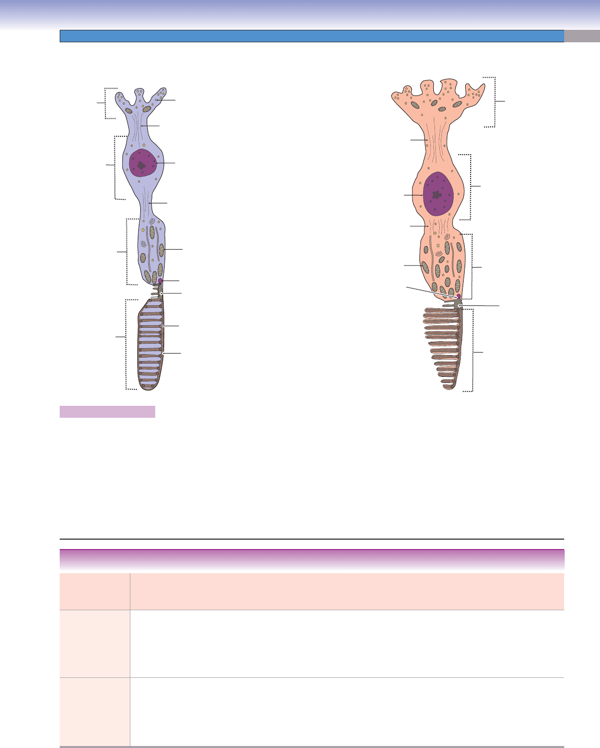
Figure 20-14A and B. A representation of rods and cones.
Rods and cones are photoreceptor cells in the retina. They are similar in structure and both have (1) a synaptic region (2) a nuclear
region (3) inner segments, and (4) outer segments. There are numerous synaptic vesicles in the synaptic regions, where the photorecep-
tors synapse with the dendrites of bipolar cells. The nuclear regions of rods and cones lie in the outer nuclear layer. The inner and outer
segments form the photoreceptor layer of the retina. The inner segments contain mitochondria, rough endoplasmic reticulum, Golgi
apparati, and other organelles that support the synthesis of proteins. The outer segments are in contact with the apical region of the
pigmented epithelium cells. Outer segments of rods are composed of a series of superimposed disks, which have individual membranes,
are stacked on each other, and are enclosed within the plasma membrane. Outer segments of cones have disks that are formed by invagi-
nations of the plasma membrane. The membranes of the disks are continuous with the plasma membrane of the cell. The inner region is
much wider than the outer region, which gives a cone appearance. Other differences between rods and cones are listed in Table 20-1.
D. Cui
Inner rod fiber
Synaptic vesicles
Synaptic
region
Nucleus
Nuclear
region
Outer rod fiber
Mitochondrion
Basal body
Disk
Plasma membrane
Modified cilium
Inner
segment
Outer
segment
A
D. Cui
Synaptic
region
Inner cone fiber
Nuclear
region
Nucleus
Mitochondrion
Basal body
Modified cilium
Outer cone fiber
Inner segment
Outer segment
B
Types of
Photoreceptor
Cells
Synaptic
Region
Nuclear
Region
Inner Segment Outer Segment Distribution in
the Retina
Main
Function
Rods Spherule
synapses with
dendrites of
one bipolar
neuron
Small,
round
nucleus
Fewer
mitochondria than
cones; synthesized
proteins passed
only to newly
forming disks
Cylindrical in shape; disk
membranes are separate
and are not connected
to the outside plasma
membrane; disks contain
rhodopsin protein
Numerous in the
peripheral retina;
none in fovea
Vision in
dim light;
motion
detection
Cones Pedicle
synapses with
dendrites of
several bipolar
neurons
Large,
ovoid
nucleus
More
mitochondria than
rods; synthesized
proteins passed
to entire outer
segment
Cone shaped; disk
membranes are connected
to the plasma membrane
and form invaginated
membranes; disks contain
iodopsin protein
Highly
concentrated in
the fovea and
macula lutea;
less numerous in
periphery
Color
vision;
perception
of fi ne detail
TABLE 20-1 Comparison of Rods and Cones
CUI_Chap20.indd 405 6/16/2010 7:38:26 PM
406
UNIT 3
■
Organ Systems
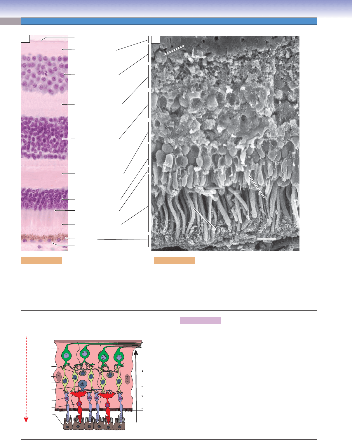
Figure 20-15A. Retinal layers. H&E, 463
There are 10 histologic layers in the retina; see Figure
20-13B and Synopsis 20-1 for details of the compo-
nents in each layer.
Ganglion cell
Ganglion cell
Cone
Cone
Rod
Rod
10 μm
Nerve fiber layer (9)
Ganglion cell layer (8)
Inner plexiform layer (7)
Inner nuclear layer (6)
Outer plexiform layer (5)
Outer limiting layer (3)
Photoreceptor layer (2)
Bruch membrane
Outer nuclear layer (4)
Pigment epithelium
layer (1)
Inner limiting membrane (10)
Ganglion cell
Cone
Rod
A
B
Figure 20-15B. Scanning electron micrograph of retinal layers.
Freeze-fracture preparation, 1,200
This freeze-fracture preparation of the peripheral retina shows the three-
dimensional shapes of the various cells in the retina. This section from
the peripheral retina has fewer ganglion cells than the macular retina
illustrated at left. Compare Figure 20-13B and C.
T. Yang
Neural
outflow
Light
Nerve fiber layer
Inner plexiform layer
Inner nuclear layer
Outer nuclear layer
Outer plexiform layer
Outer limiting layer
Photoreceptor layer
Pigment
epithelium layer
Ganglion cell layer
Inner limiting
membrane
Ganglion
cell
Müller cell
Müller cell
Amacrine
cell
Bipolar cell
Horizontal
cell
Rod cell
Cone cell
Pigment cell
C
Figure 20-15C. A representation of the functional retinal
layers.
Light passes through all retinal layers to activate the photore-
ceptor cells (rods and cones), and excess light is absorbed by
pigmented epithelial cells. The photoreceptors transduce the
light into electrochemical signals, which pass to the conduct-
ing neurons (bipolar cells, then ganglion cells). Horizontal and
amacrine cells are association neurons that have long dendrites.
The dendrites of the horizontal cells synapse with photorecep-
tors and contribute to the elaborate neuronal connections in the
outer plexiform layer. The dendrites of amacrine cells are in con-
tact with bipolar and ganglion cells and modulate signals in the
inner plexiform layer. The ganglion cells collect all visual infor-
mation and send it along their axons in the optic nerve. Müller
cells are large supporting neuroglial cells and extend from the
inner limiting membrane to the outer limiting membrane. Their
processes surround all the neuronal elements of the retina.
CUI_Chap20.indd 406 6/16/2010 7:38:26 PM
CHAPTER 20
■
Eye
407
The ganglion cells and their axons are part of the central nervous system. They cannot regenerate following severe damage.
Embryologically, the pigmented epithelium layer and the neural layers of the retina originate from different layers of the optic
vesicle. Therefore, retinal detachment may occur between the pigmented epithelium layer and the rest of the neural retina.
SYNOPSIS 20-1 Retinal Layers
(1) Pigment epithelium layer, a layer of cuboidal cells which are rich in melanin granules. This layer is important in absorbing
excess light and in esterifying vitamin A.
(2) Photoreceptor (rods and cones) layer, consists of inner and outer segments of the photoreceptor cells.
(3) Outer limiting layer, a plexus of junction complexes that joins the membranes of the photoreceptor cells and Müller
cells (retinal glial cells).
(4) Outer nuclear layer, contains nuclei of the photoreceptor cells (rods and cones).
(5) Outer plexiform layer, consists of axodendritic synapses between the axons of the photoreceptor cells and dendrites of
the bipolar and horizontal cells.
(6) Inner nuclear layer, contains nuclei of bipolar cells, horizontal cells, amacrine cells, and Müller cells.
(7) Inner plexiform layer, composed of axodendritic synapses between the bipolar cell axons, ganglion cell dendrites, and
processes of the amacrine cells.
(8) Ganglion cell layer, contains nuclei of the ganglion cells.
(9) Nerve fi ber layer, contains axons of the ganglion cells.
(10) Inner limiting membrane, the basement membrane of the Müller cells.
CLINICAL CORRELATIONS
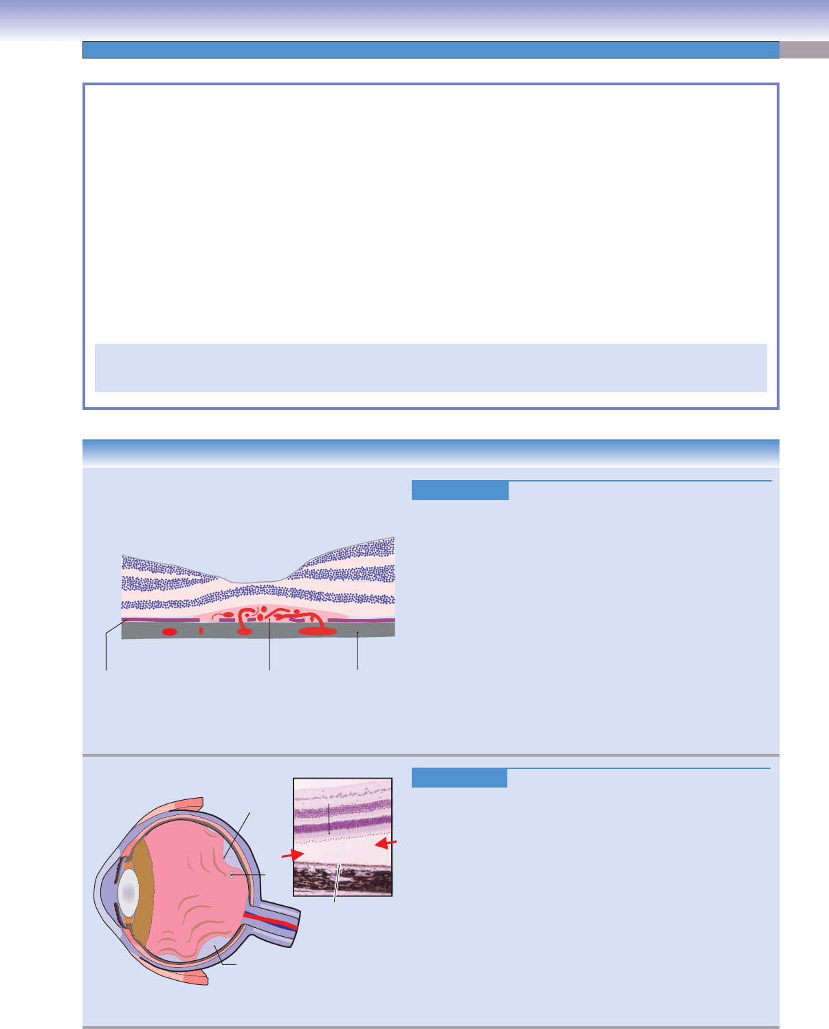
Figure 20-16A.
Age-Related Macular Degeneration.
Age-related macular degeneration (ARMD) is a degenerative eye
disease affecting the central portion of the retina. Symptoms include
blurred vision, distortion of straight lines, and worsening of color
vision. There are two forms: exudative (wet) and nonexudative (dry).
Wet ARMD is characterized by the growth of new vessels from the
choroidal circulation and serous fl uid leakage from the new vessels.
This causes detachment of the retina and macula, producing a rapid
and irreversible loss of central vision. Treatment options include
laser photocoagulation, surgery, and drugs. Dry ARMD is char-
acterized by subretinal drusen deposits (focal eosinophilic material
arising from the Bruch membrane and lying between the pigment
epithelium and the Bruch membrane), and atrophy and degeneration
of the outer retina, including the retinal pigment epithelium, Bruch
membrane, and choriocapillaris. The causes of ARMD are not well
understood, but risk factors include age, smoking, family history,
hypertension, and ethnicity (higher in non-Hispanic Caucasians).
D. Cui
Pigment epithelium
Vitreous fluid-
filled space
Detached
retina
Photoreceptor
layer
Retinal
hole
Vitreous fluid
B
Figure 20-16B.
Retinal Detachment. H&E, 62
Retinal detachment is an eye condition in which the sensory retina
separates from the underlying retinal pigment epithelium and
choroid (see red arrows). It is categorized into three major types:
(1) rhegmatogenous retinal detachment ([RRD] illustrated) is the
most common; it occurs when vitreous fl uid leaks into the subreti-
nal space through a break in the retina; (2) exudative or serous
detachment occurs in association with infl ammation or a tumor;
(3) traction detachment occurs when scar tissue on the retina’s sur-
face contracts and causes the retina to separate from the retinal pig-
ment epithelium. Retinal detachment is a medical emergency that
can lead to permanent vision loss. Time is critical for surgical reat-
tachment of the retina, which will usually preserve vision. Symptoms
include fl ashes, fl oaters, and visual fi eld distortion or loss. Treatment
options include laser photocoagulation, cryoretinopexy, vitrectomy,
and silicone oil repair.
J. Lynch
Choroid
Abnormal blood
vessels, leaking fluid
Bruch membrane
A
CUI_Chap20.indd 407 6/16/2010 7:38:28 PM
408
UNIT 3
■
Organ Systems
Lam
in
in
a
Lamina
Subarachnoid
Subarachnoid
space
space
(filled with CSF)
(filled with CSF)
Retinal nerve
fibers
(nonmyelinated)
Sclera
Sclera
Retina nerve
fibers
Neuroglial cells
Optic disk
(optic papilla)
Subarachnoid
space
(filled with CSF)
Pia mater
Optic nerve
Optic nerve
(myelinated)
(myelinated)
Optic nerve
(myelinated)
Sclera
cribrosa
cribrosa
cribrosa
A
Optic Nerve
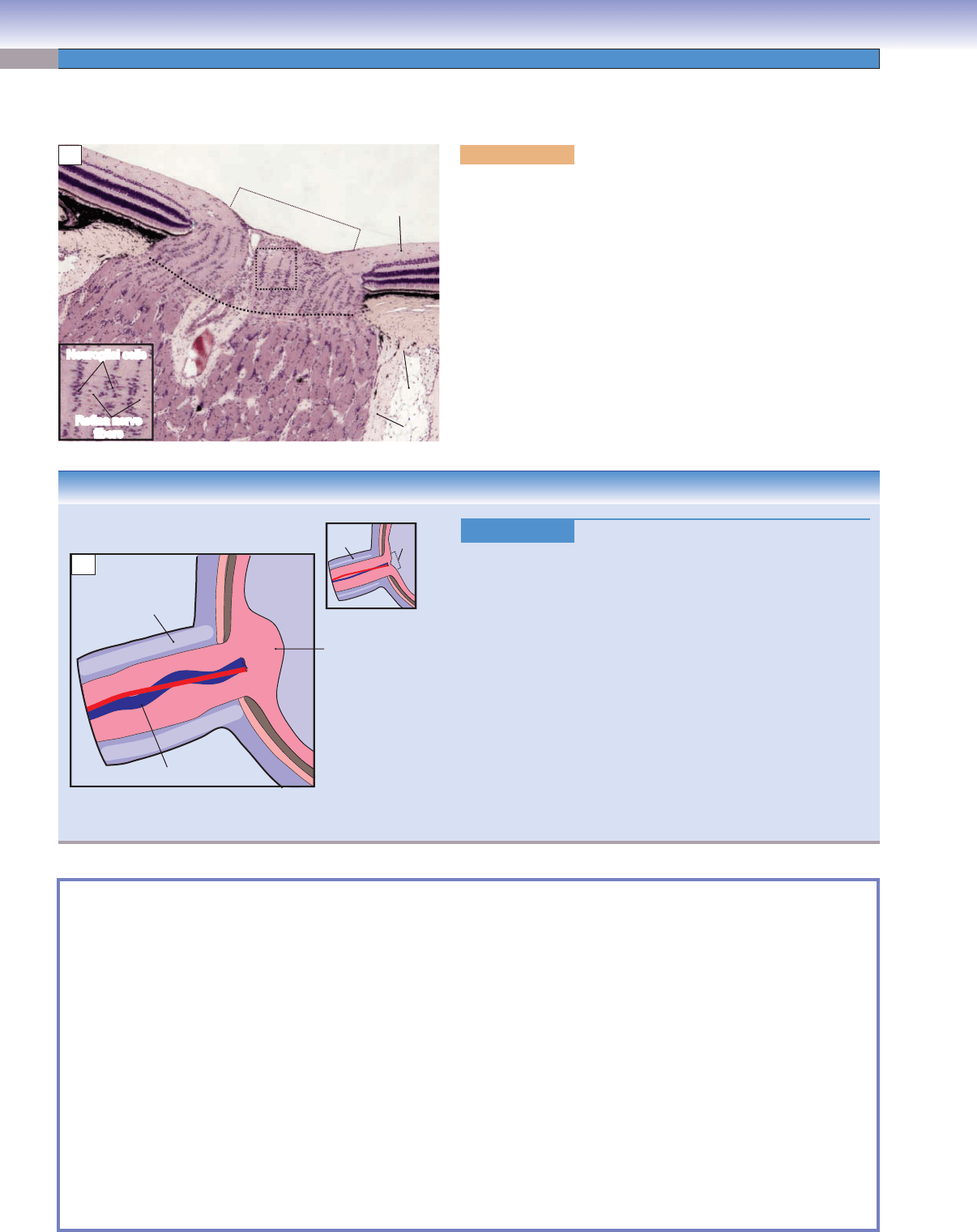
Figure 20-17A. Optic nerve and optic disk. H&E, 22; inset, 83
The optic nerve is a nerve fi ber trunk formed by the convergence of
retinal ganglion cell axons at the posterior pole of the eye. From there,
they leave the globe on their way to the brain. Each optic nerve con-
tains about one million myelinated axons and even more neuroglial
cells. The surface of the optic nerve is covered by pia mater, which is
continuous with that on the surface of the brain. The optic disk (optic
nerve papilla) is the small circular site in the retina where the retinal
nerve fi ber layer (nonmyelinated nerve fi bers) continues into the optic
nerve. The nonmyelinated nerve fi bers begin to acquire myelin at the
level of the lamina cribrosa (thin dotted line), a perforated, sievelike
region of the sclera through which optic nerve fi bers and blood ves-
sels pass. The myelinated segments of ganglion cell axons, therefore,
form the optic nerve. The neuroglial cells include oligodendrocytes,
which produce myelin for axons in the CNS, and astrocytes, which
perform several nutritive and supportive functions.
CLINICAL CORRELATION
Figure 20-17B.
Papilledema.
Papilledema is a noninfl
ammatory swelling of the optic disk. It is
produced by increased intracranial pressure that is transmitted
along the optic nerve sheath as elevated cerebrospinal fl uid pres-
sure, and it is, therefore, a sign of a potentially life-threatening
condition. The increased pressure disrupts blood circulation
within the optic nerve, causing leakage of water, protein, and
other contents into the extracellular spaces of the optic disk. The
most noticeable symptoms of increased intracranial pressure are
headache and vomiting. Clinical fi ndings include a swollen and
elevated optic disk (papilledema), engorged and tortuous retinal
veins, and focal hemorrhages. Cerebral tumors, subdural hema-
toma, malignant hypertension, and hydrocephalus are the most
common causes of increased intracranial pressure and papille-
dema. Because papilledema is a sign of many systemic intracra-
nial and spinal diseases, the correct diagnosis of the underlying
disease is essential for proper treatment.
D. Cui
Swollen and elevated
optic disk
Normal
CSF
Optic
disk
Elevated
CSF
pressure
Engorged and
tortuous central vein
B
Blepharitis ■ : Infl ammation of the eyelids and especially of their margins (Fig. 20-3C).
Ptosis
■ : Drooping or falling down, usually of the upper eyelid, because of muscle weakness or paralysis (Fig. 20-4C).
Open angle (glaucoma)
■ : Raised intraocular pressure in which obstruction to aqueous fl ow occurs at the ultrastructural
level, within the walls of the tiniest interstices of the trabecular meshwork (Fig. 20-12C).
Closed angle (glaucoma)
■ : The fl ow of aqueous fl uid is blocked by the root of the iris. This can be a generalized closure
(hyperopic eyes) or in multiple focal adhesions caused by infl ammation (peripheral anterior synechiae [Fig. 20-12C]).
Cupping of the optic disk
■ : Glaucoma destroys retinal axons as they traverse the optic disk in the walls of a central, cup-
shaped conduit. As the walls disappear, the cup enlarges (Fig. 20-12C).
Dry macular degeneration
■ : Scattered drusen damage the pigment epithelium of the macula, gradually decreasing visual
acuity (Fig. 20-16A).
Wet macular degeneration
■ : Abnormal capillaries break through the retinal pigment epithelium and leak underneath the
retina. When close to the fovea, they can decrease visual acuity (Fig. 20-16A).
Limbu
■ s: An anatomic transition zone between cornea and sclera (corneoscleral junction), which is an important landmark
for eye surgery procedures (Fig. 20-11A).
Ora serrata
■ : A denticulate border (junction) between the ciliary body and the retina; this is an important anatomic landmark
for the ophthalmologist (Fig. 20-12B).
SYNOPSIS 20-2 Clinical Terms for the Eye
CUI_Chap20.indd 408 6/16/2010 7:38:29 PM
409
21
Ear
Introduction and Key Concepts for the Ear
Figure 21-1A Overview of the Ear: Outer, Middle, and Inner Ear
Figure 21-1B Middle Ear Structures
Figure 21-2 Major Structures of the Inner Ear
Figure 21-3A Membranous Labyrinth
Figure 21-3B Cochlear Duct and Modiolus
Auditory System
Figure 21-4A Cross section of Cochlea
Figure 21-4B Scala Media and Organ of Corti
Figure 21-5 Organ of Corti and Associated Structures
Figure 21-6A Sound Transduction
Figure 21-6B Stereocilia Displacement
Figure 21-6C Auditory Hair Cells
Figure 21-7A Inner and Outer Hair Cells
Figure 21-7B Stereocilia of an Outer Hair Cell
Figure 21-7C Clinical Correlation: Sensorineural Hearing Loss
Vestibular System
Figure 21-8A Sensory Receptors
Figure 21-8B Crista Ampullaris and Macula Utriculi
Figure 21-9A Ampulla of the Semicircular Canal
Figure 21-9B Crista Ampullaris
Figure 21-9C Clinical Correlation: Ménière Disease
Figure 21-10A Macula of the Utricle
Figure 21-10B Macula of the Utricle with Otoconia
Figure 21-10C Otoconia from the Macula of the Utricle
Figure 21-11A Vestibular Hair Cells
Figure 21-11B Excitation and Inhibition in Hair Cells
CUI_Chap21.indd 409 6/2/2010 8:19:56 PM

410
UNIT 3
■
Organ Systems
Figure 21-11C Clinical Correlation: Otitis Media
Figure 21-12A Clinical Correlation: Vestibular Schwannoma
Figure 21-12B Clinical Correlation: CT and MRI of Inner Ear Structures
Synopsis 21-1 Pathological and Clinical Terms for the Ear
Introduction and Key Concepts
for the Ear
The ear is a complex structure that serves two important sen-
sory functions, hearing (through the auditory system) and bal-
ance (through the vestibular system). Sensory receptor organs
that serve the two functions are supplied by two distinct
branches of cranial nerve (CN) VIII, the acoustic branch and
the vestibular branch. The ear can be divided into three general
regions, the outer ear, middle ear, and inner ear (Fig. 21-1A).
(1) The outer ear consists of a pinna (auricle), an irregularly
shaped structure with a core of cartilage covered on both sides
by thin skin, and an external auditory meatus that conducts
sound to the middle ear. (2) The middle ear includes the tym-
panic membrane, the tympanic cavity containing the ossicles,
and the auditory tube (Fig. 21-1A,B). The tympanic membrane,
the landmark between the outer ear and middle ear, covers the
medial end of the external auditory meatus and converts sound
waves in the air to mechanical vibrations. The tympanic cavity
is an air-fi lled space that contains the ossicles, three tiny bones
that conduct the mechanical vibrations of the tympanic mem-
brane to the oval window of the cochlea. The tympanic cavity is
connected to the nasopharynx by the auditory tube (eustachian
tube), thereby allowing the air pressure on each side of the tym-
panic membrane to be equalized when the ambient air pressure
changes (e.g., by changes in altitude). (3) The inner ear consists
of structures contained within the bony labyrinth, a system of
tunnels and cavities in the petrous portion of the temporal bone,
the hardest bone in the body (Figs. 21-2 and 21-3A). The struc-
tures include the cochlear labyrinth or cochlea (Latin for “snail
shell”), which subserves hearing. It contains a spiral, fl uid-fi lled
tunnel within which a membranous tube, the cochlear duct, is
suspended (Fig. 21-3A,B). The sensory receptors that detect
sound are located in a strip of specialized epithelium, the organ
of Corti (spiral organ), in the cochlear duct. The vestibular lab-
yrinth consists of a complex group of fl uid-fi lled tunnels and
cavities in the temporal bone that contain a group of intercon-
nected membranous structures, the semicircular ducts, utricle
,
and saccule (Figs. 21-2 and 21-3A). The sensory receptors that
are involved in balance are located in specialized regions of
the semicircular ducts (rotation) and in the utricle and saccule
(static head position and acceleration).
Auditory System
Sound is normally produced by compression and rarefaction
waves in air, of various frequencies, that impinge upon the
tympanic membrane where they are converted into mechani-
cal vibrations in the ossicles. The mechanical vibrations, in
turn, are transferred to the fl uid of the vestibule at the oval
window (Fig. 21-2). This fl uid is perilymph, a sodium-rich fl uid
that is similar in composition to cerebrospinal fl uid and extra-
cellular fl uid. The resulting vibrations, or pressure waves, in
the perilymph spread into the scala vestibuli of the cochlear
labyrinth and act upon mechanoreceptors in the cochlear duct
to produce the sensation of hearing (Fig. 21-6A). The cochlear
duct is a membranous tube, triangular in cross section, that
coils inside the spiral tunnel in the cochlea (Fig. 21-3A,B). It is
suspended within the cochlear labyrinth so that it divides the
labyrinth into two tunnels, the scala vestibuli (above) and the
scala tympani (below), which are connected with each other by
a small opening, the helicotrema (Figs. 21-4A,B and 21-6A).
The cochlear duct encloses the scala media, a space contain-
ing endolymph, a potassium-rich fl uid similar in composition
to intracellular fl uid. The scala media is bounded above by the
vestibular membrane, below by the basilar membrane, and
externally by the spiral ligament and stria vascularis (Fig. 21-5).
The sensory receptors for hearing are specialized epithelial cells
(hair cells) in the organ of Corti. The hair cells get their name
from clumps of stereocilia that project from their apical sur-
faces and contact an overlying gelatinous structure, the tectorial
membrane. The organ of Corti sits on the basilar membrane
and extends its entire length, from the base of the cochlea to
the apex. It contains a single row of inner hair cells and three or
four rows of outer hair cells (Figs. 21-5 and 21-7A). In humans,
there are about 3,500 inner hair cells and 12,000 outer hair
cells, yet 95% of the afferent axons in the auditory nerve con-
tact only inner hair cells. The primary function of the inner hair
cells appears to be basic frequency and loudness discrimination,
whereas the outer hair cells appear to be primarily concerned
with the fi ne-tuning of frequency discrimination in the cochlea
(Figs. 21-6C and 21-7A,B). When sound waves cause pressure
waves to occur in the scala vestibuli and scala media, the basilar
membrane vibrates up and down, and a shearing force is gener-
ated between the surface of the organ of Corti and the tectorial
membrane. The shearing force bends the stereocilia of the hair
cells, leading to the release of neurotransmitters by the hair cells
and the initiation of action potentials in auditory nerve axons
(Fig. 21-6A,B).
Vestibular System
The sense of balance is critical to our ability to walk, run, jump,
or even just stand still with eyes closed. One important source
of neural signals that aid in controlling such behaviors is the
peripheral vestibular apparatus. This includes the vestibular lab-
yrinth, consisting of the vestibule, a cavity within the temporal
bone, and three semicircular canals, curved tunnels that connect
with the vestibule (Fig. 21-2). One canal is in approximately the
horizontal plane; the other two are in approximately the ver-
tical plane and at right angles to each other. These cavities in
the bone are fi lled with perilymph. Floating within the vestibule
are two membranous saclike structures, the utricle and saccule.
Within each of the semicircular canals is a membranous tube
called a semicircular duct, which joins the utricle at each of its
ends (Figs. 21-2 and 21-3A). The utricle, saccule, and semicir-
cular ducts contain endolymph. The semicircular ducts detect
CUI_Chap21.indd 410 6/2/2010 8:20:03 PM

CHAPTER 21
■
Ear
411
rotational movements of the head. Each duct has an enlargement
at one end where it joins the utricle. This swelling is called the
ampulla and contains the sensory receptors that are stimulated
by rotational movements (Fig. 21-9A). A short wall of connec-
tive tissue, the crista ampullaris, extends across part of each
ampulla. Hair cells, similar to those of the organ of Corti, cover
the upper surface of the crista. Their stereocilia and kinocilia
are embedded in a gel-like structure, the cupula, which blocks
the ampulla. When the head turns, the inertia of the endolymph
in the semicircular ducts causes the fl uid to push against the
cupula and defl ect the cilia of the hair cells, thereby initiating
action potentials in axons in the vestibular nerve (Figs. 21-8B
and 21-9A,B). Vestibular hair cells are also clustered in a small
region of the utricle, the macula utriculi (Figs. 21-8B and
21-10A,B). The stereocilia and kinocilia of these hair cells are
embedded in a gelatinous structure, the otolithic membrane (Fig.
21-10A). Thousands of tiny calcium carbonate crystals, otoco-
nia, are clustered on the surface of the otolithic membrane (Fig.
21-10A–C). These crystals are heavier than the surrounding
endolymph. Gravity or linear acceleration, therefore, exerts a
force on the otoconia, causing the underlying cilia to be defl ected
and consequently sending a neural signal to the central nervous
system (CNS) related to head position or acceleration. The sac-
cule contains a similar region, the macula sacculi.
CUI_Chap21.indd 411 6/2/2010 8:20:03 PM
412
UNIT 3
■
Organ Systems
A
B
Tympanic membrane
Stapes
(in oval
window)
Round
window
Tympanic
cavity
Stapes
Stapedius
muscle
Tympanic
membrane
Auditory tube
Cochlea
External auditory meatus
Semicircular canals
Temporal bone
Malleus
Malleus
Anterior malleal
ligament
Posterior incudal
ligament
Vestibular nerve
Facial nerve
Cochlear nerve
Vestibule
Incus
Cartilage
Incus
Pinna
Outer
ear
Middle
ear
Inner
ear
Insertion
of tensor
tympani
muscle
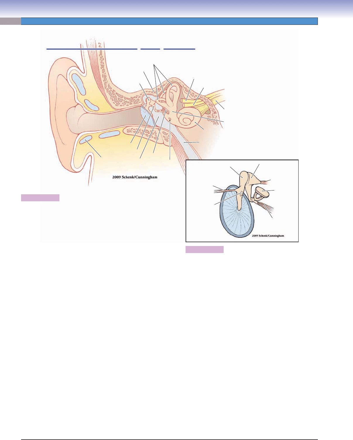
Figure 21-1A. Overview of the ear: Outer, middle, and inner ear.
The ear is divided into three regions: the outer ear, middle ear, and
inner ear. The outer ear includes the pinna and the external auditory
meatus. The pinna (or auricle) is an irregularly shaped structure of elas-
tic cartilage covered by a layer of perichondrium (connective tissue)
and thin skin. One important function of the pinna is to selectively
fi lter higher frequency sound waves and, therefore, aid in spatial local-
ization of sounds in the environment. The external auditory meatus is
a tunnel that carries sound waves to the tympanic membrane, where
the compressions and rarefactions of air are converted into mechanical
vibrations. The outer third of the meatus is lined with a continuation of
the cartilage of the pinna and a continuation of the perichondrium and
thin skin that covers the pinna. In the inner two thirds of the meatus,
the skin adheres directly to the periosteum of the temporal bone. The
middle ear is an air-fi lled cavity (tympanic cavity) that is separated from
the outer ear by the tympanic membrane. It contains three tiny bones,
the ossicles, which are the smallest bones in the body. These ossicles
transfer sound-induced movement of the tympanic membrane to the
fl uid contained in the cochlea, where the vibrations are transduced into
nerve impulses. The middle ear is connected to the posterior region of
the nasopharynx by the auditory tube (eustachian tube), which allows
the equalization of air pressure on each side of the tympanic mem-
brane. The inner ear contains the cochlea and the vestibular apparatus.
The cochlea, the snail shell–shaped organ of hearing, includes a mem-
branous tube lying within a fl uid-fi lled tunnel in the temporal bone (see
Fig. 21-2). Auditory hair cells in the cochlea are excited by vibratory
movements of the fl uid and generate action potentials in auditory nerve
fi bers. The vestibular apparatus is the body’s sensory organ for bal-
ance and consists of membranous structures contained within the three
semicircular canals and the vestibule as well as some accessory struc-
tures (see Fig. 21-2). Vestibular hair cells within the semicircular ducts
(inside the semicircular canals) detect rotational movement of the head
in three dimensions. Vestibular hair cells within the utricle and saccule
(inside the vestibule) detect static head position and linear acceleration
in the horizontal and vertical planes, respectively. The auditory and
vestibular branches of CN VIII innervate these structures.
Figure 21-1B. Middle ear structures.
The tympanic membrane (viewed here from its medial
side) is a thin, semitransparent cone-shaped sheet of col-
lagenous fi bers and fi broblasts, covered on the outer side
by a very thin layer of skin and on the inner side by the
mucosa that lines the rest of the tympanic cavity. The fi rst
of the ossicles, the malleus (hammer), is attached to the
upper half of the tympanic membrane, the incus (anvil) is
attached to the malleus by a saddle-shaped synovial joint,
and the stapes (stirrup) is attached to the incus by a ball-
and-socket synovial joint. The footplate of the stapes is
attached to the oval window of the vestibule. When sound
waves cause the tympanic membrane to vibrate, the chain
of ossicles rotates about the anterior malleal and posterior
incudal ligaments, causing the footplate of the stapes to
rock on the oval window and therefore producing waves of
compression in the fl uid that fi lls the bony labyrinth. This
lever arrangement increases the pressure on the oval win-
dow approximately 20-fold compared to the air pressure
on the tympanic membrane. The tensor tympani muscle
contracts during very loud sounds, reducing the movement
of the ossicles and the oval window.
CUI_Chap21.indd 412 6/2/2010 8:20:03 PM
CHAPTER 21
■
Ear
413
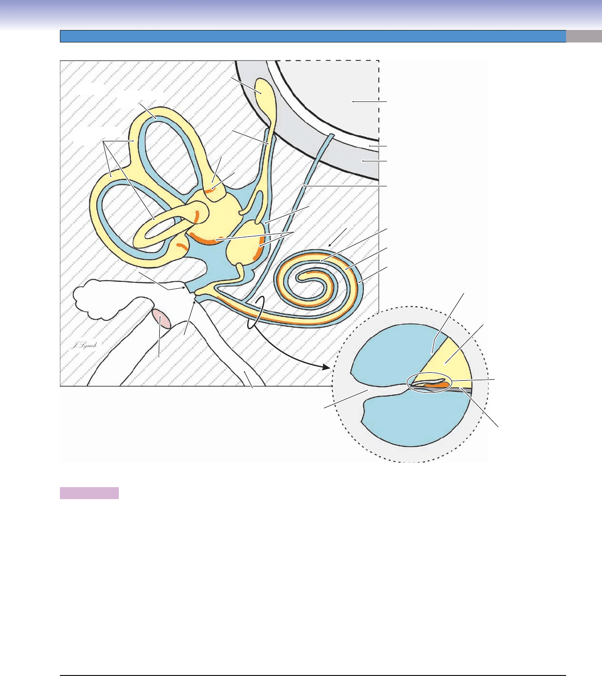
Figure 21-2. Major structures of the inner ear.
The bony labyrinth is a complex cavity in the petrous portion of the temporal bone, the hardest bone in the body. The cavity is lined
with endosteum and contains perilymph (blue shading), a clear fl uid with a composition very similar to that of cerebrospinal fl uid.
The bony labyrinth is divided into three portions, the vestibule, the cochlea (anteriorly), and the semicircular canals (posteriorly).
The membranous labyrinth, consisting of the three semicircular ducts, the utricle, and the saccule, lies within the bony labyrinth.
The walls of the membranous labyrinth are, in general, composed of a thin layer of fi brous connective tissue; a thin layer of
more delicate, more vascularized connective tissue; and an internal lining of simple epithelium. Two saclike structures, the utricle and
saccule, are found in the vestibule. A semicircular duct lies within each of the semicircular canals. A similar but more complicated
structure, the cochlear duct, lies within the bony cochlea. These membranous structures are continuous with each other and contain
endolymph (yellow shading), a fl uid with a high concentration of K
+
(potassium) ions that is unique in the body. Specialized regions
within the membranous labyrinth (indicated by thick orange lines) contain receptor cells innervated by branches of the vestibuloco-
chlear nerve (CN VIII). These include the cristae (singular, crista, from Latin for “crest”) in each of the ampullae (singular, ampulla,
from Latin for “fl ask” or “bottle”) of the semicircular ducts, the maculae (singular, macula, from Latin for “spot”) of the utricle and
saccule, and the organ of Corti (spiral organ) in the cochlear duct. The receptor cells in all these regions are specialized epithelial cells
(hair cells) that transduce movements of the basilar membrane into nerve impulses (see Figs. 21-5C, 21-7, and 21-11A).
Maculae
Maculae
Crista
Crista
Semicircular
Semicircular
canal
canal
Semicircular
Semicircular
ducts
ducts
Endolymphatic sac
Endolymphatic sac
Endo-
Endo-
lymphatic
lymphatic
duct
duct
Ampulla
Ampulla
Vestibule
Vestibule
Cochlea
Cochlea
Round
Round
window
window
Tympanic
Tympanic
membrane
membrane
Oval window
Oval window
Bone
Utricle
Ampulla
Crista
Saccule
Vestibule
Maculae
Cochlea
Mastoid
cavities
Oval window
Semicircular
ducts
Semicircular
canal
Endolymphatic sac
Endo-
lymphatic
duct
Perilymphatic duct
(cochlear duct)
Dura mater
Scala vestibuli
Cochlear duct
(scala media inside)
Scala tympani
Subarachnoid space
Brain tissue
Tympanic
membrane
Round
window
Auditory
(Eustachian)
tube
Osseous
spiral
lamina
External auditory
meatus
Scala vestibuli
(perilymph)
Bone
Scala tympani
(perilymph)
Scala media
(endolymph)
Vestibular
(Reissner)
membrane
Organ of Corti
(spiral organ)
Basilar
membrane
Cross section of cochlea
CUI_Chap21.indd 413 6/2/2010 8:20:05 PM
414
UNIT 3
■
Organ Systems
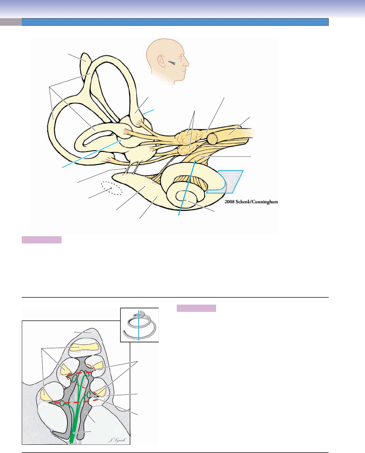
Figure 21-3A. Membranous labyrinth.
The endolymph-fi lled membranous labyrinth fl oats within the perilymph-fi lled osseous (bony) labyrinth (Fig. 21-2). This anatomically
correct drawing shows the position of the cochlea and semicircular canals within the head. The labyrinth is viewed from the direc-
tion indicated by the pointer in the small inset of the head. The position of the nerves that innervate the cochlea, cristae of the ampul-
lae, and maculae of the utricle and saccule are illustrated. The positions and planes of section of the photomicrographs that follow in
this chapter are indicated by blue lines, arrows, or boxes. The dashed oval indicates the position of the oval window in the osseous
labyrinth (Fig. 21-2).
Saccule
Facial nerve
(CN VII)
Cochlear nerve
(CN VIII)
Fig. 21-4B
Fig. 21-6
Fig. 21-9B
Ampulla
Fig. 21-3B
Fig. 21-4A
Fig. 21-8B
Fig. 21-10B,C
Vestibular nerve
(CN VIII)
Vestibular
ganglia
(of Scarpa)
Utricle
Endolymphatic sac
Semicircular
ducts
Cochlear
duct
Apex
of cochlea
Base
of cochlea
Position of
oval window
in osseous
labyrinth
Ductus reuniens
A
Cochlear
duct
Apex
of cochlea
Modiolus
Base
of cochlea
Bone
Cochlear
nerve
Osseous
spiral lamina
Spiral
ganglion
Spiral
ligament
Scala
tympani
B
Figure 21-3B. Cochlear duct and modiolus.
The membranous cochlear duct lies within a spiral-shaped tunnel
in the temporal bone. In this illustration, the cochlea has been cut
in half along the plane indicated by the single blue line in Figure
21-3A and in the inset. The spiral of the cochlear duct makes
about two and one-half turns from the base of the cochlea to its
apex. The outer bony surface of the cochlea is indicated in light
gray. A spiral, screw-shaped bony structure, the modiolus forms
the central core of the cochlea and is indicated in darker gray. A
spiral cavity within the modiolus contains the cell bodies of the spi-
ral ganglion (green) and the proximal axons of the cochlear nerve
(CN VIII). The osseous (bony) spiral lamina curves around the
modiolus like the threads of a screw (red dashed line and inset).
The central edge of the cochlear duct is attached to the spiral
lamina; the outer wall of the cochlear duct is attached to the spiral
ligament. Histological sections of these structures are shown in
Figures 21-4A and 21-4B.
CUI_Chap21.indd 414 6/2/2010 8:20:06 PM
