Cui Dongmei. Atlas of Histology: with functional and clinical correlations. 1st ed
Подождите немного. Документ загружается.

CHAPTER 19
■
Female Reproductive System
385
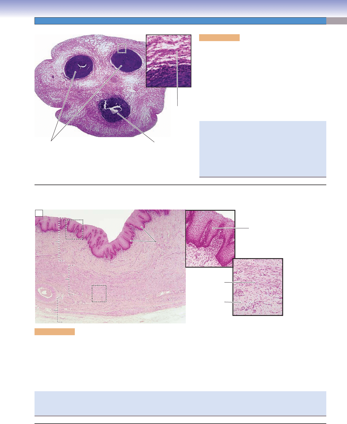
Figure 19-14A. Umbilical cord. H&E, 12; inset 79
The umbilical cord is a ropelike structure that connects the
developing fetus to the placenta. It contains two umbilical
arteries and one umbilical vein. These vessels carry oxygen
and nutrients from the mother to the fetus and waste prod-
ucts away from the fetus. The blood vessels are surrounded
by a mucous connective tissue (Wharton jelly). The umbilical
arteries carry deoxygenated fetal blood to the placenta by
way of the chorionic arteries and the chorionic villi. After
gas and nutrient exchange with the maternal blood, the
oxygenated blood is transported from chorionic veins to
the umbilical vein, which returns blood to the fetus.
A
Umbilical
arteries
Umbilical
vein
Mucous
connective tissue
(Wharton jelly)
Mucosa
Mucosa
Mucosa
Muscularis
Muscularis
Muscularis
Adventitia
Adventitia
Adventitia
Epithelium
Epithelium
Epithelium
Lumen
Lumen
Lumen
Lamina
Lamina
propria
propria
Lamina
propria
Ridges of the
Ridges of the
epithelium
epithelium
Ridges of the
epithelium
Lamina propria
Lamina propria
Lamina propria
Smooth muscle
Connective
tissue
Epithelium
B
Figure 19-14B. Vagina. H&E, 41; inset (upper) 63; inset (lower) 74
The vagina is a tubular organ that connects the cervix of the uterus to the external genitalia. The wall of the vagina consists of the mucosa,
muscularis, and adventitia. The mucosa comprises a nonkeratinized stratifi ed squamous epithelium and an underlying lamina propria
(dense irregular connective tissue with many elastic fi bers). The muscularis contains mainly longitudinal smooth muscle and some oblique
smooth muscle bundles. The adventitia layer is composed of both dense connective tissue (near the muscularis) and loose connective tissue
(outer layer). The vagina is moistened by cervical secretions, and it has many sensory nerve endings in the inferior part near the entrance. The
epithelium of the vagina undergoes minimal change during the menstrual cycle. There are numerous elastic fi bers in the connective tissue,
and ridges (folds) in the mucosa, enabling the vaginal canal to expand during sexual intercourse and during the delivery of a baby.
Funisitis is infl ammation of the umbilical cord that often
accompanies chorioamnionitis (infl ammation of the fetal
membranes). Funisitis typically occurs after 20 weeks of
gestation, often because of a bacterial infection. Neutro-
phils migrate through the umbilical vessels and may enter
the Wharton jelly. Another possible complication in preg-
nancy is an umbilical knot. In severe cases, obstruction of
blood supply can result in the fetal death.
The Papanicolaou (Pap) smear is a very important diagnostic method used for screening early signs of cervical cancer. Cells from the
epithelial surface of the vagina and cervix are collected by using a brush and spatula while the vagina is opened by a speculum. Exami-
nation of these sample cells provides valuable information for detecting precancerous changes that may require treatment.
Vagina
CUI_Chap19.indd 385 6/19/2010 12:20:56 PM
386
UNIT 3
■
Organ Systems
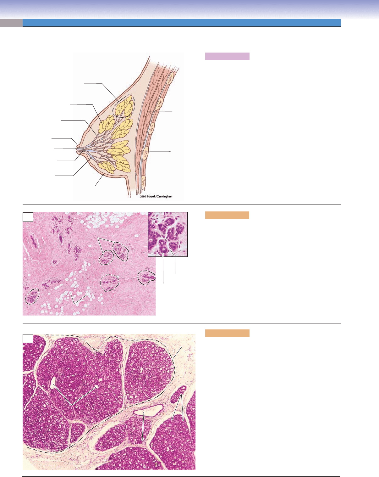
Figure 19-15A. Overview of the mammary gland.
In humans, there are two multilobed mammary glands,
one located within the connective tissue of each breast.
These exocrine glands produce milk after a pregnancy.
Each gland is composed of 15 to 25 lobes of compound
tubuloalveolar glands. Each lobe is separated from others
by dense connective tissue and adipose tissue and opens
into a lactiferous duct. The secretory alveoli produce
milk and drain it into the intralobular ducts and then to
the interlobular ducts. The interlobular ducts merge into
lactiferous sinuses from which the milk empties into the
lactiferous ducts (15–25). The female mammary glands
begin to enlarge during puberty and undergo changes at
different times based on hormone (estrogen, progester-
one, prolactin, and human placental lactogen) levels.
Mammary Glands
Lobule of
mammary gland
Interlobular
duct
Lactiferous
duct
Lactiferous
sinus
Nipple
Openings
Lobe of
mammary
gland
Pectoralis
muscles
Rib
Skin
A
Cuboidal
epithelial cell
Myoepithelial
cell
Lobules of
Lobules of
gland
gland
Lobules of
gland
Adipose tissue
Adipose tissue
Adipose tissue
Adipocytes
Adipocytes
Adipocytes
Dense irregular
Dense irregular
connective
connective
tissue
tissue
Dense irregular
connective
tissue
B
Intralobular
Intralobular
ducts
ducts
Intralobular
ducts
Alveolus
Alveolus
Lobule of
Lobule of
gland
gland
Lobule of
gland
Alveoli
Alveoli
Alveoli
Interlobular
Interlobular
duct
duct
Interlobular
duct
Connective tissue
Connective tissue
Connective tissue
C
Figure 19-15B. Inactive (resting) mammary gland.
H&E, 41; inset 359
An example of a resting mammary gland shows a large
amount of dense irregular connective tissue and adipose
tissue with small mammary gland lobules. The glandu-
lar tissue contains mainly intralobular ducts, which are
lined by cuboidal epithelial cells and underlying myo-
epithelial cells (inset). The resting mammary gland has
only a few secretory alveoli, some undeveloped intral-
obular ducts, interlobular ducts, lactiferous sinuses, and
lactiferous ducts.
Figure 19-15C. Active (during pregnancy) mammary
gland. H&E, 41
An example of a mammary gland during pregnancy
shows large lobules and a relatively small amount of
interlobular connective tissue. The glandular tissue con-
tains many proliferated alveoli and intralobular ducts.
A large interlobular duct is located within the connective
tissue shown here. When the mammary glands begin to
secrete milk (lactation), the lumina of the alveoli and the
ducts are dilated and fi lled with milk. The milk contains
many lipid droplets and proteins (caseins, lactalbumin,
and immunoglobulin A) as well as lactose, ions, vita-
mins, and water. Secretion of milk is initiated by hor-
monal changes: decrease of estrogen and progesterone
and increase of prolactin after delivery and the loss of
the placenta. The milk is released by the milk ejection
refl ex when stimulated by suckling (Fig. 19-16A).
CUI_Chap19.indd 386 6/19/2010 12:21:01 PM
CHAPTER 19
■
Female Reproductive System
387
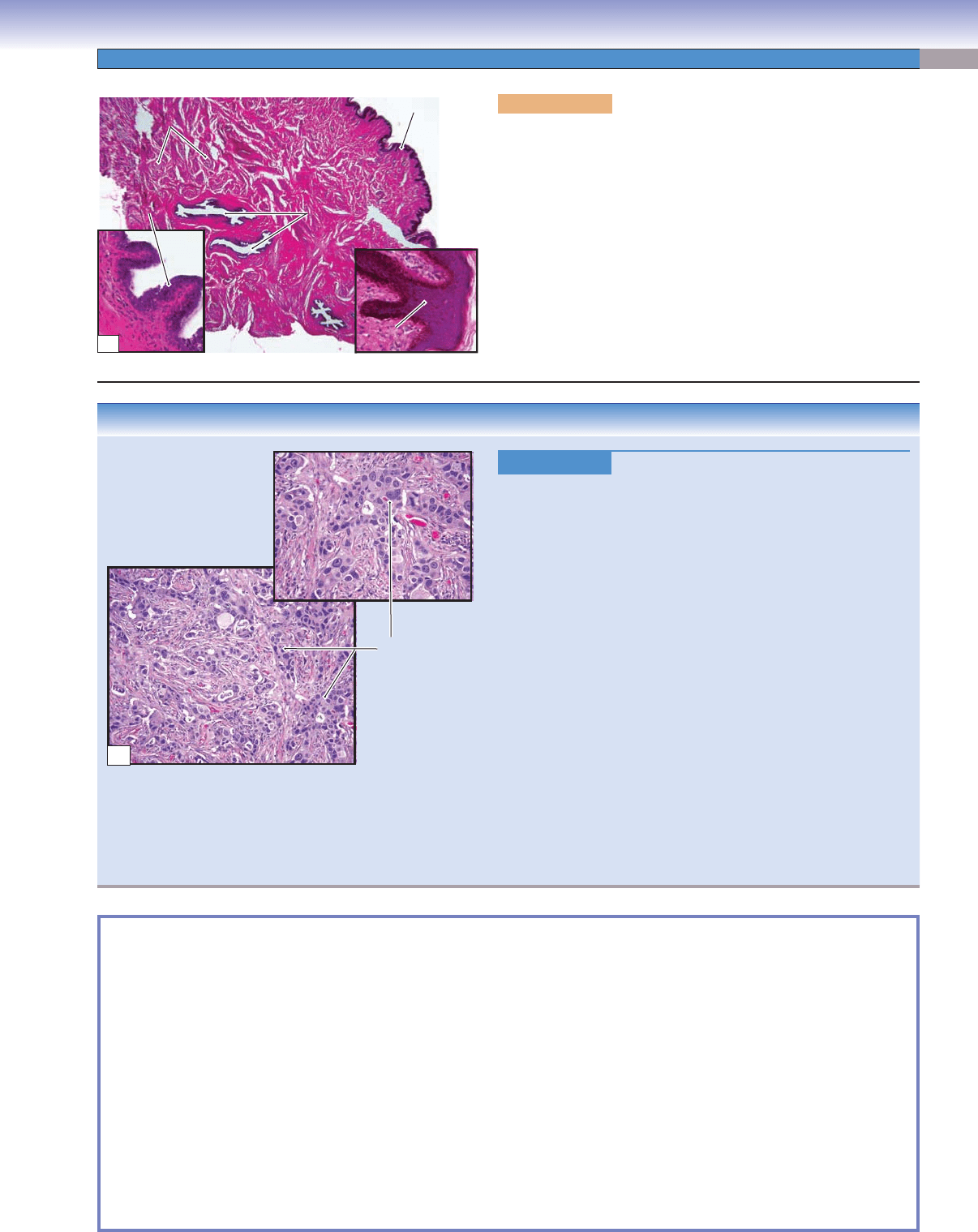
Figure 19-16B.
Adenocarcinoma of the Breast (Breast Cancer).
H&E, left (lower) 44; right (upper) 71
Infi
ltrating duct carcinoma, or invasive ductal carcinoma, is the
most common adenocarcinoma of the breast (breast cancer); it
contains no features to further classify it into special types of breast
carcinoma, such as lobular, tubular, and mucinous carcinomas.
Risk factors for the development of breast cancer include female
gender, increasing age, family history, long reproductive life, nul-
liparity, and the presence of proliferative breast lesions or ductal
hyperplasia. Approximately 5% of breast cancers are related to
specifi c gene mutations, including BRCA1 and BRCA2. Common
signs and symptoms include a palpable breast mass, bloody dis-
charge from the nipple, change in size or shape of a breast, skin
dimpling, inverted nipple, peeling of the nipple skin, and redness or
pitting of the skin over the breast. Mammograms and breast exams
are used to screen for breast cancer. Biopsy is performed on suspi-
cious lesions to determine a tissue diagnosis. Histologically, breast
cancer varies from well-formed glandular structures to sheets of
poorly differentiated cells. Histologic grading of breast cancer is
based on tubule formation, nuclear pleomorphism, and the mitotic
rate. Treatment includes surgical removal of a tumor (lumpec-
tomy), removal of the entire breast (mastectomy) and lymph nodes,
radiation therapy, chemotherapy, and hormone therapy.
Figure 19-16A. Nipple, mammary gland. H&E, 11; left inset
146; right inset 136
The nipple is a small projection at the center of the breast. It
contains 15 to 25 openings of lactiferous ducts within its connec-
tive tissue and smooth muscle bundles. It is covered by thin skin and
surrounded by the areola (pigmented skin). The nipple has many
sensory nerve endings that receive stimulation during suckling. This
stimulation results in release of oxytocin from the pars nervosa of
the pituitary; the oxytocin stimulates contraction of the myoepithe-
lial cells in the mammary gland. The contraction of the myoepithelial
cells pushes milk out of the alveoli and ducts and through the lac-
tiferous ducts to the surface of the nipple. This process is called the
milk ejection refl ex. The lactiferous ducts shown here are from the
proximal portion of the ducts near the lactiferous sinuses.
Lactiferous
Lactiferous
ducts
ducts
Lactiferous
ducts
Skin
Skin
Skin
stratified squamous
stratified squamous
epithelium
epithelium
stratified squamous
epithelium
Smooth
Smooth
muscle bundles
muscle bundles
Smooth
muscle bundles
Epithelium of
Epithelium of
lactiferous duct
lactiferous duct
Epithelium of
lactiferous duct
Connective
Connective
tissue
tissue
Connective
tissue
A
Infiltrating duct
carcinoma of the
breast
B
CLINICAL CORRELATION
SYNOPSIS 19-1 Clinical and Pathological Terms for the Female Reproductive System
Endophytic ■ : Term to describe an inward-growing process such as a neoplasm that grows on the interior of an organ
(Fig. 19-12B).
Exophytic
■ : Term to describe an outward-growing process such as a neoplasm that grows externally on an organ or within
the lumen of an organ (Fig. 19-12B).
Hemoperitoneum
■ : Blood within the peritoneal cavity because of a variety of causes including trauma, rupture of a tumor,
or rupture of an ectopic pregnancy (Fig. 19-7C).
Menorragia
■ : Refers to excessive or prolonged uterine bleeding at regular intervals during menstruation; some causes
include uterine leiomyomas, anovulation and ovarian dysfunction, hormonal imbalance, bleeding tendency, and malig-
nancy (Fig. 19-11B).
Metrorragia
■ : Refers to uterine bleeding at irregular intervals often at times between the expected menstrual periods;
causes may be similar to menorrhagia and include hormonal imbalance, malignancy, uterine polyps, and bleeding tendency
(Fig. 19-11B).
Nulliparity
■ : Term used to describe never having given birth to a child; by contrast, a woman who has given birth to two
or more children is termed multiparous (Fig. 19-16B).
CUI_Chap19.indd 387 6/19/2010 12:21:05 PM
388
20
Eye
Introduction and Key Concepts for the Eye
Figure 20-1 Overview of the Eye
Figure 20-2 Orientation of Detailed Eye Illustrations
The Eyelids
Figure 20-3A Overview of the Upper Eyelid
Figure 20-3B A Representation of the Upper Eyelid, Glands, and Muscles that Control Eyelid
Movements
Figure 20-3C Clinical Correlation: Chalazion
Figure 20-4A Upper Eyelid (Lower Part), Glands of the Eyelid
Figure 20-4B Upper Eyelid (Middle Part)
Figure 20-4C Upper Eyelid (Upper Part), Muscular Control of Upper Eyelid Movement
Tunica Fibrosa (Tunica Externa)
Figure 20-5A,B Cornea
Figure 20-5C Clinical Correlation: LASEK
Figure 20-6A Pavement Epithelium (Anterior Epithelium)
Figure 20-6B Corneal Endothelium (Posterior Corneal Epithelium)
Refractive Media of the Eye
Figure 20-7A Overview of the Lens
Figure 20-7B The Anterior Region of the Lens
Figure 20-7C The Posterior Region of the Lens
Figure 20-7D The Equatorial Region of the Lens
Figure 20-8A Lens and Zonular Fibers
Figure 20-8B A Representation of the Lens and its Function
Figure 20-8C Clinical Correlation: Cataract
CUI_Chap20.indd 388 6/16/2010 7:37:48 PM

CHAPTER 20
■
Eye
389
Tunica Vasculosa (Tunica Media)
Figure 20-9A Overview of the Iris and Nearby Structures
Figure 20-9B Iris
Figure 20-9C Constrictor Pupillae Muscle and its Function
Figure 20-9D Dilator Pupillae Muscle and its Function
Figure 20-10A Anterior Surface of the Lens
Figure 20-10B Posterior Surface of the Iris
Figure 20-11A Overview of the Ciliary Body and Nearby Structures
Figure 20-11B Ciliary Processes and the Ciliary Muscle
Figure 20-11C Ciliary Body, Transverse and Posterior Views
Figure 20-12A Posterior Portion of the Ciliary Body
Figure 20-12B Ora Serrata
Figure 20-12C Clinical Correlation: Outfl ow of Aqueous Humor and Glaucoma
Retina (Tunica Interna)
Figure 20-13A Overview of the Retina; Fovea
Figure 20-13B Macular Region of the Retina and the Retinal Layers
Figure 20-13C Peripheral Region of the Retina and Distribution of the Rods and Cones
Figure 20-14A,B A Representation of Rods and Codes
Table 20-1 Comparison of Rods and Cones
Figure 20-15A–C Retinal Layers
Synopsis 20-1 Retinal Layers
Figure 20-16A Clinical Correlation: Age-Related Macular Degeneration
Figure 20-16B Clinical Correlation: Retinal Detachment
Optic Nerve
Figure 20-17A Optic Nerve and Optic Disk
Figure 20-17B Clinical Correlation: Papilledema
Synopsis 20-2 Clinical Terms for the Eye
Introduction and Key Concepts
for the Eye
The eye is the organ of vision, perhaps the most important of the
sensory modalities. The eye converts light into nerve impulses in
a way that allows the brain to be aware of the individual’s visual
surroundings. In our study, we divide the structure of the eye
into three general categories: those structures which protect the
eye (eyelids); those structures which help form a visual image of
what the individual is looking at (cornea, lens, sclera, and asso-
ciated structures); and those structures which convert the visual
image into nerve impulses and conduct the impulses to the brain
(retina and optic nerve), where they are analyzed to produce the
sensation of vision.
The basic structure of the eye is that of a hollow sphere
with optical elements on the anterior surface that focus an
inverted image of the surroundings onto the inside of the pos-
terior wall. The primary structural element of the sphere is
the tunica fi brosa, or tunica externa, which consists of the
sclera and the cornea. Lining the inside of the tunica externa
is the tunica vasculosa, consisting of the choroid, the ciliary
body, and the iris. The eye is fi lled with a transparent liquid
(aqueous humor) and a transparent gel (vitreous body). In the
anterior portion of the eye is the lens, which is fl exible and
can adjust the focus of the image depending on the distance
from the eye to the object being viewed. The image is focused
on the retina, a layer of neurons and neural receptors that line
the internal surface of the posterior two thirds of the eye. The
axons of the ganglion cell neurons in the retina leave the eye
and form the optic nerve.
The Eyelids
The eyelids protect the eyes from injury by foreign objects and
also maintain a thin fi lm of moisture on the surface of the cornea
that prevents the cornea from drying out and becoming opaque.
Each eyelid consists of an outer layer of skin; a middle layer of
muscle, glands, and connective tissues (tarsal plate); and an inner
layer of conjunctival tissue (palpebral conjunctiva). There are
several types of glands in the eyelid that aid in keeping the cor-
nea moist, including meibomian glands, glands of Zeis, glands
of Moll, and accessory lacrimal glands (Figs. 20-3 and 20-4).
Several muscles are associated with the eyelids. These
include (1) the orbicularis oculi muscle, a circular sheet of stri-
ated muscle that is innervated by the facial nerve (cranial nerve
CUI_Chap20.indd 389 6/16/2010 7:37:54 PM

390
UNIT 3
■
Organ Systems
[CN] VII) and functions to close the eyelids; (2) the levator palpe-
brae superioris muscle, a thin fl at striated muscle that originates
in the orbit, passes forward, and inserts into the upper eyelid. It
is innervated by the oculomotor nerve (CN III) and is responsible
for opening the eyelid and holding it open; and (3) the superior
tarsal muscle (Müller muscle), a bundle of smooth muscle that
arises from the interstitia of the levator muscle and inserts on
the upper end of the tarsal plate and superior conjunctiva of
the eyelid. The superior tarsal muscle is innervated by sympa-
thetic nerve fi bers from the superior cervical ganglion and helps
to raise the upper eyelid.
Tunica Fibrosa (Tunica Externa)
The outermost structures of the eye are the cornea and sclera
(Fig. 20-1).
THE CORNEA is a transparent tissue that covers the anterior
sixth of the eye (Figs. 20-5 and 20-6). The cornea contains no
blood vessels and aids in focusing the visual image onto the
retina. It consists of fi ve layers. The thickest layer, the stroma,
comprises 90% of the thickness of the cornea and consists
of collagen fi bers and fi broblasts embedded in an extracel-
lular matrix (Fig. 20-6B). The anterior surface of the cornea
is covered by a thin layer of pavement epithelium (stratifi ed
squamous epithelium) resting on the Bowman membrane. The
posterior surface of the cornea is covered by a layer of corneal
endothelium (simple squamous epithelium) that is only one cell
thick and rests on the Descemet membrane (Figs. 20-5A and
20-6B).
THE SCLERA is a tough, thin structure consisting of dense,
irregular, opaque connective tissue that comprises the posterior
fi ve sixths of the outer surface of the eyeball (Figs. 20-1, 20-11A,
and 20-12A). The cornea and sclera are continuous with each
other at the limbus (Fig. 20-11A). The extraocular muscles,
which move the eyes in their orbits, insert in the sclera. The con-
junctival tissue, which covers the inner surfaces of the eyelids,
also attaches to the sclera.
Refractive Media of the Eye
THE LENS is a transparent, fl exible, biconvex structure that
is suspended from the ciliary processes by zonular fi bers. The
curvature of the lens can be changed by contraction or relax-
ation of the ciliary muscles (under control of parasympathetic
nerve fi bers of the oculomotor nerve) so that the image of
nearby or distant objects can be focused on the retina. The
lens has three components: the lens capsule, the subcapsular
epithelium, and the lens fi bers (Figs. 20-7 and 20-8). The lens
capsule is a transparent basement membrane that surrounds
the entire lens. Immediately beneath it, on only the anterior
surface of the lens, is a single layer of squamous cells, the sub-
capsular epithelium (Fig. 20-10A). In the region of the equator
of the lens, proliferating epithelial cells become elongated, are
displaced toward the center of the lens, and lose their nuclei.
They are then called lens fi bers and comprise the major bulk of
the lens (Fig. 20-10A).
THE AQUEOUS HUMOR is a thin, watery, transparent fl uid
that is produced continuously by the ciliary body and fi lls the
anterior chamber. It exits the anterior chamber in the region
of the angle of the anterior chamber. It is produced by the
ciliary processes, which are rich in capillaries (Figs. 20-11C and
20-12C).
THE VITREOUS BODY is a transparent gelatinous substance
that fi lls the eye between the posterior surface of the lens and the
retina (Fig. 20-1). Its composition is predominantly water with
small amounts of collagen and hyaluronic acid. The surface of
the vitreous body is covered by a layer of condensed vitreous
fi bers called hyaloid membrane. It is in contact with the poste-
rior lens capsule, the zonular fi bers, the posterior portion of the
ciliary epithelium (pars plana), the retina, and the optic nerve
head (Fig. 20-1). The vitreous body is important in maintaining
the transparency and shape of the eye.
Tunica Vasculosa (Tunica Media)
The tunica vasculosa (sometimes called the uveal tract) lies just
internal to the tunica externa and consists of the iris (anteriorly),
the ciliary body, and the choroid (posteriorly).
THE IRIS is a thin diaphragm of tissue in the anterior cham-
ber, composed of a highly vascularized, loose connective tissue
stroma, two groups of contractile elements, the anterior iridal
border, and the posterior iridal border. The posterior iridal bor-
der contains two layers of pigmented epithelium, the anterior
iridal epithelium (anterior pigmented epithelium) and the pos-
terior iridial epithelium (posterior pigmented epithelium). Two
groups of muscle fi bers regulate the diameter of the pupil, the
circular hole in the center of the iris, and adjust the amount of
light entering the eye (Fig. 20-9). The circular constrictor pupil-
lae muscle (smooth muscle fi bers) reduces the size of the pupil
under the infl uence of parasympathetic nerve fi bers; the radial
fi bers of the dilator pupillae muscle (myoepithelial cells) act to
increase the size of the pupil under the infl uence of sympathetic
nerve fi bers.
THE CILIARY BODY lies interior to the anterior margin of
the sclera, between the choroid and the iris. It is composed of
two concentric rings of tissue, the pars plicata and the pars
plana, and includes epithelial tissue, a stroma of connective tis-
sue, and smooth muscle fi bers (Figs. 20-9A and 20-11). The
muscles of the ciliary body control the curvature of the lens
and, therefore, function to focus the visual image on the retina.
The epithelium of the ciliary body has two layers, a pigmented
layer and a nonpigmented layer. The latter secretes the aqueous
humor, which fi lls the anterior chamber of the eye and leaves
the anterior chamber in the region of the anterior chamber
angle (Fig. 20-12C).
THE CHOROID is a highly vascularized tissue containing
some collagen fi bers that is loosely attached to the overlying
sclera (Fig. 20-13A). The inner surface of the choroid adheres
tightly to the pigment epithelium layer of the retina. The inner-
most layer of the choroid is the choriocapillaris, which supplies
oxygen and nutrients to the outer layers of the retina. Bruch
membrane delineates the junction between the choriocapillaris
and the retinal pigment epithelium (Fig. 20-13B).
Retina (Tunica Interna)
The retina consists of a thin sheet of neurons that covers the
inner surface of the posterior two thirds of the eye and a layer
of cuboidal epithelial cells that sit on the choroid and contain
CUI_Chap20.indd 390 6/16/2010 7:37:54 PM

CHAPTER 20
■
Eye
391
melanin (retinal pigment epithelium). In general, the retina can be
divided into an optic (neural) retina and a non-optic (nonneural)
retina. The neural retina, often referred to as simply “retina,”
contains neural elements and has the visual functions described
below. The nonneural retina has no neural elements and no visual
function. It is the anterior continuation of the pigmented layer,
which covers the surface of the ciliary body and the posterior
surface of the iris.
Unlike the rest of the eye, the retina develops as a part of
the central nervous system (CNS). It contains fi ve types of neural
elements: photoreceptor cells (rods and cones), bipolar cells,
horizontal cells, amacrine cells, and ganglion cells (Fig. 20-15).
The retina can be divided into 10 layers, some of which contain
the nuclei of cells and others of which contain cell processes and
synapses (Figs. 20-13 and 20-15).
The retina is not homogeneous throughout its extent. Over
most of the retina, the focused light of the visual image must
pass through all of the neural layers, as well as small blood
vessels, before reaching the photoreceptors. This degrades the
image to a certain extent. However, in the fovea centralis, a small
region near the posterior pole of the eye, the superfi cial layers are
displaced to the side, and light strikes the photoreceptors directly
(Figs. 20-1 and 20-13A). The visual image in this region is per-
ceived with the greatest detail. The area immediately surrounding
the fovea centralis is the macula lutea (Fig. 20-13B). This is the
thickest region of the retina and contains a high concentration of
both cones (for color vision) and rods (for low-light vision [Fig.
20-14]). More peripherally, the retina becomes thinner. There are
fewer ganglion cells, fewer cones, and a relatively higher propor-
tion of rods (Fig. 20-13C). These changes cause visual acuity to
be reduced but sensitivity to low light levels to increase.
Optic Nerve
The ganglion cells are the output cells of the retina (Fig. 20-15C).
The axons of ganglion cells travel in the nerve fi ber layer to the
optic disk, where the nerve fi bers exit the eye and form the optic
nerve (Fig. 20-17A). There are no photoreceptors in the optic
disk; this absence produces a small blind spot in the visual fi eld.
The appearance of the optic disk when viewed through an oph-
thalmoscope is an important diagnostic aid.
CUI_Chap20.indd 391 6/16/2010 7:37:54 PM
392
UNIT 3
■
Organ Systems
I. Tunica Fibrosa (Tunica Externa)
A. Cornea
1. Pavement epithelium (anterior corneal epithelium)
2. Bowman membrane
3. Corneal stroma (corneal substantia propria)
4. Descemet membrane
5. Endothelium (posterior corneal epithelium)
B. Sclera
1. Episclera (blood vessels and adipose tissue)
2. Sclera proper
II. Tunica Vasculosa (Tunica Media)
A. Choroid
1. Bruch membrane
2. Choriocapillaris
3. Choroid propria
B. Iris
1. Pigment epithelium
2. Stroma
3. Constrictor pupillae muscle
4. Dilator pupillae muscle
C. Ciliary body
1. Ciliary processes
2. Ciliary epithelium
3. Ciliary muscle
III. Refractive Media of the Eye
A. Lens (biconvex, fl exible, transparent structure)
1. Lens capsule
2. Subcapsular epithelium
3. Lens fi bers
4. Zonular fi bers
B. Aqueous humor (transparent fl uid occupying the space
between the lens and cornea)
C. Vitreous body (refractive gel fi lling the interior of the
globe posterior to the lens)
IV. Retina (Tunica Interna)
A. Fovea
B. Macula
C. Peripheral retina
V. Optic nerve
Pupil
Aqueous
humor
Cerebrospinal
fluid
Central retinal
artery and vein
Optic nerve
Optic disk
(Optic nerve papilla)
Iris
Ciliary processes
Ciliary muscle
Ora serrata
Retina
Choroid
Choroid
Sclera
Sclera
Extraocular muscles
Retina
Ciliary body
Zonular fibers
Anterior chamber
Posterior chamber
Cornea
Macula
Fovea
Lens
Vitreous body
D. Cui
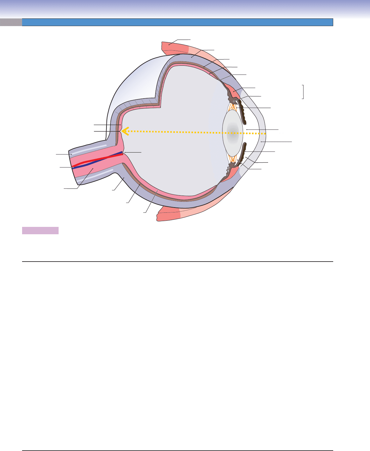
Figure 20-1. Overview of the eye.
The yellow dashed line indicates light coming in and projecting on the fovea.
Structures of the Eye
CUI_Chap20.indd 392 6/16/2010 7:37:54 PM
CHAPTER 20
■
Eye
393
D. Cui
Fig. 20-13A
Fig. 20-13C
Fig. 20-17A,B
Fig. 20-13B
Fig. 20-5A,B
Fig. 20-6A,B
Fig. 20-9B,C,D
Fig. 20-11A,B,C
Fig. 20-7B
Fig. 20-10A
Fig. 20-7C
Fig. 20-7D
Fig. 20-8A
Fig. 20-12B
Fig. 20-12A
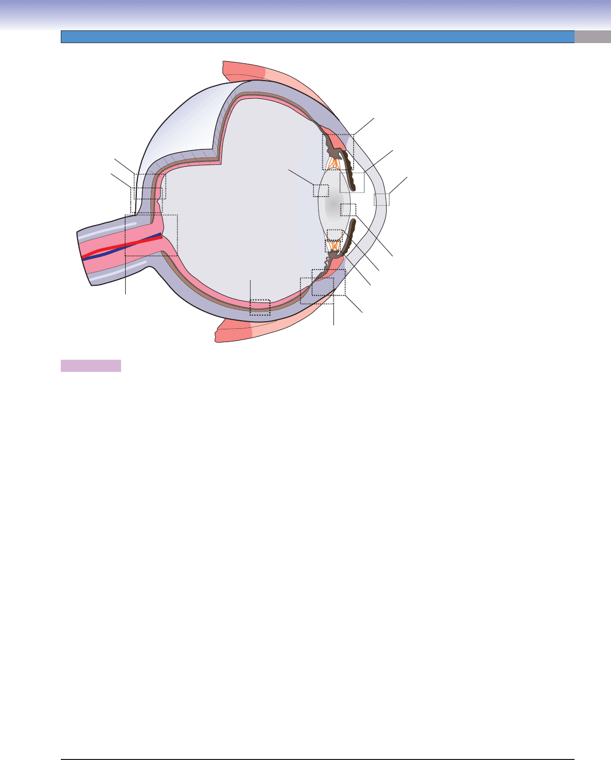
Figure 20-2. Orientation of detailed eye illustrations.
Structures of the Eye with Figure Numbers
Eyelid
Figure 20-3A
Figure 20-3B
Figure 20-3C
Figure 20-4A
Figure 20-4B
Figure 20-4C
Cornea
Figure 20-5A
Figure 20-5B
Figure 20-5C
Figure 20-6A
Figure 20-6B
Lens
Figure 20-7A
Figure 20-7B
Figure 20-7C
Figure 20-7D
Figure 20-8A
Figure 20-8B
Figure 20-8C
Figure 20-10A
Iris
Figure 20-9A
Figure 20-9B
Figure 20-9C
Figure 20-9D
Figure 20-10B
Ciliary Body
Figure 20-11A
Figure 20-11B
Figure 20-11C
Figure 20-12A
Figure 20-12B
Figure 20-12C
Retina
Figure 20-13A
Figure 20-13B
Figure 20-13C
Figure 20-14A
Figure 20-14B
Figure 20-15A
Figure 20-15B
Figure 20-15C
Figure 20-16A
Figure 20-16B
Optic Nerve
Figure 20-17A
Figure 20-17B
CUI_Chap20.indd 393 6/16/2010 7:37:54 PM
394
UNIT 3
■
Organ Systems
Tarsal
Plate
Tendon of the
levator
palpebrae
muscle
Palpebral
conjunctiva
Lid margin
Eyelash
follicles
Skin
Meibomian
(Tarsal)
glands
Superior
tarsal muscle
Orbicularis
oculi muscle
A
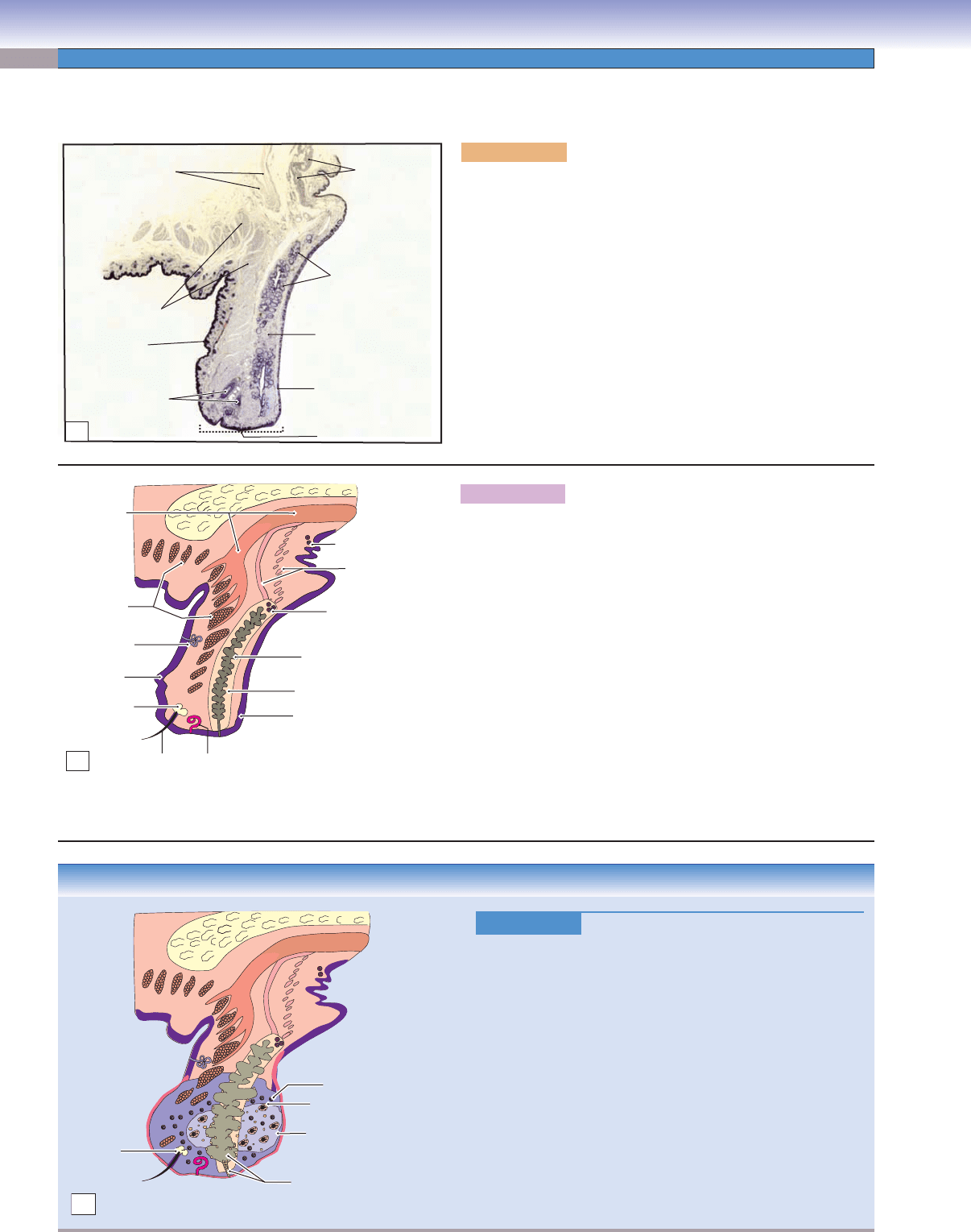
Figure 20-3A. Overview of the upper eyelid. H&E, 7.6
A low-power photomicrograph of the upper eyelid is shown. The
eyelids contain an outer layer of skin; a middle layer of muscles,
glands, and tarsal plate; and an inner layer of conjunctival tissue
(palpebral conjunctiva). Eyelids cover and protect the eye from
the environment, injury, and intense light. They also maintain a
smooth corneal surface by spreading a fi lm of lacrimal fl uid (tears)
evenly over the cornea to moisten the eye. The skin of the eyelids is
thin, loose, and delicate and, therefore, may permit extreme swell-
ing. The internal lid is covered by a palpebral conjunctiva, a layer
of stratifi ed low columnar epithelium. It is continuous with the
bulbar conjunctiva where it covers the sclera of the eyeball. The
tarsal plate is a dense fi broelastic tissue, which provides fl exible
support. The tarsal glands (meibomian glands) are embedded in it.
The eyelashes are located in the anterior margins of the eyelids.
Eyelid
Levator
palpebrae
superioris
muscle and
its tendon
Meibomian
(tarsal) gland
Palpebral
conjunctiva
Tarsal plate
Superior
tarsal muscle
Accessory lacrimal
gland (of Krause)
Accessory lacrimal
gland (of Wolfring)
Orbicularis
oculi
muscle
Sweat
gland
Skin
Gland
of Zeis
Eyelash
Gland
of Moll
D. Cui
B
Figure 20-3B. A representation of the upper eyelid, glands, and
muscles that control eyelid movements.
Several types of glands in the eyelids include (1) meibomian (tarsal)
glands, sebaceous glands that produce a lipid-rich substance; (2) glands
of Zeis, modifi ed sebaceous glands associated with the follicles of the
eyelashes; (3) glands of Moll, modifi ed sweat glands, associated with
eyelash follicles; and (4) accessory lacrimal glands, serous glands
that contribute to tears. The muscles associated with upper eyelids
are the (1) orbicularis oculi muscle, a circular sheet of striated muscle
which functions to close the eyelids; (2) levator palpebrae superioris
muscle, a thin, fl at, striated muscle that arises from the apex of the
orbit and inserts into the posterior surface of the orbicularis oculi
muscle and the skin of the upper eyelid, and which opens the eyelid;
and (3) superior tarsal muscle (Müller muscle), smooth muscle that
arises from the interstitia of the levator muscle and inserts on the
upper end of the tarsal plate and superior conjunctiva of the lid. It
joins the levator palpebrae superioris muscle in raising the upper
eyelid. (For innervation, see text of Fig. 20-4C.)
Figure 20-3C.
Chalazion.
Chalazion is a chronic eyelid infl
ammatory lesion that results
from the obstruction of the ducts of either the Zeis or meibo-
mian glands, or both. Trapped sebaceous secretions leak into
the surrounding tissue and cause a granulomatous infl am-
mation. This is frequently associated with blepharitis and
occasionally becomes secondarily infected. Early symptoms
and signs include eyelid swelling and erythema. In time, it
changes into a fi rm nodule within the eyelid or the tarsal
plate. Histologic examination reveals granulation tissue char-
acterized by focal aggregation of epithelium-like (epithelioid)
cells, lymphocytes, giant cells of Langerhans, and yellow
lipid-laden macrophages. Treatment options include warm
compresses applied to the outer lid until acute symptoms dis-
appear, topical antibiotics, and surgical incision if the lesion
is large and disturbs vision.
Obstructed meibomian gland
Lipid-laden macrophage
Nodule
Lymphocyte
Gland
of Zeis
D. Cui
C
CLINICAL CORRELATION
CUI_Chap20.indd 394 6/16/2010 7:37:55 PM
