Creagh D., Bradley D. (Eds.) Physical Techniques in the Study of Art, Archaeology and Cultural Heritage. Volume 2
Подождите немного. Документ загружается.


grains in oxidised silver gelatin emulsions are often surrounded by clouds of smaller parti-
cles. This confirmed the theory of Torigoe and co-workers that the consequence of image
oxidation is the formation of smaller colloidal particles causing yellow discoloration to the
photograph (Torigoe et al., 1984).
Second, he confirmed the results of Feldman, showing that mirrored areas are charac-
terised by colloidal particles at the top surface of the emulsion. Hendriks believed that
these particles were proof of an upward migration of the silver ions. He added that the
presence of transfer images in the baryta layer of deteriorated silver gelatin prints, already
noticed by Weyde in the 1950s (Weyde, 1955), was proof of a downward migration of the
silver ions.
1
He thought the silver mirroring particles to be made of a very thin layer of
elementary silver, centred on a silver sulphide nucleus (Hendriks, 1984). He indicated the
following as compounds responsible for silver mirroring:
(a) Compounds present either in the image layer or in the support as a result of careless
processing;
(b) Atmospheric gases such as sulphur dioxide, hydrogen sulphide, oxides of nitrogen,
peroxides, and ozone; and
(c) Compounds present in the filing enclosures in contact with the photograph.
Images published by Hendriks provide strong evidence that the first step to image dete-
rioration is the oxidation of the image grains. Image grains lose their integrity, and smaller
particles are formed within the emulsion or at the emulsion surface. Nevertheless, Hendriks
did not explain why the ions or the colloidal particles would migrate towards the emulsion
surface. And he did not explain why, in some cases, small particles are formed within the
emulsion. These result in macroscopic yellowing discoloration, and in other cases the parti-
cles are formed at the top surface of the emulsion, resulting in silver mirroring.
An attempt to answer these crucial questions and the question of the chemical nature of
silver mirroring was made by Nielsen and Lavedrine (Nielsen, 1993; Nielsen and
Lavedrine, 1993). They published transmission electron micrographs of historically and
artificially mirrored photographs showing that surface particles were present only in the
mirrored regions and that smaller particles are found underneath the top layer of closely
packed mirroring particles. The concentration and size of the smaller particles decrease as
the distance from the surface increases. This particle distribution is considered as a proof
for the migration of silver salts towards the surface, although no driving force for such a
migration is given. The close-fitting mosaic of the uppermost particles is indicated as a
proof of their gradual growth from smaller nuclei. They did not define the chemical compo-
sition of silver mirroring because only qualitative scanning electron microscope-energy
162 G. Di Pietro
1
“Transfer images” (also called “ghost images”) are silver-based images formed spontaneously, during process-
ing, on the baryta layer of prints processed with developing solutions contaminated with fixing solutions. They
were reported for the first time by Weyde (1955). They are formed in wet emulsions when the fixer dissolves
the not-exposed silver halide grains and the resulting silver ions are reduced back to silver by the developer. The
reduction of the silver ions by the developer is normally not possible in solution but it can take place when the
ions are attached to particles present in the baryta layer. This mechanism is at the base of diffusion transfer photo-
graphic processes, as in Polaroid photographs.
dispersive X-rays (SEM-EDX) analysis was performed. The analysis detected the presence
of both silver and sulphur.
Finally, it is important to discuss an article written in 1988 that proposes a theory of
silver mirroring formation drastically different from the ideas developed in the twentieth
century. The theory presented by Barger and Hill (1988) is based on the fact that both surface
roughness measurements and scanning electron micrographs showed that the mirrored areas
have a higher surface roughness in comparison with non-mirrored areas. Moreover, their
SEM-EDX analysis detected, in the mirrored areas, the presence of silver. These results,
combined with the relevant difference between the reflectance spectrum of mirrored emul-
sions and of silver sulphide or of silver films, prompted them to propose a non-chemical
mechanism of silver mirroring formation. The bluish appearance of mirrored photographs
would result from a shrinking of the gelatin around the image particles. Such a rough surface
would scatter the light differently and, therefore, would acquire a bluish tone.
2.2. Open questions on silver mirroring
The established model for silver mirroring formation among the community of photo-
graphic conservators is the oxidation–migration–re-aggregation model. The work of
Nielsen and Lavedrine has clearly demonstrated that particles are present at the top surface
of the emulsion only in the visually mirrored areas. This is in contradiction with the gelatin
shrinking model of Burger and Hill.
Nevertheless, the oxidation–migration–re-aggregation model leaves two main questions
to be answered:
1. What is the chemical composition of silver mirroring and, therefore, which are the
compounds responsible apart from oxidant compounds?
2. Which is the driving force for the formation and/or aggregation of particles at the emul-
sion surface and, therefore, why is there the formation of silver mirroring in certain
cases and yellowing discoloration in others?
Answering the first question is crucial to understanding which compounds are actually
responsible for the formation of silver mirroring. If the silver mirroring particles are made
of elementary silver (Ag), then the compounds responsible for silver mirroring, apart from
oxidant compounds, have to be searched for among silver-reducing substances (e.g. alde-
hydes), while if they are made of silver sulphide (Ag
2
S), they have to be searched for
among sulphur containing substances (e.g. hydrogen sulphide (H
2
S)). This question will
be answered by analysing the chemical composition of the silver mirroring layer with
spectroscopic techniques.
Answering the second question is crucial to distinguishing the formation of silver mirror-
ing, which has been shown microscopically to be a layer of colloidal particles clustered at
the top surface of the emulsion, from the other corrosion-based degradation forms of black-
and-white photographs, which microscopically consist of small particles aggregated within
the emulsion bulk. This question will be answered by analysing the particle shape and
the density of the particle distribution underneath the mirroring layer. The particle shape
and density are properties inherited by the mechanism of formation of silver mirroring.
Investigations into the Degradation of Photographic Materials 163
A surface layer of particles, like silver mirroring, can result either from particles formed
within the emulsion bulk and then migrating towards the emulsion surface or from parti-
cles formed directly at the emulsion surface. These two mechanisms lead to two different
trends for the particle shape.
In the first case, the particle shape is predicted to be spherical at great distances from
the surface and ellipsoidal close to the surface, as colloidal particles cannot escape from
the emulsion and cluster once their spatial density is sufficiently high.
In the second case, particles would be formed by the reaction between silver ions, results
of the oxidation of the image grains, and an external compound, and, as the reaction has
no preferred direction, their shape is predicted to be spherical at any distance from the
surface.
2.3. Experiments
The following experiments were performed on silver gelatin glass negatives belonging to
the Cueni study collection, a collection of about 150 glass plate negatives of the Swiss
painter and amateur photographer Adolf Cueni active in the Basel region in the years
1910–1920s. The Laboratory of Image and Media Technology of the University of Basel
presently owns the plates.
2.3.1. Choice of spectroscopic techniques for the analysis of the
chemical composition of silver mirroring
The spectroscopic methods suited to determine the chemical composition of silver mirror-
ing have to fulfil two prerequisites.
The first prerequisite is that they have to distinguish between compounds present in the
mirroring layer, of thickness on the order of hundreds of nanometres, and compounds pres-
ent in the emulsion bulk, of thickness of the order of 50 µm.
The second prerequisite is that they have to be able to detect silver sulphide. The simple
detection of sulphur is not sufficient to draw valid conclusions. Indeed, sulphur
compounds can arise from the protein cysteine, one of the constituent proteins of the gela-
tin, and from the incomplete removal of sodium thiosulphate (Na
2
S
2
O
3
), the usual fixing
agent. In order to arrive at a conclusion, it is fundamental either to determine the amount
of sulphur and silver in the mirroring layer (if the particles are made of silver sulphide,
their amount has to be in the ratio 2:1) or to detect the presence of silver sulphide directly
(e.g. detection of silver sulphide crystal structures).
Spectroscopic analyses are based on the detection of the radiation emitted by a sample
excited by an incoming beam. The emitted radiation is characteristic of the elements and,
in some cases, of the compounds present in the sample.
In spectroscopic equipments, samples of mirrored emulsions can be analysed using one
of the following arrangements:
1. emulsion layer perpendicular to the incoming radiation (flat samples);
2. emulsion layer parallel to the incoming radiation (cross section samples); and
3. powder of emulsion scratched off from the mirroring layer (powder samples).
164 G. Di Pietro

The fulfilment of the first prerequisite depends on both the properties of the incoming
radiation beam and on the arrangement used.
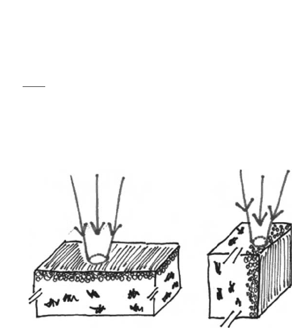
Flat and cross section sample analyses give reliable results when the penetration depth
and area of the incoming beam are smaller than the silver mirroring thickness, respectively
(Fig. 6). Powder sample analyses are in every case reliable but require highly sensitive
spectroscopic techniques due to the small amount of sample analysed.
It is therefore necessary to evaluate initially the penetration depth of the radiation used
by the X-ray techniques available to the author, X-rays and electrons, into photographic
emulsions.
When X-rays penetrate into a material, they are absorbed by different mechanisms (scat-
tering, photoelectric effect, and pair production), so that the intensity I (-) of the incom-
ing beam decreases with the distance z (cm) travelled in the material according to a simple
exponential law:
(1)
where m (cm
2
g
-1
) is the mass attenuation coefficient and r (g cm
-3
) is the material
density. The penetration depth pd (cm) is defined as the distance at which the incoming
beam has reduced its intensity to e
-1
of the initial value:
(2)
The mass attenuation coefficient depends on the energy of the incoming beam, and it is
tabulated for most of the materials (Weast, 1985).
pd =
×
1
µρ
Iz I()=
−
0
e
mr
z
Investigations into the Degradation of Photographic Materials 165
(
a
)(
b
)
Fig. 6. (a) Flat samples: the emulsion layer is perpendicular to the incoming radiation.
(b) Cross-sectional samples: the emulsion layer is parallel to the incoming radiation.

If the sample is composed of two materials A and B present in volume fractions j(A)
and j(B), the penetration depth will be:
(3)
Assuming that an emulsion is made of 10% (by volume) of silver (m(Ag) = 218 cm
2
g
-1
,
r(Ag) = 10.5 g cm
-3
) and 90% of gelatin (m(gel) = 9.8 cm
2
g
-1
as for water, r(gel) = 1.29 g cm
-3
),
the penetration depth calculated for an incoming X-ray beam of 8.041 keV (energy of the
most common X-ray source in X-ray diffraction (XRD) instruments) is of the order of
1 mm. The penetration depth in a mirrored emulsion will be smaller because the emulsion
is covered by a layer of particles. Nevertheless, even the penetration depth in pure silver is
on the order of 4 µm, about 20 times bigger than the thickness of the silver mirroring.
Therefore, the flat sample arrangement is not suitable for XRD experiments.
On the other hand, X-ray equipment capable of focussing the beams are often not available
in research laboratories, especially systems that can focus the X-ray beam to a diameter
smaller than few microns. See Creagh (Chapter 1) for information on focussed beams in
microspectroscopy at synchrotron radiation sources. In addition, the cross section arrange-
ment of the specimen is not suited to this approach.
When electrons penetrate into a material, they lose their energy with different
processes dependent on their energy value. If their kinetic energies are in the range of kilo
electron volts, the main mechanism is ionisation or excitation of the atoms present in the
material. The amount of energy loss per unit of travelled path is proportional to the elec-
tron’s density in the material, such that materials made of heavy elements will stop elec-
trons much faster than light element materials. The electron’s penetration depth is
estimated calculating the path travelled by the electrons before they stop. This is called
the continuous slowing down approximation (CSDA) range, and it is tabulated for most
materials.
For starting electron energies of 20 keV, the typical energy of the incoming electrons in
an SEM X-ray microanalysis apparatus, the penetration depth is about 6 mm in photo-
graphic gelatin and 1.5 mm in silver, in both cases more than the thickness of the silver
mirroring layer. Therefore, the flat sample arrangement is not suited for SEM experiments.
If the electron kinetic energy is on the order of a few hundreds of electron volts, the
CSDA range is not valid. Few hundreds of electron volts are the typical energy of the elec-
trons emitted in X-ray photoelectron spectroscopy (XPS) equipments. This energy is so
small that uniquely the electrons emitted from atoms at a maximum distance of 10 nm
from the sample surface can escape and be detected (Grunthaner, 1987). Therefore, the flat
sample arrangement can give reliable results in XPS experiments.
In conclusion, the methods fulfilling the first requisite are XRD on powder samples and
XPS on flat samples. Both methods also satisfy the second requisite because XRD detects
crystalline compounds (and, therefore, directly the presence of silver sulphide), while XPS
provides quantitative results of the sample atomic composition.
The combination of these X-ray analyses has been used to determine the chemical
composition of the edge silver mirroring present on four glass negatives belonging to the
Cueni study collection. Three out of the four plates examined (numbers 1, 2, and 4) were
processed negatives, while the number 3 was a historical non-processed glass negative.
pd
AAABBB
=
××+××
1
µρϕµρϕ
() () () () () ()
166 G. Di Pietro

Small, approximately squared samples were cut out from the plates with a diamond knife,
cutting from the glass side. Every sample underwent only one type of analysis.
Table 1 summarises the experiments performed.
2.3.1.1. X-ray diffraction (XRD)
Principles of the technique. The XRD analysis detects the crystal structures present in a
sample. Every crystal in the sample has a set of characteristic distances between the planes
on which the atoms are located. When an X-ray beam of wavelength l hits the atoms, the
rays reflected from the atoms located on two parallel planes combine additively if the
distance d between the two planes satisfies the so-called Bragg’s law:
nl = 2d sin(q) (4)
where n is an integer number and q is the angle between the incident (or the reflected)
beam and the perpendicular to the plane.
By changing the angle q, the distances between the planes are scanned. Peaks of intensity of
the diffracted radiation are present in correspondence of the distances satisfying the relation (4).
In the case where the diffracted radiation is detected with a scintillation detector, the
spectrum is a graph of the intensity of the diffracted X-ray towards the angle q.
In the case where the diffracted radiation is recorded on a photographic film, the spec-
trum consists of circles located around the direction of the incoming beam of radii simply
related to the characteristic distances.
Measurements on powder samples. The XRD analysis on powder samples was performed
at the XRD facilities of the Netherlands Institute for Cultural Heritage, Amsterdam, in collab-
oration with Mr. P. Hallebeek. The XRD instrument is composed of an X-ray generator
(Philips PW 1010) using a Cuka X-ray source at wavelength l = 1.5406 Å and energy
E = 8.041 keV, using a Debey–Scherrer powder camera for which the recording medium was
Investigations into the Degradation of Photographic Materials 167
Table 1. Spectroscopic analyses
Samples Analysis
Plate 1
1a XRD powder
Plate 2
2a XRD powder
2b XRD flat
2c XPS flat
2d SEM-EDX
Plate 3
3a XRD flat
3b XPS flat
Plate 4
4a SEM-EDX

double-coated CEA Reflex 25 film. The sample, consisting of few powder grains, is fixed on
the tip of a glass fibre with cedar oil and it is continuously rotated during the measuring time
(order of a few hours) to cover all the possible mutual positions between the incoming beam
and the crystal planes within the sample. This instrument is able to detect the crystal compo-
sition of extremely minute amount of sample, of dimension of the order of 0.5 mm
2
.
The silver mirroring was scratched off the plates with a scalpel under a loupe, with care-
ful attention paid to removing only the mirroring layer and not the underlying emulsion.
The total amount of material was of the order of few powder grains.
Measurements on flat samples. The XRD analysis on flat samples was performed at the
XRD facility of the Institute of Inorganic Chemistry of the University of Zürich, in collab-
oration with Prof. H. Berke. The XRD diffractometer is a Kristalloflex instrument
produced by Siemens using a Cuka X-ray source at wavelength l = 1.5406 Å and energy
E = 8.041 keV. The diffracted rays are collected with a scintillation detector. The relative
position of the sample and the detector was changed in steps of 2q = 0.05∞ using a meas-
uring time of 0.3 s for every step.
The samples have approximate dimensions of 2 cm ¥ 1 cm and they are hold flat in
aluminium holders. In order to prevent contributions in the diffracted beam from the side
areas not presenting silver mirroring, these areas were covered with self-adhesive tape.
2.3.1.2. X-ray photoelectron spectroscopy (XPS)
Principles of the technique. XPS is a spectroscopic technique based on the measurement
of the binding energies (E
b
) of the core electrons of the atoms contained in a material. It is
able to detect all the atoms of the periodic table other than hydrogen.
The sample is bombarded with monochromatic X-rays of energy E
X
higher than the
core-electron-binding energies; the atoms present in the sample absorb the X-rays and
eject their core electrons with a kinetic energy E
k
satisfying the relation:
E
k
= E
x
-E
b
(5)
Since E
x
is known and E
k
is measured in the experiment, the core-electron-binding ener-
gies E
b
are calculated and used to identify the atoms.
Shifts in the measured core-electron-binding energy can be used to determine the
molecular composition of the sample. XPS is a real surface analysis because only electrons
ejected from a maximum distance of 10 nm from the sample surface have enough kinetic
energy to escape and be detected.
An XPS spectrum is a graph of the number of emitted electrons against the detected
energies. Peaks are present in correspondence to the binding energies of the core electrons
of the atoms contained in the material. As the area A
i
of the peaks is proportional to the
amount of atoms ejecting the electrons, the percentage atomic composition M
i
of the
sample can be calculated as follows:
(6)
where C
i
is the photoionisation cross section for the atomic core electron i.
M
A
C
A
C
i
i
i
i
i
=×
∑
1
168 G. Di Pietro
If the sample is not conductive, the ejected electrons can accumulate above the sample
surface, and they can shift the position of the peaks eventually, making the peak determi-
nation impossible.
Measurements on flat samples. The measurements were performed at the XPS labora-
tory, Institute of Physics of the University of Basel, in collaboration with Prof. P. Oelhafen.
The samples, with dimensions of approximately 10 mm ¥ 20 mm, were fixed with metal
screws on a metal holder and inserted into the spectrometer where an air pressure of about
10
-9
mbar is reached in about 10 h. In spite of the high vacuum attained during the meas-
urement, no damage was visible on the samples. The samples were then bombarded with
X-ray and the spectra recorded in a few minutes.
Due to high surface-charging effects, it was possible to record spectra only from the
silver mirroring areas close to the metal screws. In these areas, the surface charging was
minimised because the electrons could diffuse to the metal holder and dissipate.
2.3.2. Size and shape distributions of the silver mirroring particles
The size and shape distributions of the silver mirroring particles, belonging to the edge
mirroring present on a historical non-processed glass plate, was analysed, making use of
transmission electron microscopy (TEM). The TEM micrographs were later digitised, and
the digital images were analysed with image analysis software. Software from the
Mathematica
®
libraries was used in the statistical analysis of the data.
2.3.2.1. Transmission electron microscopy (TEM)
Principle of the technique. A TEM basically consists of a column of vacuum where elec-
trons, emitted by a heated tungsten filament, are accelerated to a high voltage (200 keV) and
then focussed by electron lenses (condenser lenses) on the specimen. The specimen is a thin
section of material (thickness on the order of 70 nm) partially absorbing the electrons. The
transmitted beam is then enlarged and focussed by electron lenses (imaging system), so that
an enlarged image is formed on the viewing screen, and a plate covered with phosphors
fluoresces when hit by the electrons. If a permanent image of the sample is required, the
screen is removed and the electrons can directly hit a photographic film, which is subse-
quently developed and printed. For a more in-depth description of TEM, see Hayat (2000).
The electron absorption by the specimen is dependent on the sample thickness and
composition (dense areas absorb more electrons). This determines an amplitude contrast
and, therefore, an image.
As the electrons have to pass through the sample, the sample has to be 100 nm thick
(maximum). Thin slices can be obtained by cutting the sample with an ultramicrotome.
Normally, a soft sample cannot be cut straightaway but it has to be embedded in a harder
material first. Sample embedding is a crucial factor in TEM.
Measurements. Small portions (dimension 1 mm ¥ 2 mm) of mirrored emulsion of a histor-
ical non-processed glass negative were removed by immersing the plate in a solution of water
and alcohol, and using a knife blade and tweezers under the loupe. The samples were then
embedded in a resin following a standard embedding method reported in Kejser (1995). The
resin was a standard mixture of epoxy embedding medium, hardener (DDSA and MNA), and
Investigations into the Degradation of Photographic Materials 169

accelerator (BDMA), all of which were produced by Fluka. They were laid flat on a resin
drop and, after removing the excess water on the emulsion, were covered with a second
drop of resin and inserted into an oven at 60∞ for about 8 h. Later, slices of 70 nm thick-
ness were cut in the cross-sectional direction with an ultramicrotome. The slices were
transferred onto a grid and were then placed under the microscope. Images with magnifi-
cation on the order of 10 000 times were taken. As the negative was a non-processed plate,
long electron exposures had to be avoided in order to avoid the development of the silver
bromide grains physically. The TEM micrographs were digitised, and the digital images
were analysed with the software NIH Image
®
. The software Mathematica
®
was used in the
statistical analysis of the data.
After embedding, slices of thickness 70 nm were cut in the cross-sectional direction
with an ultramicrotome. The slices were transferred onto a grid, and they were placed
under the microscope available at the Interdivisional Electron Microscopy Laboratory of
the University of Basel (LEO EM 912), where images with magnification on the order of
10 000 were taken. As the negative was a non-processed plate, long electron exposures
had to be avoided in order to avoid the development of silver bromide grains physically.
2.4. Results
2.4.1. Results on the chemical composition of silver mirroring
The results of the analysis of the chemical composition of silver mirroring are presented
in Table 2. The XRD analysis on powder samples has determined that the silver mirroring
scratched off from plates 1 and 2 is composed of 100% silver sulphide (Ag
2
S). No other
crystalline compounds were detected. In the case of detection of a single compound, the
error in the mass composition of the sample attained by the instrument used in this meas-
urement is on the order of 5–10%.
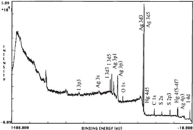
The XPS analysis revealed in sample 2c (Fig. 7) the presence of the following elements:
carbon C (40%), oxygen O (11%), silver Ag (29%), sulphur S (14%), iodine I (4%), and
mercury Hg (2%). The percentages refer to the relative atomic composition and are calcu-
lated from the spectra using formula (6). In sample 3b, carbon C (49%), oxygen O (10.5%),
170 G. Di Pietro
Table 2. Results of the analysis of the chemical
composition of silver mirroring
Sample XRD powder XPS
1a Ag
2
S
2a Ag
2
S
2c Ag
2
S
O, C, I, Hg
3b Ag
2
S
O, C, Br, I,
Hg

silver Ag (25%), sulphur S (11%), bromine Br (3%), iodine I (1%), and mercury Hg (0.5%)
were detected.
In both samples, the main component is carbon, arising from the collagen contained in
the gelatin emulsion. It is followed by silver and sulphur. The ratio between the amounts
of silver and sulphur in the mirrored areas ranges between 2.07 and 2.27, almost stoichio-
metric for silver sulphide Ag
2
S. Moreover the S(2p) peak has, in both cases, a negative
shift (ranging from –4.5 to –6.3 eV), suggesting that sulphur is in the S
2-
state (Hammond
et al., 1975). This indicates that silver sulphide is present in the mirroring areas. The
amount of oxygen detected probably arises from the collagen present in the emulsion;
indeed, the absence of the O(1s) peak at 529 eV excludes the presence of silver oxide
(AgO) or silver dioxide (Ag
2
O). The presence of bromine in sample 3b is consistent with
the fact that plate 3 is a non-processed negative.
The XRD analysis on flat samples uniquely determined the presence of silver (Ag) crys-
tal structures in sample 2b. In sample 3a, both silver and silver bromide (AgBr) structures
were identified, consistent with the fact that plate 3 is a non-processed negative.
2.4.2. Results on the size and shape distribution of the silver mirroring particles
The samples were difficult to cut because of the softness of the specimen core. Although
the slices often broke apart after cutting, it was possible to obtain some sections suitable
for the analysis of particle distribution (Fig. 8).
The micrograph shown in Fig. 8 was digitised and used to analyse the variation of area,
density, and sphericity of the small silver mirroring particles residing underneath the main
layer. With the help of Adobe
®
Photoshop
®
, the upper silver mirroring particles were
Investigations into the Degradation of Photographic Materials 171
Fig. 7. XPS spectrum of sample 2c.
