Creagh D., Bradley D. (Eds.) Physical Techniques in the Study of Art, Archaeology and Cultural Heritage. Volume 2
Подождите немного. Документ загружается.

182 G. Di Pietro
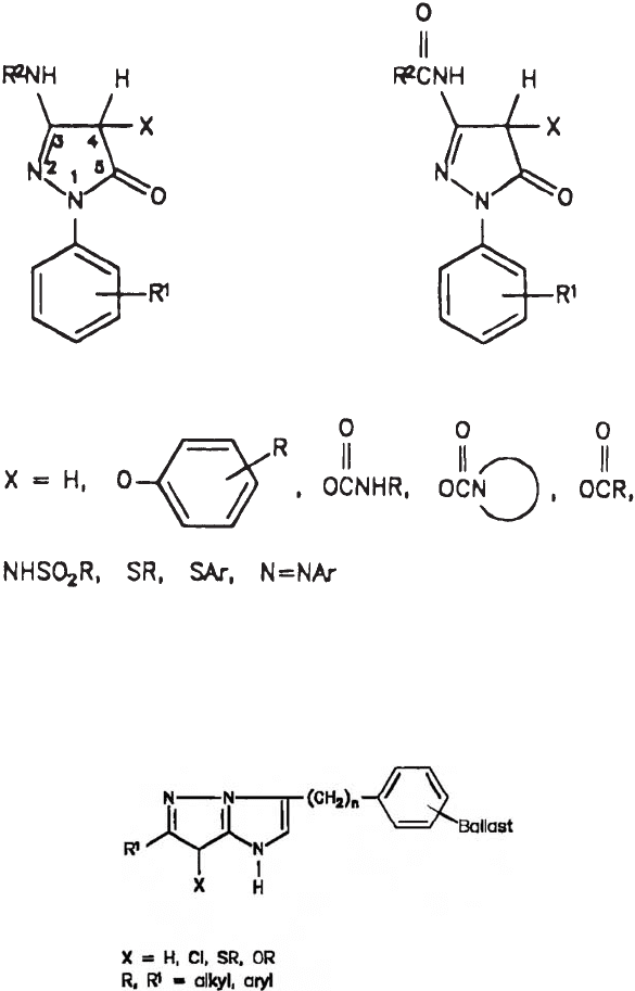
Fig. 13. Two couplers of the pyrazolinone class: 3-arylamino substituted (left) and
3-acylamino substituted (right). R, R
1
, and R
2
are various organic moieties (from Theys
and Sosnoysky, 1997). Reprinted with permission from: Theys, R.D., Sosnovsky, G., 1997.
Chemistry and color photography. Chem. Rev. 97, 83–132. © 1997 American Chemical
Society.
Fig. 14. Magenta-forming pyrazolotriazole couplers (from Theys and Sosnoysky, 1997).
Reprinted with permission from: Theys, R.D., Sosnovsky, G., 1997. Chemistry and color
photography. Chem. Rev. 97, 83–132. © 1997 American Chemical Society.
photographic processes, the colour couplers are ballasted in the emulsion and are not
washed away after processing the film. This means that the unused couplers will stay in
the emulsion and can undergo reactions leading to colour products. This is the reason why
chromogenic processes (processes in which the colour couplers are not incorporated into the
emulsion but are precipitated in the three layers during the processing steps, for example,
Kodakchrome) have traditionally a high dark stability.
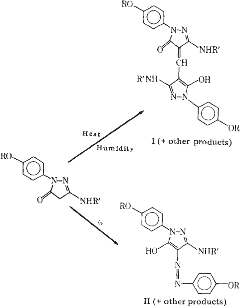
An important reaction related to magenta dye instability is the reaction involving the
residual magenta coupler shown in Fig. 16 and resulting in the yellowing of the non-
image areas. This reaction leads to a yellow-coloured compound, either in the dark or in
the presence of light. The product is methynylbis coupler in the dark and the azo-dye in
the light. This problem was solved with the substitution of two of the ortho positions of the
1-phenyl ring (Tuite, 1979).
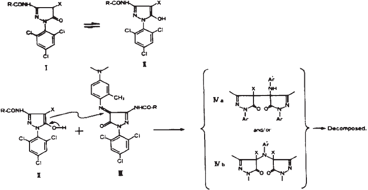
Another important reaction occurs between the residual magenta coupler (3-acylamino
pyrazolone couplers) and the magenta dye to form a variety of colourless products. Using
aldehydes in the final processing bath solved this problem. Indeed, the cross-linked
couplers do not react with the magenta dye. Later, magenta couplers (anilino pyrazolone)
were devised, which showed much lower tendency to react with their magenta dyes and
were used in paper prints (Tuite, 1979). Nevertheless, the 3-acylamino pyrazolone couplers
were the most common couplers used in camera-use films such as colour negatives, colour
reversal films, and colour films for motion pictures at least up to 1988 (Sakanoue and
Furutachi, 1988) because of their image quality. Sakanoue and Furutachi (1988) found that
the degree of dye decomposition relates to the pKa value of the coupler and that the use of
low-pKa couplers could eliminate the necessity of formaldehyde from the processing solu-
tion. They proposed a fading mechanism, described in Fig. 17.
A third important class of reactions involving magenta dyes is the reaction with resid-
ual thiosulphate (from the fixing bath) (Miyagawa and Shirai, 1985; Kurosaki et al.,
1988). In these articles, the authors report the reductive fading pathway, the structures of
the dye, and the colourless adduct.
Investigations into the Degradation of Photographic Materials 183
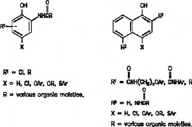
Fig. 15. Cyan-forming phenol (right) and naphthol (left)-type couplers (from Theys and
Sosnoysky, 1997). Reprinted with permission from: Theys, R.D., Sosnovsky, G., 1997.
Chemistry and color photography. Chem. Rev. 97, 83–132. © 1997 American Chemical
Society.
The result of the innovations on magenta couplers is that none of the modern (post-
1979) Kodak colour photographic materials that use traditional processing are magenta
dye limited for dark stability. They can be limiting for light stability. Adding an UV
absorbing layer in the prints in front of the magenta layer circumvents this problem.
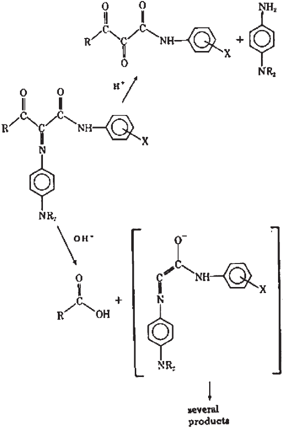
The yellow dye is the least stable dye in the dark because of hydrolytic reactions occur-
ring both in acidic and alkaline environments. The acidic hydrolytic attack occurs at the
azomethine linkage, while alkaline hydrolysis occurs at the keto linkage (Tuite, 1979). The
final processing pH of a film is chosen to minimise these two reactions. The reaction
shown in Fig. 18 is considered the most important dark fading reaction occurring when a
film suffers from vinegar syndrome.
Indeed, yellow fading was monitored for years at Kodak as an indication of the onset of
the degradation of the cellulose triacetate base (vinegar syndrome). Tuite (1979) reports
that the dark fading characteristic of the yellow dye for 10% dye loss ranges from 3 years
for Kodak Ektachrome 40 Movie Film (type A, EM-25 process) to 32 years for Kodak
184 G. Di Pietro
Fig. 16. Reactions involving residual magenta coupler (from Tuite, 1979). Reprinted
with permission of IS & T: The Society for Imaging Science and Technology sole ©
owners of the Journal of Applied Photographic Engineering.
Ektachrome 50 Professional Film 6118 (Daylight, E-8 process). As far as light stability is
concerned, the largest single improvement came from the simple change from a benzoyl
group to a pivalyl group on the yellow coupler (Tuite, 1979).
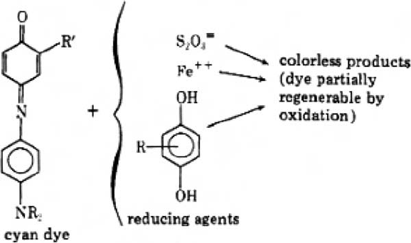
The cyan dye is the dye most stable to light. The principal undesired dark reaction is the
reduction of the residual cyan coupler to the colourless leuco form of the dye (Fig. 19). Other
reducing agents that have not been removed completely can also cause this or similar reac-
tions: e.g. retained thiosulphate from the fix bath, ferrous ion from a poorly regenerated bleach
fix bath, and ballasted hydroquinones that are used as incorporated interlayer scavengers.
This problem was solved by a change in the colour-developing agent from CD-2 to
CD-3 and by a change in the dye structure, namely the use of phenol-type couplers with
amide group in the 2.5 position of the ring (Tuite, 1979; Theys and Sosnovsky, 1997).
The masking dyes that gives the orange colour to negatives is very stable in the dark
(Bergthaller, 2002c).
Photochemical degradation involves absorption of light by a dye to generate both
singlet- and triplet-excited states. Aerial oxidation of the excited molecules forms undesir-
able by-products. Stabilisation of the dye involves intercepting both the actinic radiation
with a UV absorber and one or more of the reactive intermediate with a quencher such as
nickel dibutyldithiocarbamate. Bergthaller (2002c) proposes a degradation trail common
for both dark fading and photo fading. He assumes that under appropriate conditions the
detachment of the developer fragment from a dye cloud could be started more readily from
a sequence of single electron transfer and proton transfer to the azomethine bond. This idea
is based on the fact that, in many cases, increased resistance of dyes to dark stability has
resulted from efforts directed towards improved light stability.
Investigations into the Degradation of Photographic Materials 185
Fig. 17. Proposed mechanism for the reaction between magenta 3-acylamino pyrazolone
coupler and magenta (from Sakanoue and Furutachi, 1988)
The atmospheric constituents, which most often cause colorants to fade, are
oxides of sulphur and nitrogen, and ozone. Atmospheric oxygen can also be a signifi-
cant contributor to the fading of certain types of yellow and magenta dyes. In contrast,
it was found that oxygen could inhibit the fading of cyan dyes (Theys and Sosnovsky,
1997).
186 G. Di Pietro
Fig. 18. Yellow dye hydrolysis under acid (top) and alkaline (bottom) conditions (from
Tuite, 1979).
3.4. Experimental analysis of dyes
3.4.1. Choice of spectroscopic technique and sample preparation
A colour film has a layered structure, where each layer contains only one type of dye.
Figure 2 shows the cross-sectional structure of typical print and negative stocks. Each layer
has dimensions in the range of 5–10 µm. The “China girl” image in the leader of the movie
film was chosen as the appropriate test film because it is closest to the surface and, there-
fore, more susceptible to environmental conditions, and it is on all the films, and it is not
part of the film content.
To analyse the dyes present in each layer of the “China girl” image on the film leader,
it is possible to follow three different strategies:
∑ Use an analytical technique that can select the layer non-destructively, e.g. using a
confocal microscope associated with an FTIR spectrometer.
∑ Cut the film in the cross section and analyse the layers. This is achievable if the analytical
technique has a detection area of diameter of 5 µm or less.
∑ Dissolve the dyes and separate them either with thin layer chromatography (TLC) or
high-performance liquid chromatography (HPLC). The dyes can then be analysed with
Fourier transform infrared spectrometry (FTIR), mass spectrometry (MS), or with other
techniques.
The first strategy was attempted with the Raman Confocal Microscope at the University
of Canberra. This was unsuccessful because the surface of the film gave a very high fluo-
rescence signal that masked the signal from the underlying layers.
Investigations into the Degradation of Photographic Materials 187
Fig. 19. Reaction between cyan dye and reducing agents (from Tuite, 1979). Reprinted
with permission of IS & T: The Society for Imaging Science and Technology sole ©
owners of the Journal of Applied Photographic Engineering.
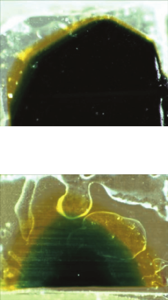
The second strategy was performed using the microtome available at the Electron
Microscopy Unit of the Australian National University. This does not attempt to investi-
gate the cross section of the film directly. A small piece of film (approximate dimensions
of 2 mm ¥ 2 mm) was cut with a scalpel and mounted flat on a resin capsule using super-
glue. Care was taken to stick the film base to the resin capsule and not the film emulsion
side. The capsule was then fixed on the microtome, and thin sections were cut with a glass
knife as precisely as possible parallel to the base. As it is not possible to have sections paral-
lel to the base on the whole film surface, the cut will be slanted, and it will expose the three
colour layers (Figs. 20 and 21). The slanted section can be used for analysis and imaging.
The thin sections can be either discarded or kept for further analysis. In the second
case, it is important to collect specimens continuously. This can be achieved by float-
ing them in isopropyl alcohol. This solvent is contained in a so-called boat built and
attached on purpose to the glass knife. It is difficult to retrieve thin sections flat because
they tend to curl up. It is essential that the boat be completely full and the sections
(which tend to sink) be lifted using a small loop made by a hair (human or animal)
attached to a bamboo stick.
TLC coupled with FTIR has been used at the Research School of Chemistry of the
Australian National University. To separate the dyes with TLC, it is necessary to
dissolve them, leaving a piece of film in a vial with ethanol for few days. The dissolu-
tion process is aided by little heating and stirring. The ethanol is then allowed to evap-
orate, and diethyl ethanol is added to the vial. Diethyl ethanol is used because of the
higher solubility of the dyes in this solvent. It cannot be used initially because of its
high evaporation rate. A few millilitres of this solution is spread following a line on a
large silica TLC plate of dimension 200 mm ¥ 200 mm (DC-Alufolien Kieselgel60
produced by Merck) using a glass capillary. The use of large TLC plates is necessary if
sufficient compounds are to be collected for further analysis. The TLC plate is then
immersed in a tray containing the mobile phase (100 ml of a solution of diethyl ether
and petroleum spirit in diethyl ether, in proportion 40:60 for the prints and 90:10 for
the negatives) and left there for the time necessary for the mobile phase to rise to the
top of the plate (~90 min). This procedure produces a plate with separate lines of the
different dyes. It is noticeable that fresh print film gave rise to 3 lines (magenta, cyan,
and yellow), while aged print films gave rise to more lines (usually the three main
colour lines followed by a second much fainted line of the same colour). This is due to
the fact that ageing has caused breakdown of some of the dye molecules. The dyes can
be scratched from the plate and dissolved in diethyl ether. After the silica is decanted,
the remaining liquid is dried and mixed with KBr (potassium bromide) to produce
pellets that can be analysed with FTIR. Unfortunately, the fainter lines, which proba-
bly contain the dye degradation products, did not yield sufficient material to produce
a pellet and could not be analysed with FTIR. The dyes were analysed using Raman
spectroscopy (RS) and FTIR.
3.4.2. Raman spectroscopy
The Renishaw 2000 Raman Microscope at the University of Canberra was used with the
laser beam perpendicular to the film surface, in an attempt to use the confocal charac-
teristics of the microscope and to detect signals from the three different layers.
188 G. Di Pietro
Investigations into the Degradation of Photographic Materials 189
Fig. 20. Reflection light microscopy photograph (¥64) of the slanted section of the
Kodak print film 5384 (~1980). Notice the black-coloured top layer (resulting from
the superposition of the magenta cyan and yellow layer) and the exposed cyan and
yellow layers.
Fig. 21. Reflection light microscopy photograph (¥64) of the slanted section of the Kodak
print film 5384mod (or 5386) (~1995). Notice the exposed cyan and yellow layers.
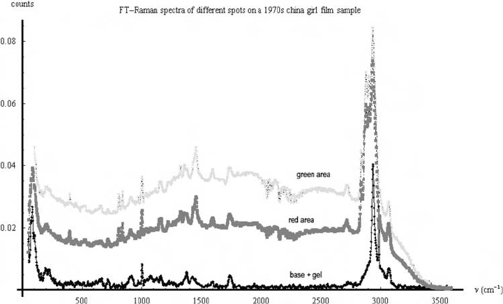
Fluorescence remained a problem, however. One way of circumventing the fluorescence
problem is by using a much higher excitation wavelength. Unfortunately, the tests made
using the Bruker IFS 100-Fourier transform Raman spectrometer with excitation wave-
length of 1064 nm at the University of Sydney were also not successful, because of the
limited optical throughput of the instrument. In this geometry (the system does not have
a microscope and requires much higher sample masses), the signal was dominated by the
cellulose acetate base, and the dyes were not visible (Fig. 22). It is important to notice
that Fourier transform Raman spectroscopy has been used successfully to identify photo-
graphic dyes present in effluent streams. In this case, the dyes are first concentrated
using solid-phase extraction (SPE) and then analysed in the FT-Raman apparatus
(Bristow et al., 1998).
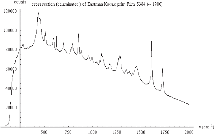
The Renishaw 785 nm Raman microscope was then used to analyse the cross section
(Fig. 23). Cross section geometry has two advantages: it maximises the scattering volume,
and it allows the spatial separation of the three coloured layers. Cross sections can be
produced either with the methods explained earlier or by briefly immersing the emulsion
in liquid nitrogen and then cracking and tearing it. This causes the emulsion to become
delaminated from the base. The former procedure (microtoming) is far more reproducible
and precise than the latter. Still, the delamination procedure has the advantage of being
quick and simple and does not require any special apparatus.
190 G. Di Pietro
Fig. 22. FT Raman spectra of different spots on a 1970s China girl film sample. The
spectra from the red and green spots have the same peaks as the spectra from the
cellulose acetate base and gelatin spot.
A pronounced fluorescence shoulder still dominates the spectra, but characteristic
Raman peaks are also visible. The fluorescence shoulder can be removed by background
subtraction.
A more elegant way to get rid of the fluorescence signal is to use a technique depend-
ent on the subtraction of “fixed pattern non-uniformity” and known as subtracted shifted
Raman spectroscopy (SSRS) (Bell et al., 1998). The basic idea comes from shifting the
spectrum of a sample by a small amount of about 15 cm
-1
, by changing the angle of the
diffraction grating in the spectrometer. This overcomes the noise-limiting non-uniformity
of adjacent pixels in a CCD detector. The two spectra are subtracted, and the Raman peaks
appear as derivative waveforms. Curve fitting is then applied, and the original Raman spec-
trum is reconstituted without the fluorescence background.
The microtomy method explained earlier has not only the advantage of being repeatable
and more precise, it also offers the possibility, through the slanted cut, to expose an area
of a single layer that is large enough (about 10 µm) to be independently detected in the
Raman microscope. Figure 24 shows the Raman spectra of each single layer in the Kodak
print film 5384.
This was compared with spectra from a recent Kodak film stock (5384 or 5386). As Fig. 25
shows, the two spectra are different, a proof that RS can identify the differences in dyes.
Unfortunately, in both cases, the spectra from the cyan and magenta layer do not show
different characteristic peaks, and the spectra of the yellow layer are dominated by fluo-
rescence. This hints to the fact that the peaks shown here refer only to the main backbone
Investigations into the Degradation of Photographic Materials 191
Fig. 23. Raman spectra of the cross section (delaminated) of Eastman Kodak print film
5384 (~1980).
