Biermann Ch. Handbook of Pulping and Papermaking
Подождите немного. Документ загружается.

624 27. HARDWOOD ANATOMY
Buttresses of trees growing in swamps or flooded
areas contain wood that is much lighter in weight
than that from upper portions of the same trees;
this wood should be separated from the heavier
wood to assure material of uniform strength.
Tupelo is cut principally for lumber, veneer,
pulpwood, and some railroad ties and slack coo-
perage. Lumber goes into boxes, pallets, crates,
baskets, and furniture.
Color, Wood of the different tupelos is quite
similar in appearance and properties. The heart-
wood is light brownish gray and merges gradually
into the lighter colored sapwood, which is general-
ly several inches wide.
Macroscopic structure. Black tupelo is
diffuse—porous with small pores like sweetgum.
The vessels are numerous and in radial multiples
which may be irregularly distributed. Growth
rings are inconspicuous to moderately distinct.
Rays are slightly coarser than in sweetgum but are
numerous and closely spaced; they are inconspic-
uous against the background color, but appear to
occupy half of the surface (in the corss section, x).
The longitudinal parenchyma occur in scattered
paratracheal—scanty and apotracheal—diffuse
arrangements, but are very difficult to see. See
sweetgum for its distinction from black tupelo.
Microscopic structure. Intervessel pitting of
black tupelo is opposite, pits are nearly square in
outline, and there are up to 6 pits per row.
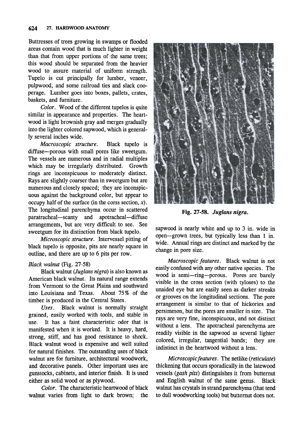
Black walnut (Fig. 27-58)
Black walnut
(Juglans nigra)
is also known as
American black walnut. Its natural range extends
from Vermont to the Great Plains and southward
into Louisiana and Texas. About 75% of the
timber is produced in the Central States.
Uses. Black walnut is normally straight
grained, easily worked with tools, and stable in
use.
It has a faint characteristic odor that is
manifested when it is worked. It is heavy, hard,
strong,
stiff,
and has good resistance to shock.
Black walnut wood is expensive and well suited
for natural finishes. The outstanding uses of black
walnut are for furniture, architectural woodwork,
and decorative panels. Other important uses are
gunstocks, cabinets, and interior finish. It is used
either as solid wood or as plywood.
Color. The characteristic heartwood of black
walnut varies from light to dark brown; the
Fig. 27-58. Juglans nigra.
sapwood is nearly white and up to 3 in. wide in
open—grown trees, but typically less than 1 in.
wide. Annual rings are distinct and marked by the
change in pore size.
Macroscopic
features. Black walnut is not
easily confused with any other native species. The
wood is semi—ring—porous. Pores are barely
visible in the cross section (with tyloses) to the
unaided eye but are easily seen as darker streaks
or grooves on the longitudinal sections. The pore
arrangement is similar to that of hickories and
persimmon, but the pores are smaller in size. The
rays are very fine, inconspicuous, and not distinct
without a lens. The apotracheal parenchyma are
readily visible in the sapwood as several lighter
colored, irregular, tangential bands; they are
indistinct in the heartwood without a lens.
Microscopic features. The netlike (reticulate)
thickening that occurs sporadically in the latewood
vessels
(gash
pits) distinguishes it from butternut
and English walnut of the same genus. Black
walnut has crystals in strand parenchyma (that tend
to dull woodworking tools) but butternut does not.
ANATOMY OF HARDWOOD SPECIES 625
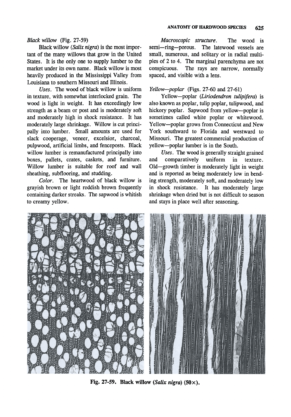
Black willow (Fig. 27-59)
Black willow
{Salix nigra)
is the most impor-
tant of the many willows that grow in the United
States. It is the only one to supply lumber to the
market under its own name. Black willow is most
heavily produced in the Mississippi Valley from
Louisiana to southern Missouri and Illinois.
Uses. The wood of black willow is uniform
in texture, with somewhat interlocked grain. The
wood is light in weight. It has exceedingly low
strength as a beam or post and is moderately soft
and moderately high in shock resistance. It has
moderately large shrinkage. Willow is cut princi-
pally into lumber. Small amounts are used for
slack cooperage, veneer, excelsior, charcoal,
pulpwood, artificial limbs, and fenceposts. Black
willow lumber is remanufactured principally into
boxes,
pallets, crates, caskets, and furniture.
Willow lumber is suitable for roof and wall
sheathing, subflooring, and studding.
Color, The heartwood of black willow is
grayish brown or light reddish brown frequently
containing darker streaks. The sapwood is whitish
to creamy yellow.
Macroscopic structure. The wood is
semi—ring—porous. The latewood vessels are
small, numerous, and solitary or in radial multi-
ples of 2 to 4. The marginal parenchyma are not
conspicuous. The rays are narrow, normally
spaced, and visible with a lens.
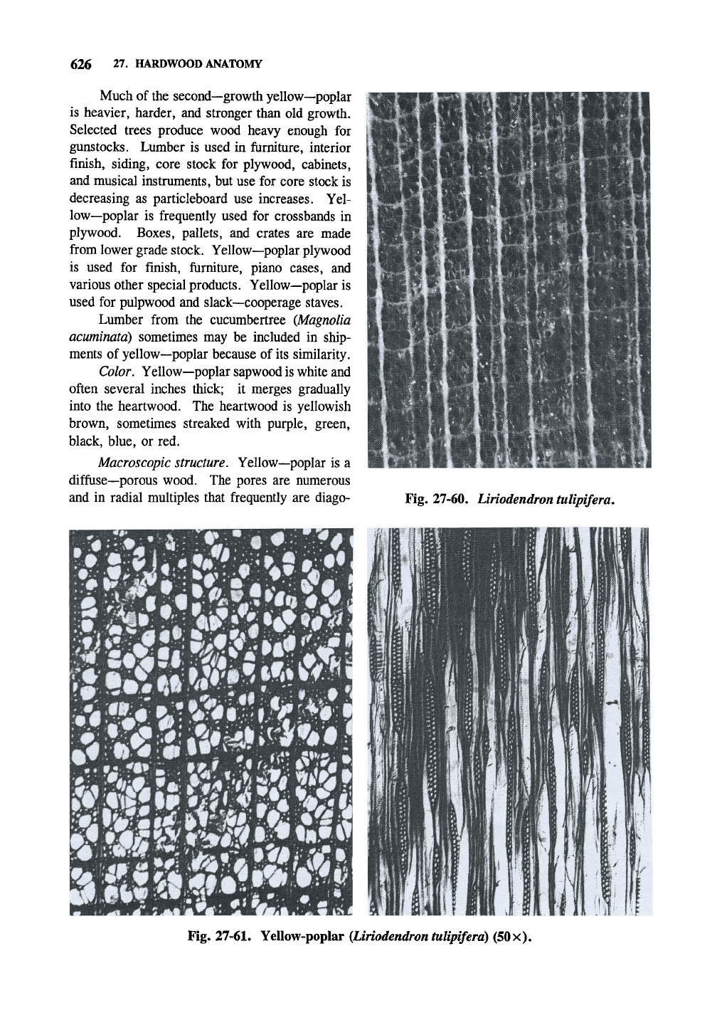
Yellow—poplar (Figs. 27-60 and 27-61)
Yellow—poplar (Liriodendron tulipifera) is
also known as poplar, tulip poplar, tulipwood, and
hickory poplar. Sapwood from yellow—poplar is
sometimes called white poplar or whitewood.
Yellow—poplar grows from Connecticut and New
York southward to Florida and westward to
Missouri. The greatest commercial production of
yellow—poplar lumber is in the South.
Uses. The wood is generally straight grained
and comparatively uniform in texture.
Old—growth timber is moderately light in weight
and is reported as being moderately low in bend-
ing strength, moderately soft, and moderately low
in shock resistance. It has moderately large
shrinkage when dried but is not difficult to season
and stays in place well after seasoning.
Fig. 27-59. Black willow {Salix nigra) (50X).
626 27. HARDWOOD ANATOMY
Much of the second—growth yellow—poplar
is heavier, harder, and stronger than old growth.
Selected trees produce wood heavy enough for
gunstocks. Lumber is used in furniture, interior
finish, siding, core stock for plywood, cabinets,
and musical instruments, but use for core stock is
decreasing as particleboard use increases. Yel-
low—poplar is frequently used for crossbands in
plywood. Boxes, pallets, and crates are made
from lower grade stock. Yellow—poplar plywood
is used for finish, furniture, piano cases, and
various other special products. Yellow—poplar is
used for pulpwood and slack—cooperage staves.
Lumber from the cucumbertree (Magnolia
acuminata) sometimes may be included in ship-
ments of yellow—poplar because of its similarity.
Color. Yellow—poplar sapwood is white and
often several inches thick; it merges gradually
into the heartwood. The heartwood is yellowish
brown, sometimes streaked with purple, green,
black, blue, or red.
Macroscopic structure. Yellow—poplar is a
diffuse—porous wood. The pores are numerous
and in radial multiples that frequently are diago-
%'•*
»« fiii
IK
*If''«
Z*4
V^ b
Fig. 27-60. Liriodendron tulipifera.
Fig.
27-61.
YeUow-poplar
(Liriodendron tulipifera)
(50x).
ANATOMY OF HARDWOOD SPECIES 627
nal.
Rays are normally spaced, much fewer than
in sweetgum, and visible to the unaided eye.
Longitudinal parenchyma are marginal, forming a
continuous white line of several rows in the outer
portion of the latewood. The vessel distribution is
variable, frequently being less numerous in the
outer latewood.
Microscopic structure. Intervessel pitting is
opposite, pits are rectangular in outline.
Similar woods. Yellow—poplar can usually
be distinguished from other woods by its greenish
tinge. Sweetbay (magnolia) is very similar, but
can be separated by use of a microscope.
Non—North American Species
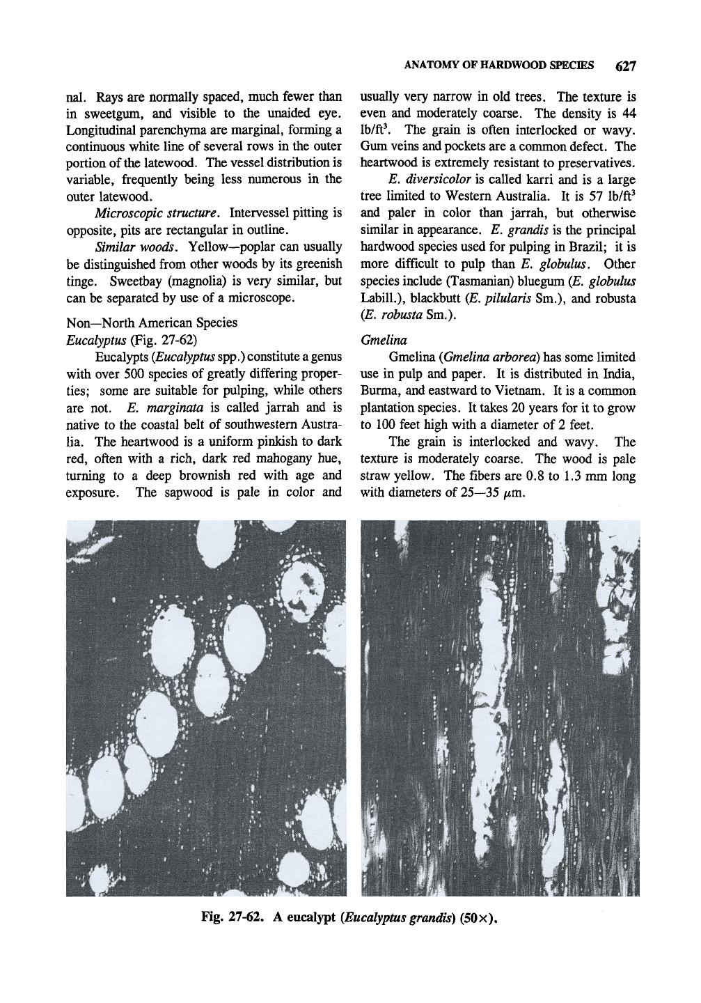
Eucalyptus (Fig. 27-62)
Eucalypts
(Eucalyptus
spp.) constitute
a
genus
with over 500 species of greatly differing proper-
ties;
some are suitable for pulping, while others
are not. £*. marginata is called jarrah and is
native to the coastal belt of southwestern Austra-
lia. The heartwood is a uniform pinkish to dark
red, often with a rich, dark red mahogany hue,
turning to a deep brownish red with age and
exposure. The sapwood is pale in color and
usually very narrow in old trees. The texture is
even and moderately coarse. The density is 44
lb/ft\ The grain is often interlocked or wavy.
Gum veins and pockets are a common defect. The
heartwood is extremely resistant to preservatives.
E. diversicolor is called karri and is a large
tree limited to Western Australia. It is 57 Ib/ft^
and paler in color than jarrah, but otherwise
similar in appearance. E. grandis is the principal
hardwood species used for pulping in Brazil; it is
more difficult to pulp than E. globulus. Other
species include (Tasmanian) bluegum (E. globulus
Labill.),
blackbutt (E, pilularis Sm.), and robusta
{E. robusta Sm.).
Gmelina
Gmelina
(Gmelina arborea)
has some limited
use in pulp and paper. It is distributed in India,
Burma, and eastward to Vietnam. It is a common
plantation species. It takes 20 years for it to grow
to 100 feet high with a diameter of 2 feet.
The grain is interlocked and wavy. The
texture is moderately coarse. The wood is pale
straw yellow. The fibers are 0.8 to 1.3 mm long
with diameters of 25—35 iim.
Fig. 27-62. A eucalypt (Eucalyptus grandis) (50
x).
28
WOOD FIBER ANATOMY
AND
IDENTIFICATION
28,1 FIBER ANALYSIS
Introduction
The references
for
this chapter
are
found
on pages 537—540
of
Chapter
25.
It
is
useful
to
look
at
wood
in
terms
of its
fiber anatomy
and
properties, since this
is how it
is used
by the
pulp
and
paper industry.
There are many excellent references available
for fiber analysis
(see the
Annotated Bibliography
in Chapter
25), and
much
of the
information
presented
in
earlier chapters
can be
used. This
chapter gives
an
overview
of
fiber morphology
from wood
and
shows what features
to
look
for in
fibers for their identification. Information
is
also
given
to
help determine pulping
and
bleaching
methods used
to
prepare
the
pulp.
It
is
ftindamental that
the
properties
on
paper
depend
on the
fiber properties
and the
method
of
fiber preparation (pulping, bleaching,
and
fiber
processing).
It is
difficult
to
separate wood prior
to pulping (latewood from earlywood, fast growth
wood from slow growth wood, compression wood
from normal wood, juvenile wood from mature
wood, etc.),
so
variability
of
wood must
be
over-
come
by
varying
the
processing
of the
wood
and
pulp.
The
availability
of
and price
of
wood
are as
important
as the
properties
of the
wood
itself.
Fiber anatomy considerations
Identification
of
fibers
is
difficult since there
is much less information available than
in the
wood; identification
is
often down
to a few
members within
a
genus, rather than
to a
particu-
lar species. Other species, like Douglas—fir with
spiral thickening,
are
unique
and can be
identified
as
to the
species.
The
situation
is
complicated
by
the fact that many papers
can
contain two, three,
or more types
of
fibers, especially
if the
paper
contains appreciable amounts
of
recycled fiber.
Panshin
and de
Zeeuw (1980,
pp.
657—666)
in-
clude
a key for
fiber identification.
It
is
important to have authentic fiber samples
of contributing species
on
hand
for
comparison
with unknown samples.
For
example,
if one is
using
a lot of
"precommercial" thinnings with
a
high percentage of juvenile wood, fiber length will
be shorter than published values.
It takes
a
large investment
of
time
to
become
proficient
at
fiber identification,
and
many refer-
ences containing micrographs should
be
used.
This chapter allows
one to
visualize
the
major
differences
in
pulps that contribute
to the
paper-
making properties
and is
useful
for
simple identifi-
cations, such
as
determining when various places
in
a
mill
are
affected
by the
changeover
of a
digester line from
one
species
to
another.
In mechanical pulps
one can
learn about
the
relative morphology
of
the contributing species
by
looking
for
fiber bundles.
Weight factors
When attempting quantitative analysis
of
fiber
mixtures,
the
number
of a
particular type
of
fiber
is multiplied by
its
weight factors since some types
of fibers appear
to be
present
in
larger amounts
than others, such
as
those with
low
surface areas.
Weight factors
are
given
in T 401
[based largely
on
Graff, J.H.,
Paper Trade
J.
110(2):37(1940)].
The weight factors
for
coastal Douglas—fir
and
loblolly pine
are 1.40, for
most hardwoods
0.50,
and
for
bleached straw
0.35. The
higher
the
relative surface area
(per
given mass
of
pulp),
the
lower
the
weight factor. Parham
and
Gray (1990)
give weight factors
on
page
35
[largely from
D.W. Einspahr
and J.D.
Hankey, Tappi 61(12):
86-87(1978)].
One
could determine one's
own
weight factors
for
a particular application. Weight
factors
are a
crude method
of
taking number
averages
and
converting them
to
weight averages.
Fiber
pitting
Pitting,
in
general,
is
very useful
in the
determination
of
isolated fibers. However,
one
must keep
in
mind that pulping operations
and
refining
are
bound
to
affect
the way a pit
looks
under
the
microscope. Intervessel pitting
of
hardwoods
and ray
cross—field pitting
in
soft-
woods
are the
principal means
of
identification.
Ray cross—field pitting shows whether
ray
paren-
chyma
and or
tracheids
are
present;
it
also indi-
cates
the
height
of the
rays.
628
FTOER ANALYSIS 629
Fiber and wood staining
Chemical stains can help with the analysis by
identifying the type of pulping method used to
generate the fibers. This might be more important
in brown papers that might have some recycled
newsprint rather than in white printing papers, but
these might have some bleached CTMP pulp.
Hoadley (1990) references examples of
species determination of wood using chemical
stains including boxelder and other maples, red
gum and black tupelo, elms, pond and sand pines,
spruces and pines, and other groups. Separation
of red and white oaks and red and sugar maples is
discussed in Panshin and de Zeeuw (1980). East-
ern spruce and balsam fir are separated by the
method of Kutscha et al. in Wood Sci. and Tech.
12:293-308(1978). Separation of heartwood and
sapwood is referenced (Kutscha and Sachs, 1962).
Iodine is used to detect the presence of
starch, which might be present in sapwood but not
heartwood or in paper such as in the detection of
forged currency. It is used with iodide to form
the soluble
I3'
species in water (0.3% I2 and 1.5%
KI in water). Safranin O is a general stain for
cellulosic materials (red). It is used as a 0.5—1
%
solution in water or 50% ethanol.
Pulp stains are described in detail in Graff
(1940),
Isenberg (1967), and TAPPI Standard
T401 om-82. Graff (1940) describes three types
of stains. (1) Spot stains or groundwood reagents
using chemicals such as aniline sulfate, phloroglu-
cinol & hydrochloric acid, and/?—nitroaniline for
detection of mechanical pulps in paper. The first
class is said here to be of limited value. (2) The
iodine/iodide metallic salt stains including the
Herzberg stain, the "A" stain, and the "C" stain;
these give qualitative information about fibers. (3)
Aniline dyes for the determination of
the
degree of
cooking, bleaching, and purity of pulps.
Phloroglucinol is specific for lignin, causing
it to turn red. It is used as a 2% solution in
18.5%
HCl. Isenberg (1967) suggests mixing 1 g
in 50 mL of EtOH to which is added 25 mL of
concentrated HCl; this is made up immediately
before use. One use is to show mechanical pulps
in white papers.
Mechanical softwood and hardwoods are
differentiated with the use of 2% aniline sulfate
made acidic (1 drop concentrated H2SO4 per 50
mL) followed by 0.02% methylene blue after
removal of the first dye by blotting. Softwoods
are yellow and hardwoods are bluish green. The
Maule reaction can also be used.
Mechanical pulp bleached with dithionite no
more than a few months old is determined by
detecting traces of SO2 or sulfite as H2S liberated
by stannous chloride. Several stains are available
to determine the level of bleaching (or cooking) of
pulps,
such as Bright stain. These can be used to
check the uniformity of bleaching (and pulping) in
the mill (Isenberg, 1967). Stains even exist for
the identification of dirt, although scanning elec-
tron microscopy (SEM-EDDAX) page 184) has
made many of these methods somewhat obsolete.
Other
techniques
Monosaccharide analysis (although tedious)
will give a good indication of the ratio of hard-
woods to softwoods since galactose and mannose
come from softwoods. Dissolving pulps will have
low levels of hemicelluloses, of course.
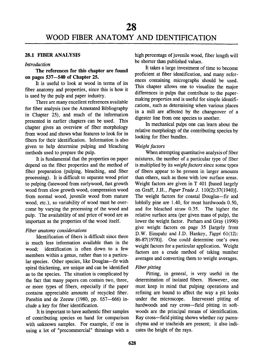
28.2 SOFTWOOD FIBER
Fibers over 2 mm long are most often soft-
wood fibers. Be sure to use Table 26-3 (page
550) as a general guide to softwood fiber analysis,
and Table 26-4 (page 552) as a summary of some
important softwood fiber properties. Fig. 28-1
shows six types of softwood fibers.
Parham and Gray (1990) claim that Doug-
las—fir, redwood, baldcypress, podocarp,
parana—pine, sugar pine, white pine, and hard
pines should
be
distinguishable under most circum-
stances, although these separations require a high
degree of skill. Spruce, larch, and hemlock may
not be able to be distinguished from each other as
with true—fir and western redcedar. If the
source of
the
pulp is known then regional informa-
tion can be used (i.e., European or American;
eastern or western, etc.).
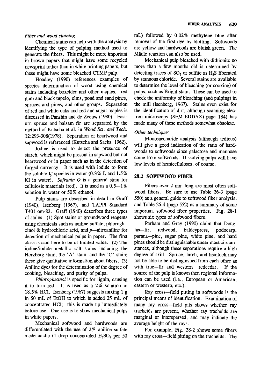
Ray cross—field pitting in softwoods is the
principal means of identification. Examination of
many ray cross—field pits shows whether ray
tracheids are present, whether ray tracheids are
marginal or interspersed, and may indicate the
average height of the rays.
For example. Fig. 28-2 shows some fibers
with ray cross—field pitting on the tracheids. The
Douglas—fir
Southern pine
Eastern white pine
Sitka spruce Western hemlock Western cedar
Fig. 28-1. Various softwood fibers isolated in the laboratory (60x).
SOFTWOOD FIBER 631
western white pine shows the fenestriform ray
cross—field pitting that is characteristic of the
white pines and the small pits are from ray
tracheids. Ponderosa pine has pinoid ray cross—-
field pitting with pits that are variable is size and
up to 7 pits per cross—field. The Sitka spruce
fiber shows piceoid ray cross—field pitting of
uniform size with 3 or more pits per cross—field
common.
28.3 HARDWOOD FIBER
Be sure to use Table 27-1 (page 590) as a
general guide to hardwood pulp analysis. Whole
fibers less than 2 mm long usually originate from
hardwoods. Hardwood fibers are accompanied by
vessels or vessel fragments depending on the
pulping process. The fibers shown in this chapter
were isolated by a laboratory pulping process that
leaves the vessels largely intact. Furthermore,
parenchyma cells have not been removed as occurs
to various degrees in pulp processing. Fig. 28-3
shows fibers from hardwoods.
Hardwood libriform fibers or fiber tracheids
are not helpful in species determination unless
helical thickening is observed. Vascular or
vasicentric tracheids [the shape of vasicentric
tracheids is observed in red oak (upper right) in
Fig.
28-3],
indicate the presence of certain spe-
cies.
However, vessel elements offer much
information based on their overall size, shape,
intervessel pitting, and ray contact pitting. The
intervessel pitting of Populus (observe black
Cottonwood in
Fig.
28-3) and Salix genera are easy
to recognize . Ring—porous woods may be diffi-
cult to determine since the large vessels are gener-
ally fragmented during processing.
Western white pine Ponderosa pine Sitka spruce
Fig. 28-2. Portions of fiber tracheids of three softwoods (600x).
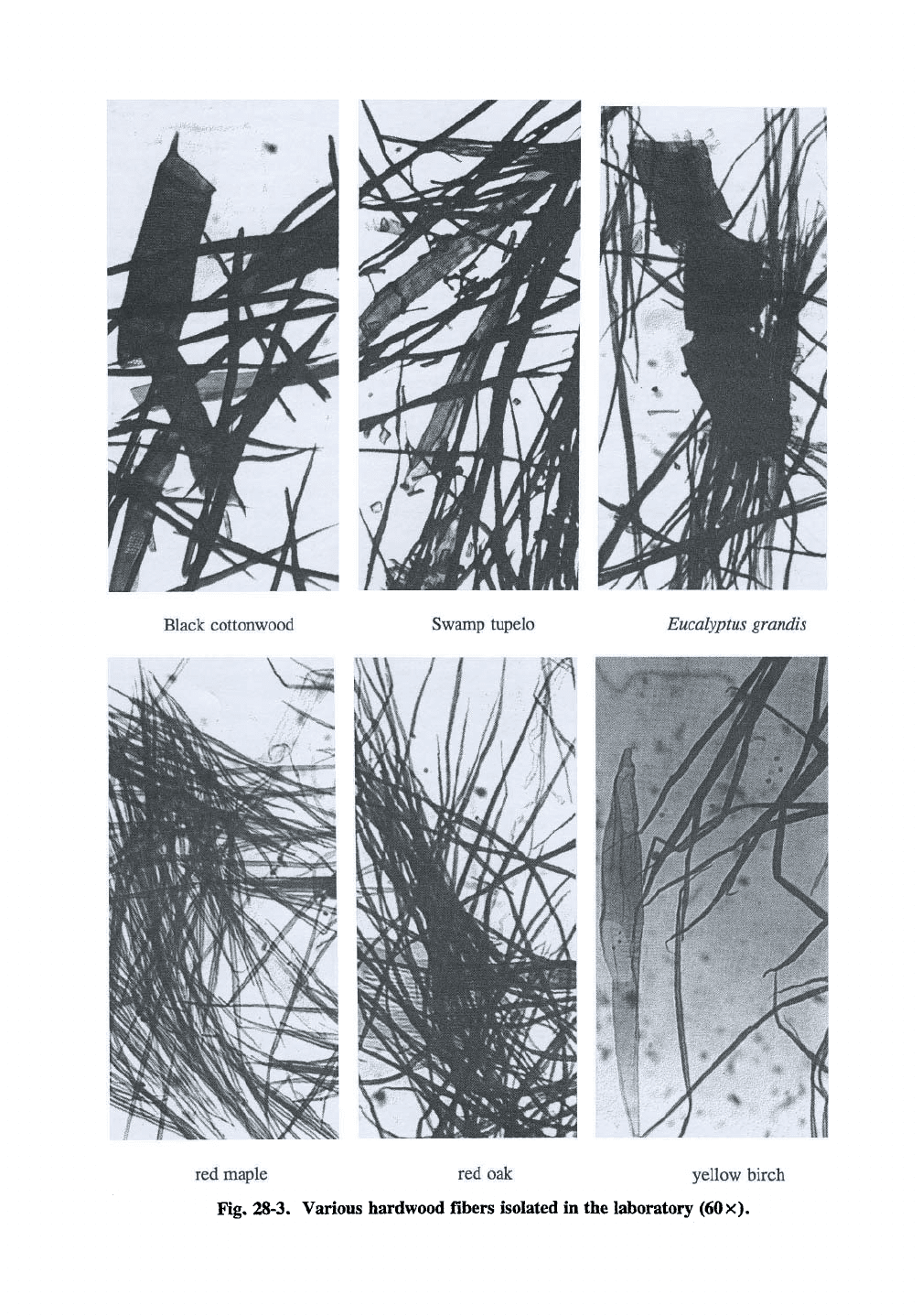
Black Cottonwood
Swamp tupelo
Eucalyptus grandis
red maple red oak yellow birch
Fig, 28-3. Various hardwood fibers isolated in the laboratory (60x).
29
NONWOOD FIBER USE IN PULP AND PAPER
29.1 INTRODUCTION
Nonwood fiber is increasingly being used
worldwide as a fiber source for papermaking. Ac-
cording to Atchison (1988), major nonwood pulp
production includes (in millions of metric tons per
year) straw, 5.4 (over 80% produced in China);
sugar cane bagasse, 2.1; bamboo, 1.4; reeds,
1.6; cotton linters, 0.4; and other, 1.0. This
chapter provides some background information on
the topic. See pages 47-50 for more information.
Taxonomy
All seed plants (members of the phylum
Spermaphyta) are divided into two subphyla:
Gymnospermae (gymnosperms, those with naked
seeds) and Angiospermae (angiosperms, those with
seeds enclosed within the ovary of the flower).
Angiospermae are arranged into two groups:
the monocots
{monocotyledons)
and dicots (dicoty-
ledons),
Monocotyledoneae have one leaf in the
seed (a seed leaf is a cotyledon; a peanut consists
of two cotyledons with a small embryo in the
middle so peanuts are dicots) and include mostly
herbaceous plants such as grasses, palms, lilies,
and corn. A few plants with a wood—like stem
such as bamboo that are used in pulp and paper
are members of this class.
Dicotyledoneae have two leaves in the seed
and include
kenaf,
hemp, flax, and the hardwoods.
The numerous Dicotyledoneae are further divided
into subclasses, orders, suborder, families, and
some subfamilies.
Monocots have parallel veins in their leaves,
and dicots have branching veins in the leaves.
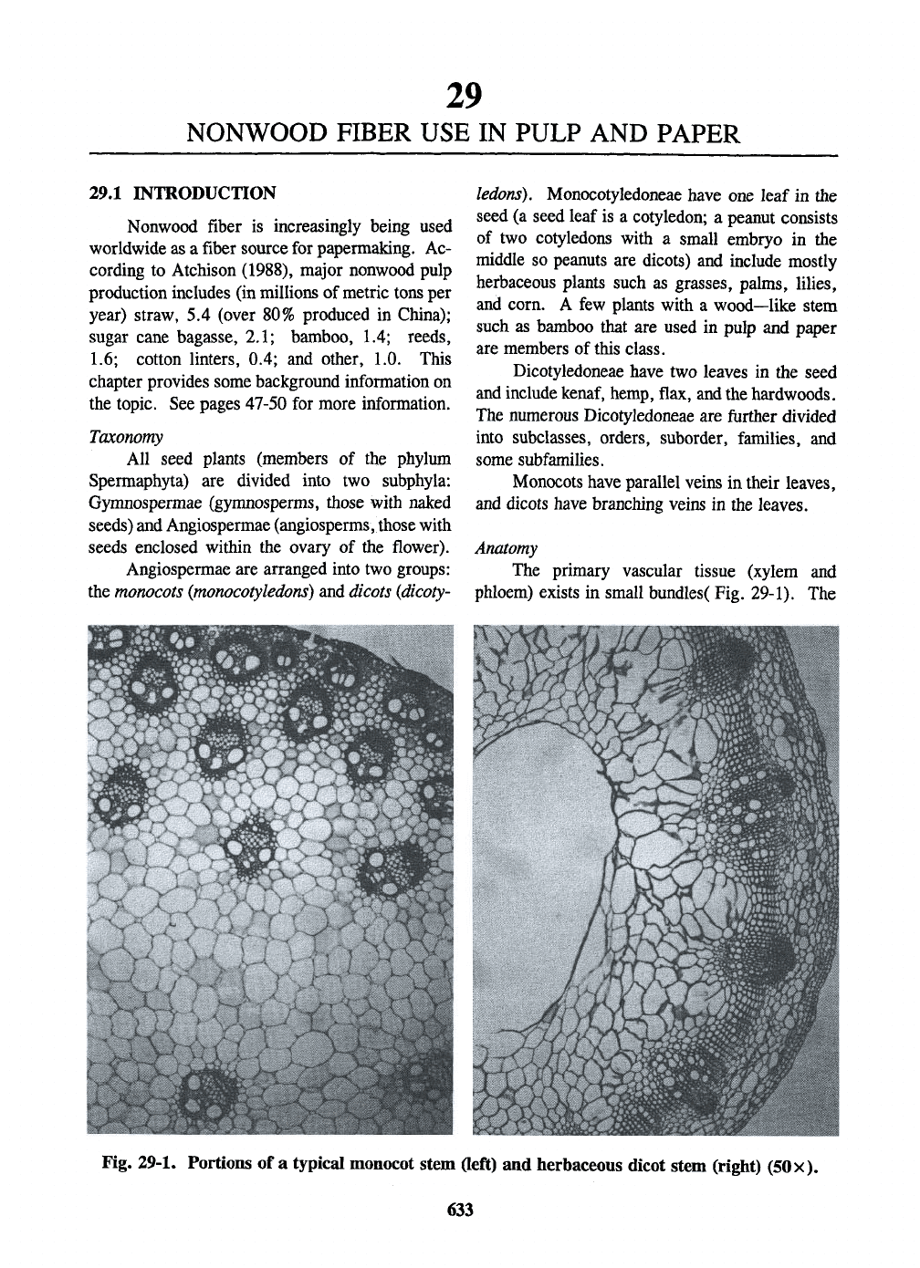
Anatomy
The primary vascular tissue (xylem and
phloem) exists in small bundles( Fig. 29-1). The
Fig. 29-1. Portions of a typical monocot stem Geft) and herbaceous dicot stem (right) (50x).
633