Bergaya F. Handbook of Clay Science
Подождите немного. Документ загружается.


these studies is within error for coordination number determinations, the interatomic
distances are different . Contributions from wave vectors at higher k influence the fit
to wave vectors at lower k, resulting in different R
eff
. Interestingly, incorporating
more shells at higher R*, and ignoring any contributions of multiple-scattering
events, improve the fit on R
eff
. This can be seen by comparing the results in Table
12.3.1 with those of Vantelon et al. (2003).
F. Polarised EXAFS Spectroscopy
Application of P-EXAFS to layer silicat es is unique in that it allows all out-of-plane
contributions to the EXAFS spectrum to be systematically removed, and accurate
structural information about the in-plane contributions only (and vice versa) to be
obtained. A backscattering phase shif t between a heavy (e.g., Fe, Mn, Ni, etc.) and a
light (e.g., Al, Mg) atom enhances quantification of the heterogeneous nature of
octahedral and tetrahedral cation occupancies in smectites and other layered min-
erals. P-EXAFS also has the inherent ability to increase spectral resolution enabling
differentiation of atomic shells separated by only 10–20 pm (Manceau et al., 1998).
In powder EXAFS, the resolution power in the R* range is limited by low signal-to-
noise ratios in the high k region. On the other hand, the P-EXAFS method increases
the signal-to-noise ratio at high k, thereby improving the resolving power in R*.
For powdered systems (random disorder of the particles) P-EXAFS spectra taken
at several angles will show little, if any, variation in amplitude (Manceau, 1990). For
self-supporting clay mineral films where the layers are aligned, changes in the film
angle (a) with respect to the X-ray beam will alter the amplitude (pleochroism).
Backscattering atoms situated out-of-plane (tetrahedral Si, Al or Fe, structural ox-
ygen) from the target absorber (e.g., octahedral Fe) will contribute positive
pleochroism (amplitude enhancement) and those atoms within the film plane (e.g.,
Al, Mg, Fe) will contribute negative pleochroism to the EXAFS amplitude with
increasing a.
Exactly how the P-EXAFS amplitude varies with a for Garfield nontronite
(Manceau et al., 1998) is shown in Table 12.3.2 and Fig. 12.3.7A .InTable 12.3.2, the
term b is the average angle between the absorber and the backscattering atoms of a
particular shell, with respect to the film plane normal, and N
cryst
the number of
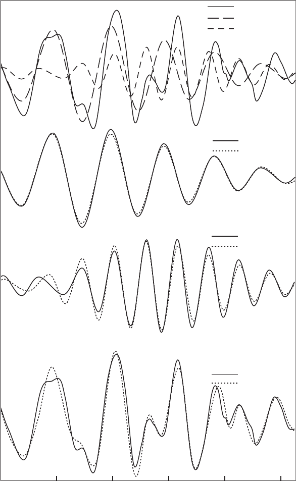
Fig. 12.3.6. Analysis of ligand (FeyO
1
) and nearest cation (FeyM
1
) contributions to the k
3
weighted Fe K EXAFS spectra of ferruginous smectite SWa-1, collected on an oriented film at
a ¼ 351 (equivalent to a powder EXAFS). The FeyO
1
and FeyM
1
shells were isolated from
the Fourier transform at R* ranges of 0.8–2.05 A
˚
and 2.05–3.2 A
˚
, respectively, and back-
transformed (top panel). The FeyO
1
(upper middle panel) and FeyM
1
(lower middle panel)
shells were fit to the structural parameters displayed. A fit of the total contribution of the
FeyO
1
and FeyM
1
shells, as well as higher R* shells, to the EXAFS is shown in the bottom
panel. See Table 12.3.1 for fitting parameters of this latter fit.
Chapter 12.3: X-ray Absorption Spectroscopy806

2
4 6 8 10 12
Wavevector k (Å
-1
)
k
3
χ(k)
Fe K EXAFS
Fe-O
1
shell
Fe-M
1
shell
Fe K EXAFS
FEFF Fit
k
3
χ(k)
Fe-O
1
shell
FEFF Fit
k
3
χ(q)
2 O d = 201 pm
4 O d = 202 pm
SWa-1
Thin film
α = 35˚
Fe-M
1
shell
FEFF Fit
k
3
χ(q)
2.1 Fe d = 305 pm
0.9 Al d = 303 pm
4 Si d = 237 pm
See Table 1 for Fit
12.3.1. Synchrotron-Based Techniques 807

atoms of the shell of the idealised structure. The relationship between N
cryst
and the
effective number of neighbouring atoms (N
eff
) actually seen by P-EXAFS at a given
film angle, is given by (Manceau et al., 1999)
w
ij
k; aðÞ¼
N
eff
N
cryst
w
iso
ij
kðÞ ð9Þ
In Eq. (9), w
ij
(k,a) is the amplitude determined by P-EXAFS for an atomic pair as
a function of reciprocal k-space and film angle a and w
ij
iso
(k) the amplitude deter-
mined on perfectly random systems. N
eff
for each film angle is given by
N
a
eff
¼ 3N
cryst
cos
2
bsin
2
a þ
sin
2
bcos
2
a
2
"#
ð10Þ
Note that at a ¼ 01
N
0
eff
¼
3N
cryst
sin
2
b
2
ð11Þ
and at a ¼ 901
N
90
eff
¼ 3N
cyrst
cos
2
b ð12Þ
From knowledge of the crystallographic angle, b, between the absorbing and the
backscattering atoms, as well as the idealised number of neighbours, N
cryst
,itis
possible to calculate the effective number of neighbours, N
eff
a
, observed at any angle
in a P-EXAFS experiment. Alternatively, b canbecalculatedfromknowledgeofN
eff
a
determined from P-EXAFS spectra or from simulations. The ability to predict N
eff
Table 12.3.2. Angular dependence of some atomic shell contributions to P-EXAFS spectra of
Garfield nontronite. Modified after Manceau et al. (1999a), with R* calculated using FEFF.
The b angle is measured with respect to film normal. See text for definitions
Shell R* (pm) ob>(1) N
cryst
N
eff
0
N
eff
20
N
eff
35
N
eff
60
N
eff
80
N
eff
90
FeyO
1
197, 204 57 6 6.3 6.2 6.0 5.6 5.4 5.3
FeyFe
1
304 90 3 4.5 4.0 3.0 1.1 0.1 0
Fey(Si,Al)
1
326 32 4 1.7 2.5 4.0 7.3 8.4 8.6
FeyO
2
345 11 2 0.1 0.8 2.0 4.4 5.6 5.8
FeyO
3
374, 382 73 6 8.2 7.4 6.0 3.2 1.7 1.5
FeyO
4
402, 419 37 4 2.2 2.8 4.0 6.3 7.5 7.6
Fey(Si,Al)
2
449 52 4 3.7 3.8 4.0 4.3 4.5 4.5
FeyNa 510 15 2 0.2 0.8 2.0 4.2 5.4 5.6
FeyFe
2
528 90 6 9 7.9 6.0 2.3 0.3 0
FeyFe
3
609 90 3 4.5 4.0 3.0 1.1 0.1 0
Chapter 12.3: X-ray Absorption Spectroscopy808

0°
20°
35°
60°
90°
12.5105 7.52.5
Wavevector k (Å
-1
)
k
3
χ(k)
B
O
1
O
2
O
3
O
4
Fe
1
Si
1
9
6
3
0
0 153045607590
N
eff
Film angle α
A
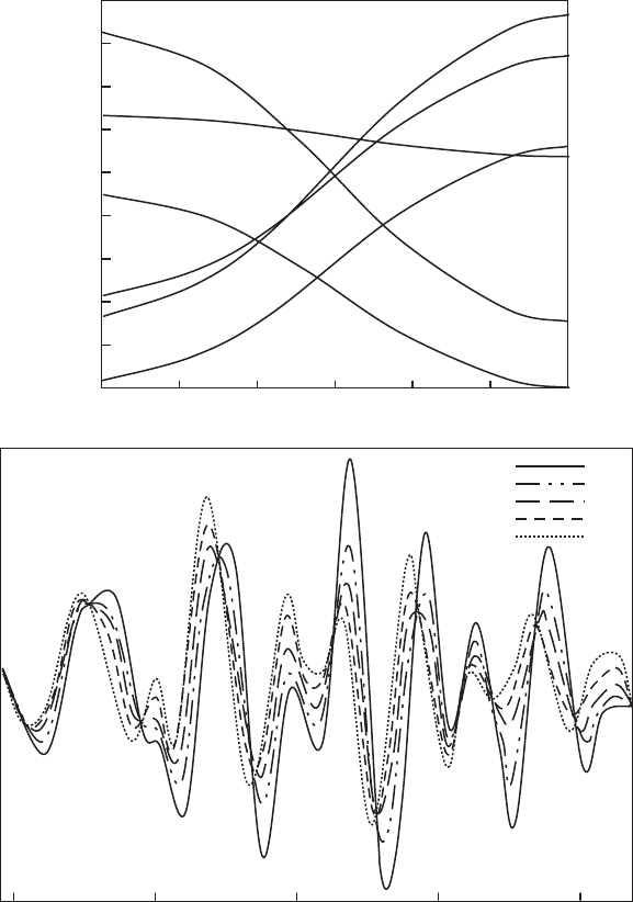
Fig. 12.3.7. (A) Variation of N
eff
as a function of film angle a for Fe K P-EXAFS spectra. (B)
Polarised k
3
weighted Fe K EXAFS spectra of ferruginous smectite SWa-1. The a ¼ 901
spectrum was calculated from a linear extension of the experimental amplitude as a function of
cos
2
a. Isobestic points occur where the EXAFS amplitude (k
3
w(k)) is invariant regardless of a,
and separate in-plane contributions, where k
3
w(k) increases with increasing a, from out-of-
plane contributions, where k
3
w(k) decreases with increasing a.
12.3.1. Synchrotron-Based Techniques 809
from structural information provides better fits of the spectra, as it decreases the number
of fitting variables, or at least puts realistic limits upon them.
Table 12.3.2 shows that for shells whose atoms lie at shallow angles with respect to
thefilmnormal(e.g.,FeyO
2
,FeyO
4
and Fey(Si,Al)
1
), large increases in N
eff
a
occur
with increasing a. For those shells having atoms residing at steep angles with respect to
thefilmnormal(e.g.,FeyFe
1
,FeyFe
2
,FeyFe
3
and FeyO
3
), N
eff
a
decreases by
differing amounts: N
eff
a
diminishes to 0 for the FeyFe
1
,FeyFe
2
and FeyFe
3
shells at
a ¼ 901, but to 1.5 for O
3
. For shells with atoms lying near b ¼ 551,littlechangeoccurs
in N
eff
a
as a function of a.Thus,b 551 is a critical angle where N
eff
a
is invariant for all
values of a.Atafilmangleofa 351, the effective number of neighbours is approx-
imately equal to the crystallographic number (N
eff
35
E N
cryst
), a fact demonstrated by
Manceau et al. (1988) for single crystal and powdered mica. Thus, a 351 is another
critical angle in that P-EXAFS spectra collected at a 351 will be identical to powder
EXAFS spectra.
The in-plane contributions can be completely removed from an a ¼ 901 EXAFS
spectrum, whi ch can then be used to estimate the magnitude of out-of-plane con-
tributions. This estimate is then used to remove any out-of-plane contributions from
the a ¼ 01 spectrum to obtain an estimate of in-plane contributions only. Trans-
mission measurements at a ¼ 901 are not easily obtained experimentally—it should
be possible to use glancing angle or wide angle X-ray scattering (GAXS or WAXS)
analyses—and P-EXAFS data are usually collected at several angles of the self-
supporting film with respect to the X-ray beam (Figs. 12.3.7B and 12.3.8A).
Processing P-EXAFS data requires careful ampli tude normalisation because the
amount of sample probed increases with increasing a (it doubles from a ¼ 01 to 601).
The normalised a ¼ 01,201,351, y ,601 spectra for a given sample are then used to
calculate an a ¼ 901 spectrum by linear regression of the k
3
w(k) (or k
1
w(k)) amplitude
against cos
2
a, for all points in reciprocal (k) space (Manceau et al., 1998). The
resulting calculated a ¼ 901 spectrum is then processed in the same way as for other
EXAFS spectra, the difference being that the a ¼ 901 contributions are isolated from
the forward FTs and subtracted from those isolated from the a ¼ 01 spectrum to
obtain a partial EXAFS spectrum containing only in-plane contributions.
Recall that the FeyM
1
shell in a powder EXAFS spectrum of a smectite is a
composite of in-plane (octahedral cations) and out-of-plane (ligand and tetrahedral
cations) contributions. The difference of the resulting in-plane FeyM
1
partial
P-EXAFS spectra from that obtained on powder samples where the out-of-plane
scattering events contribute, is striking (Fig. 12.3.8B) and, as was shown abov e,
influences the resulting analysis (Table 12.3.1 and discussion in Section 12.3.2D). As
the subtraction process removes the out-of-plane contributions, a phase shift to
lower wave vectors is observed in the partial FeyM
1
P-EXAFS spectrum relative to
the partial FeyM
1
powder EXAFS spectrum.
The a ¼ 901 contributions are normally only calculated for a limited k(q) range,
but there is no reason, other than increased complexity, that the entire spectrum
could not be processed and analysed in a similar fashion. Detailed analysis of highly
Chapter 12.3: X-ray Absorption Spectroscopy810

Wavevector k (Å
-1
)
Powder EXAFS
SWa-1
Fe-M
1
shell
P - EXAFS
k
3
χ
χ
(k)
3 5 7 9 11 13
046
FT (k
3
χ(k)
R* (Å)
0°
20°
35°
60°
90°
Fe-O
1
Fe-O
2
Fe-Fe
1
Fe-Fe
2
Fe-Si
1
Fe-Si
2
Fe-O
3
Fe-O
4
A
Fe-Na
B
2
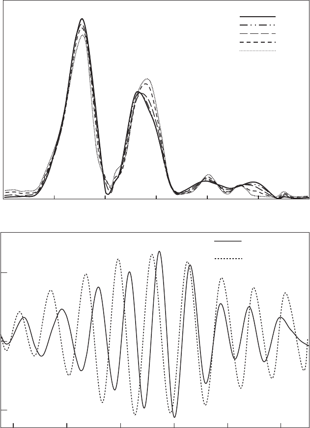
Fig. 12.3.8. (A) The resulting Fourier transforms of k
3
weighted Fe K P-EXAFS in Fig.
12.3.7B. Assignments to shells based on Manceau et al. (2000a). (B) Comparison of the
FeyM
1
shell isolated from the a ¼ 351 spectrum of ferruginous smectite SWa-1 before
(powder EXAFS) and after (P-EXAFS) subtracting the tetrahedral contributions using the
procedures described in Section 12.3.3.
12.3.1. Synchrotron-Based Techniques 811
textured self-supporting nontronite films (Manceau et al., 1998, 2000a, 2000b) in-
dicates that P-EXAFS spectra returns the appropriate values calculated from theory.
Interestingly, Schlegel et al. (1999a, 1999b, 2001a) and Da
¨
hn et al. (2002a, 2002b,
2003) recently collected P-EXAFS data at angles as high as a ¼ 801. Given the
uncertainties associated with measurement and extraction of EXAFS amplitudes, as
well as the dispersion inherent in self-supporting films (Manceau et al., 1998, 1999;
Schlegel et al., 1999a, 1999b; Manceau et al., 2000a, 2000b; Schlegel et al., 2001a;
Da
¨
hn et al., 2002a, 2002b, 2003) this measuremen t is essentially equivalent to one
calculated from an a ¼ 901 spectrum (compare the N
eff
values at a ¼ 801 and a ¼ 901
in Fig. 12.3.7A).
For further information on experimental set-up, sample preparation and spectral
analysis of P-EXAFS data, the reader is referred to the papers by Manceau et al.
(1998, 1999, 2000a) and Manceau and Schlegel (2001). Poor alignmen t of individual
particles within self-supporting films remains a potential limitation of P-EXAFS as
this will result in dispersion, amplitude dampening for out-of-pl ane features, am-
plitude augmentation of in-plane features and loss of linearity in the pleochroic
relation. However, for typical smectite self-supporting films, the error arising from
this condition is either small enough to be neglected or can be corrected using charts
prepared by Manceau and Schlegel (2001).
12.3.2. XAFS STUDIES ON SMECTITE STRUCTURE
A. Structural Refinement by P-EXAFS
It is only recently that P-EXAFS was applied to investigate the structures of fine-
grained and poorly crystalline layer silicates (Manceau et al., 1998, 1999a; Schlegel
et al., 1999a; Manceau et al., 2000a, 2000b; Schlegel et al., 2001a; Da
¨
hn et al., 2002a,
2002b, 2003), although it was earlier used to study the location of Fe in micas and
chlorites (Manceau et al., 1988, 1990a; Manceau, 1990; Waychunas and Brown,
1990; Dyar et al., 2002) and to determine bond angles in graphite intercalates
(Bonnin et al., 1986). Heald and Stern (1977) were the first to use anisot ropic X-ray
absorption to study single crystals. Manceau et al. (1988) applied P-EXAFS to
examine Fe distribution in chlorite and biotite, observing that the second peak
(FeyM
1
) in the FT occurred at shorter R* distance when the electric vector was in-
plane with the octahedral sheet than when it was out-of-plane. Direct comparison of
the back-transformed partial EXAFS spectra with that of biotite showed that about
25% of the total Fe was located in interlayer sites of the chlorite.
P-EXAFS was extended to structural refinements of nontronite (Manceau et al.,
1998, 1999a, 2000a, 2000b) and phyllomanganates (Manceau et al., 1999a) and more
recently to determine the location of adsorbed metals on hectorite (Schlegel et al.,
1999a, 1999b, 2000) and montmorillonite (Da
¨
hn et al., 2002a, 2002b, 2003) surfaces.
P-EXAFS improved our understanding of sorption sites (Hazemann et al., 1992;
Chapter 12.3: X-ray Absorption Spectroscopy812
O’Day et al., 1994a; Schlegel et al., 1999a, 1999b, 2001a, 2001b; Da
¨
hn et al., 2002a,
2002b, 2003). The technique is also potentially capable of assessing site occupancies
of Al and Mg in Fe-poor beidellites, montmorillonites and saponites, where
27
Al
NMR fails (Muller et al., 1997; Manceau et al., 2000a; Vantelon et al., 2003).
P-EXAFS has yet to be applied to obtain structural refinements of other layered
minerals such as palygorskites, sepiolites and layered double hydroxides. In addition,
P-EXAFS would be suitable for assessing trace metal substitution (e.g., Zn or Mn
for Al) in smectites formed under various geochemical conditions or exposed to
different weathering regimes.
The structures of some nontronites and ferruginous smectite were refined by
Manceau et al. (1998, 1999a, 2000a) using a combination of P-EXAFS and mod-
elling. Distance-valence least-squares (DVLS) modelling of the c2/m symmetry of
nontronite enabled the contribution of atomic scattering paths (from FEFF) to
partial EXAFS spectra to be determinded (Manceau et al., 1998). The DVLS and
FEFF method was found to produce calculated spectra in excellent agreement with
experimental spectra. In addition, Manceau et al. (1999a) provided a method to
determine crystallographic angles associated with the layered structure. Manceau
et al. (2000a) found that Fe(III) predominantly occupies M2 (cis) octahedra in
agreement with earlier electron diffraction studies (Tsipursky and Drits, 1984). For
the Fe-poor smectites, Vantelon et al. (2003) recently observed that modelling pow-
der EXAFS spectra in which Fe(III) cations occupied both M1 (trans) and M2
octahedra provided good agreement with experimental spectra, but attempts at fur-
ther refinement of the montmorillonite structure using P-EXAFS have yet to be
published.
B. Dioctahedral vs. Trioctahedral Structural Types
Trioctahedral smectites may readily be distinguished from dioctahedral smectites by
XRD (see Chapter 12.1) and IR spectr oscopy (see Chapter 12.6). Since the b unit cell
parameter is longer in trioctahedral smectites, the d(060) (or d(06-33)) line in the
XRD pattern is close to 0.153 nm as compared with E0.149 nm for dioctahedral
smectites (Brindley and Brown, 1980). The OH stretchi ng region in the IR spectrum
of trioctahedral smectites is shifted by about 100 cm
–1
to higher energies relative to
dioctahedral smectites of similar composition, and also shows considerable
pleochroism under plane polarisation (Farmer, 1974).
P-EXAFS is arguably the best XAFS technique for distinguishing tri- from di-
octahedral smectites because the out-of-plane contributions from the tetrahedral
sheet can be isolated and removed (Manceau et al., 1988, 1998; Manceau, 1990). In
fact, if the tetrahedral Si and Al contributions are not accounted for in powder
EXAFS analysis of smectites, the contribution from octahedral Al and Mg would be
overestimated. This is due to overlap of the backscattering phases for these atoms
at the two different interatom ic distances typically associated with octahedra and
tetrahedra (Fig. 12.3.5)(Manceau et al., 1988). As a result, the in-phase difference
12.3.2. XAFS Studies on Smectite Structure 813
between MySi
(tet)
and MyAl
(oct)
shells due to differences in interatomic distances
(MySi
(tet)
E316–328 pm; MyAl
(oct)
E300–310 pm) is cancelled. If the octahedral
sheet contai ns heavy atoms, these atoms would be underestimated if the tetrahedral
contributions were not fully accounted for. The same would also occur if the con-
tributions from lighter octahedral atoms are not taken into account, or the lighter
atoms themselves are underestimated. As will be shown in Sections 12.3.1C and
12.3.1D, much progress was made in dealing with complex chemistries by combining
P-EXAFS with other methods.
In powder EXAFS, trioctahedral character is indicated by the amplitude and
position of the first cation shell (Muller et al., 1997). This shell could be used a priori
(Manceau et al., 1998) to distinguish between the two groups of minerals since an
absorber would be surrounded by six other octahedral cations in a trioctahedral
structure, but only by three other cations in a dioctahedral structure. However, when
Al, Mg and different oxidation states of Fe are present, misinterpretations can easily
be made. Manceau (1990) therefore recommended that EXAFS be used in a sup-
portive role for such studies. The first cation shell in the FT of the Fe K P-EXAFS
spectra is shifted to slightly lower R*(30 pm) and is only 50–60% as intense for
biotite (trioctahedral mica) compared with nontronite (dioctahedral smectite), de-
spite there being only 3Fe
3+
nearest octahedral neighbours in nontronite compared
with 4Fe
2+
and 2Al or Mg nearest octahedral neighbours in biotite (Manceau,
1990). The magnitude of amplitude change, as a function of film angle, is greater in
biotite than in nontronite. Two strongly pleochroic shells, in the 380–620 pm R*
range are observed for biotite, but not for nontronite. This indicates increased co-
herence in both interatomic distances and crystallographic angles in the trioctahedral
structure as compared with its dioctahedral counterpart. A third cation shell
(FeyFe
3
) at about 580 pm is observed for biotite (Manceau et al., 1998), the am-
plitude of which is increased by constructive inter ference between outgoing and
incoming scattered waves in co-linear multiple-scattering events (O’Day et al.,
1994c). These co-linear scattering paths do not exist to any great extent in dioc-
tahedral structures (Manceau et al., 1998). In a dioctahedral structure, tetrahedral
sheet rotation and tilting results in a splitting of FeyO
3
and Fe yO
4
interatomic
distances into two dist inct ranges (Table 12.3.2), thus diminishing the overall signal
of these ligand shells due to interference. These and add itional ligand shells at R*as
high as 600 pm also interfere with octahedral cation backscattering contributions
(Fig. 12.3. 5 ).
C. Tetrahedral Cation Distributions in Smectites
EXAFS Spectroscopy
Since Bonnin et al. (1985) first applied Fe K pre-edge XAFS to determine the
amount of tetrahedral Fe in Garfield nontronite, XAFS studies of site occupancy in
smectites become more common. Bonnin et al. (1985) found that the amount of
tetrahedral Fe in nontronites, if present, was less than about 5% of total Fe, in
Chapter 12.3: X-ray Absorption Spectroscopy814
accord with IR, Mo
¨
ssbauer (Bonnin et al., 1985) and P-EXAFS (Manceau et al.,
1998) analyses. Using Fe K pre-edge and P-EXAFS analyses, Manceau et al. (2000a)
assumed that the Garfield nontronite had nil tetrahedral Fe and used it as their
reference octahedral Fe(III) mineral. They found that the Panamint Valley nontro-
nite and the ferruginous smectite SWa-1 also contain ed nil tetrahedral Fe, but that
the German nontronite NG-1 contained about 17% Fe in tetrahedral sites.
Given the uncertainties associated with pre-edge analyses (Manceau and Gates,
1997), Gates et al. (2002) applied Fe K powder EXAFS, in conjunction with IR and
XRD methods, to two ferruginous smectites and 12 nontronites to determine the
distribution of Fe in tetrahedral and octahedral sites. The first peak in the FT of Fe
K EXAFS spectra contains all the contributions of the ligan d first shell. A nearly
p-difference in phase exists between the
IV
FeyO(d ¼ 195 pm) and
VI
FeyO
(d ¼ 202 pm) waveforms (Manceau et al., 2000a) in the reverse transform of this
peak. The FeyO contributions to the EXAFS spectra associated with tetrahedral
Fe interfere systemat ically with those contributions associated with octahedral Fe
(Fig. 12.3.9). This finding was used by Gates et al. (2002) to estimate tetrahedral Fe
in nontronites. For nontronites with significant Al (Al
2
O
3
X5–12% ignited basis),
least-squares fitting of linear combinations of the
IV
FeyO(d ¼ 195 pm) and
VI
FeyO(d ¼ 202 pm) waveforms to experimental data yielded tetrahedral Fe con-
tents within 3% of total Fe. However, the method was less sensitive (>5% of total
Fe) for nontronites with very low total Al contents. For example, the Spokane
nontronite with o3% Al
2
O
3
(ignited basis) was estimated to have 12% of total Fe
occupying tetrahedral sites (Fig. 12.3.9B) by this method. Although this value falls
within the error of the method, it is unrealistically low because the tetrahedral cation
composition woul d be unfilled. Near-IR or XRD analyses are more reliable over the
entire composition range studied (Gates et al., 2002), but for the majority of sampl es,
the XAS method would be highly useful. Obviously, refinements in d(
VI
FeyO) and
d(
IV
FeyO) for each individual nontronite would improve the ability of this powder
EXAFS technique in determining tetrahedral Fe.
Pre-Edge Spectroscopy
The effect of incorporating
IV
Fe into the structure of nontronite is depicted in
Fig. 12.3.3B (Manceau et al., 2000a; Gates et al., 2002). The German nontronite,
NG-1, was found by other methods to have a s much as 17% of total Fe in tet-
rahedral sites. As such, it displays considerably enhanced pre-edge amplitude com-
pared to the Garfield nontronite, despite being chemically similar. The pre-edge
amplitude is enhanced for the Spokane nontronite and the South Australian non-
tronite, NAu- 2, as well. For all three nontronites, the resolution of the t
2g
-e
g
splitting of energy states is lost relative to Garfield nontronite, suggesting that
IV
Fe
with a smaller t
2g
-e
g
splitting is present in these samples.
The pre-edge amplitude for Spokane nontronite suggests an unrealistically low
(Gates et al., 2002) estimate of
IV
Fe (10%). The diminished amplitude of the
Spokane nontronite is likely related to a high symmetry about the octahedral sites.
12.3.2. XAFS Studies on Smectite Structure 815
