Berg J.M., Tymoczko J.L., Stryer L. Biochemistry
Подождите немного. Документ загружается.

IV. Responding to Environmental Changes 34. Molecular Motors 34.2. Myosins Move Along Actin Filaments
Figure 34.14. Sliding-Filament Model. Muscle contraction depends on the motion of thin filaments (blue) relative to
thick filaments (red). [After H. E. Huxley. The mechanism of muscular contraction. Copyright © 1965 by Scientific
American, Inc. All rights reserved.]
IV. Responding to Environmental Changes 34. Molecular Motors 34.2. Myosins Move Along Actin Filaments
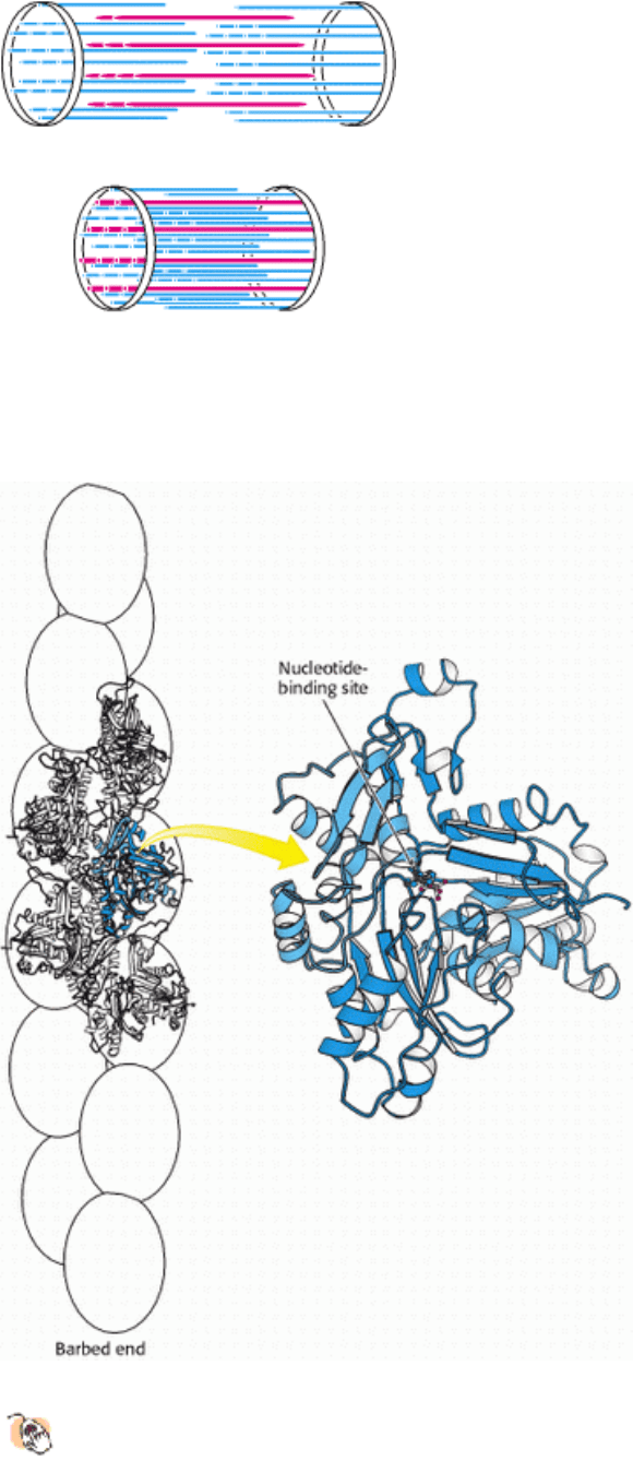
Figure 34.15. Actin Structure.
(Left) Five actin monomers of an actin filament are shown explicitly, one in blue.
(Right) The domains in the four-domain structure of an actin monomer are identified by different shades of blue.
IV. Responding to Environmental Changes 34. Molecular Motors 34.2. Myosins Move Along Actin Filaments
Figure 34.16. Actin and Hexokinase.
A comparison of actin (blue) and hexokinase from yeast (red) reveals structural
similarities indicative of homology. Both proteins have a deep cleft in which nucleotides bind.
IV. Responding to Environmental Changes 34. Molecular Motors 34.2. Myosins Move Along Actin Filaments
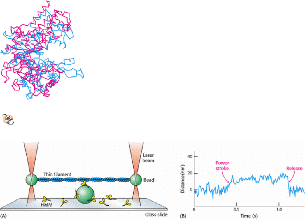
Figure 34.17. Watching a Single Motor Protein in Action. (A) An actin filament (blue) is placed above a heavy
meromyosin (HMM) fragment (yellow) that projects from a bead on a glass slide. A bead attached to each end of the
actin filament is held in an optical trap produced by a focused, intense infrared laser beam (orange). The position of these
beads can be measured with nanometer precision. (B) Recording of the displacement of the actin filament induced by the
addition of ATP. [After J. T. Finer, R. M. Simmons, and J. A. Spudich. Nature 368(1994):113.]
IV. Responding to Environmental Changes 34. Molecular Motors 34.2. Myosins Move Along Actin Filaments
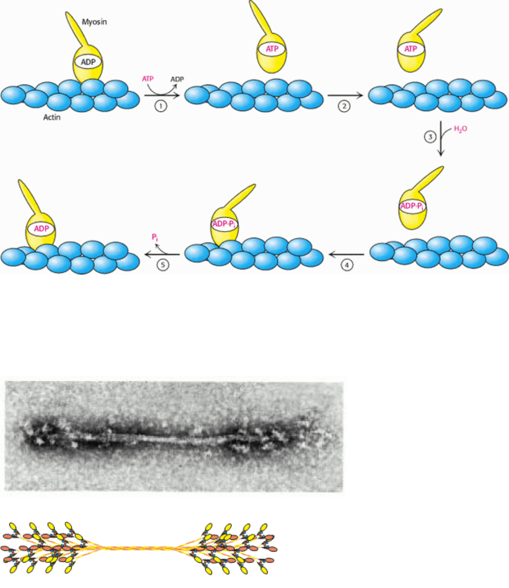
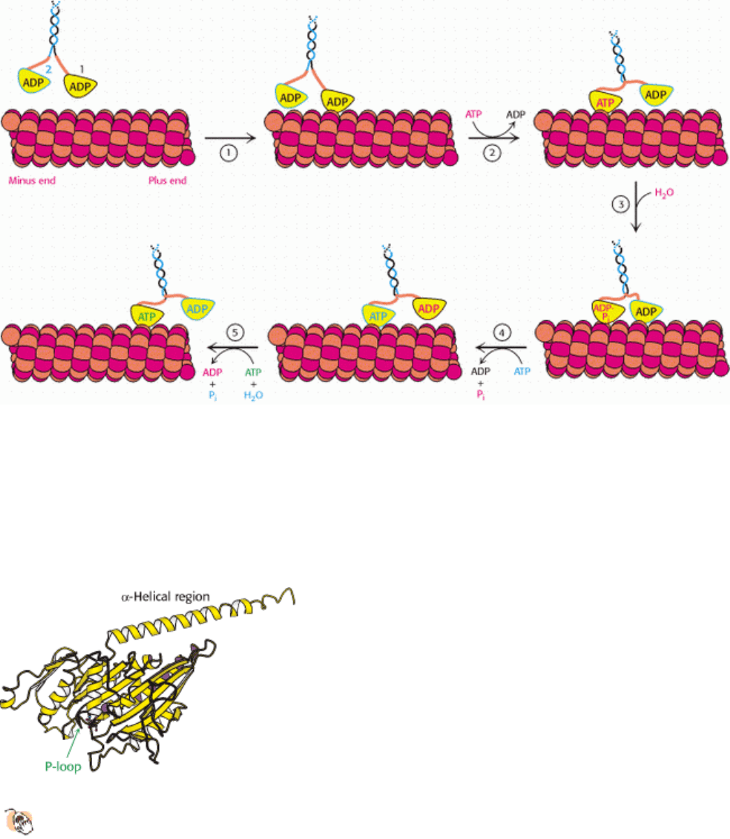
Figure 34.18. Myosin Motion Along Actin. A myosin head (yellow) in the ADP form is bound to an actin filament
(blue). The exchange of ADP for ATP results in (1) the release of myosin from actin and (2) substantial reorientation of
the lever arm of myosin. Hydrolysis of ATP (3) allows the myosin head to rebind at a site displaced along the actin
filament (4). The release of Pi (5) accompanying this binding increases the strength of interaction between myosin and
actin and resets the orientation of the lever arm.
IV. Responding to Environmental Changes 34. Molecular Motors 34.2. Myosins Move Along Actin Filaments
Figure 34.19. Thick Filament. (A) An electron micrograph of a reconstituted thick filament reveals the presence of
myosin head domains at each end and a relatively narrow central region. (B) A schematic view shows how myosin
molecules come together to form the thick filament. [Part A courtesy of Dr. Hugh Huxley.]
IV. Responding to Environmental Changes 34. Molecular Motors 34.2. Myosins Move Along Actin Filaments
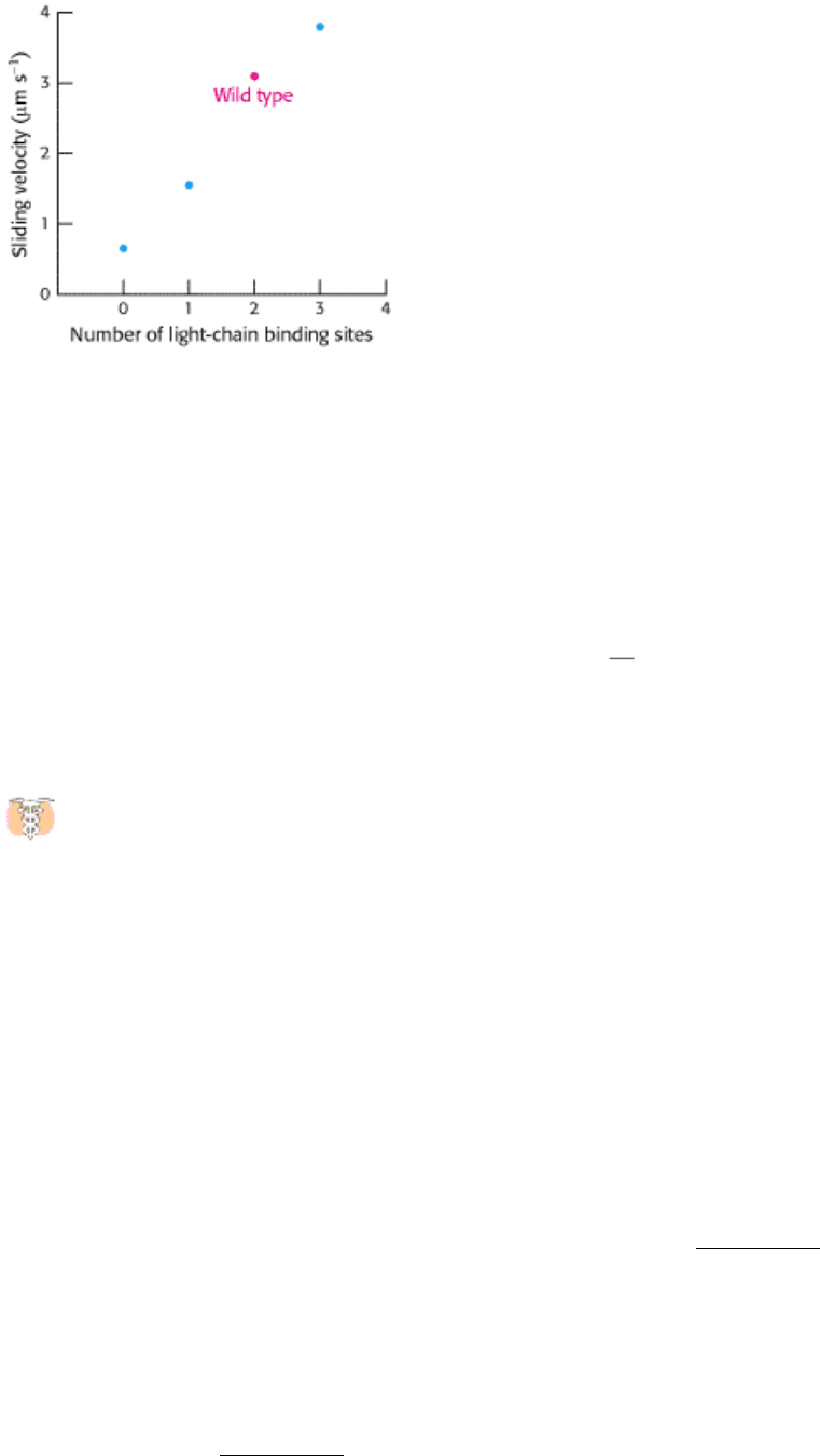
Figure 34.20. Myosin Lever Arm Length. Examination of the rates of actin movement supported by a set of myosin
mutants with different numbers of light-chain binding sites revealed a linear relation; the greater the number of light-
chain binding sites (and, hence, the longer the lever arm), the faster the sliding velocity. [After T. Q. P. Uyeda, P. D.
Abramson, and J. A. Spudich. Proc. Natl. Acad. Sci. USA 93(1996):4459.]
IV. Responding to Environmental Changes 34. Molecular Motors
34.3. Kinesin and Dynein Move Along Microtubules
In addition to actin, the cytoskeleton includes other components, notably intermediate filaments and microtubules.
Microtubules serve as tracks for two classes of motor proteins
namely, kinesins and dyneins. Kinesins moving along
microtubules usually carry cargo such as organelles and vesicles from the center of a cell to its periphery. Dyneins are
important in sliding microtubules relative to one other during the beating of cilia and flagella on the surfaces of some
eukaryotic cells.
Some members of the kinesin family are crucial to the transport of organelles and other cargo to nerve endings at
the periphery of neurons. It is not surprising, then, that mutations in these kinesins can lead to nervous system
disorders. For example, mutations in a kinesin called KIF1Bβ can lead to the most common peripheral neuropathy
(weakness and pain in the hands and feet), Charcot-Marie-Tooth disease, which affects 1 in 2500 people. A glutamine-to-
leucine mutation in the P-loop of the motor domain of this kinesin has been found in some affected persons. Knockout
mice with a disruption of the orthologous gene have been generated. Mice heterozygous for the disruption show
symptoms similar to those observed in human beings; homozygotes die shortly after birth. Mutations in other kinesin
genes have been tentatively linked to a predisposition to schizophrenia. In these disorders, defects in kinesin-linked
transport may impair nerve function directly, and the decrease in the activity of specific neurons may lead to other
degenerative processes.
34.3.1. Microtubules Are Hollow Cylindrical Polymers
Microtubules are built from two kinds of homologous 50-kd subunits, α- and β-tubulin, which assemble in an helical
array of alternating tubulin types to form the wall of a hollow cylinder (Figure 34.21). Alternatively, a microtubule can
be regarded as 13 protofilaments that run parallel to its long axis. The outer diameter of a microtubule is 30 nm, much
larger than that of actin (5 nm). Like actin, microtubules are polar structures. One end, termed the minus end, is anchored
near the center of a cell, whereas the plus end extends toward the cell surface.
Microtubules are also key components of cilia and flagella present on some eukaryotic cells. For example, sperm propel
themselves through the motion of flagella containing microtubules. The microtubules present in these structures adopt a
common architecture (Figure 34.22). A bundle of microtubules called an axoneme is surrounded by a membrane
contiguous with the plasma membrane. The axoneme is composed of a peripheral group of nine microtubule pairs
surrounding two singlet microtubules. This recurring motif is often called a 9 + 2 array. Dynein drives the motion of one
member of each outer pair relative to the other, causing the overall structure to bend.
Microtubules are important in determining the shapes of cells and in separating daughter chromosomes in mitosis. They
are highly dynamic structures that grow through the addition of α- and β-tubulin to the ends of existing structures. Like
actin, tubulins also bind and hydrolyze nucleoside triphosphates, although for tubulin the nucleotide is GTP rather than
ATP. The critical concentration for the polymerization of the GTP forms of tubulin is lower than that for the GDP forms.
Thus, a newly formed microtubule consists primarily of GTP-tubulins. Through time, the GTP is hydrolyzed to GDP.
The GDP-tubulin subunits in the interior length of a microtubule remain stably polymerized, whereas GDP subunits
exposed at an end have a strong tendency to dissociate. Marc Kirschner and Tim Mitchison found that some
microtubules in a population lengthen while others simultaneously shorten. This property, called dynamic instability,
arises from random fluctuations in the number of GTP- or GDP-tubulin subunits at the plus end of the polymer. The
dynamic character of microtubules is crucial for processes such as mitosis, which require the assembly and disassembly
of elaborate microtubule-based structures.
The structure of tubulin was determined at high resolution by electron crystallographic methods (Figure 34.23). As
expected from their 40% sequence identity, α- and β-tubulin have very similar three-dimensional structures.
Further analysis revealed that the tubulins are members of the P-loop NTPase family and contain a nucleotide-binding
site adjacent to the P-loop. Tubulins are present only in eukaryotes, although a prokaryotic homolog has been found.
Sequence analysis identified a prokaryotic protein called FtsZ (for filamentous temperature-sensitive mutant Z) that is
quite similar to the tubulins. The homology was confirmed when the structure was determined by x-ray crystallography.
Interestingly, this protein participates in bacterial cell division, forming ring-shaped structures at the constriction that
arises when a cell divides. These observations suggest that tubulins may have evolved from an ancient cell-division
protein.
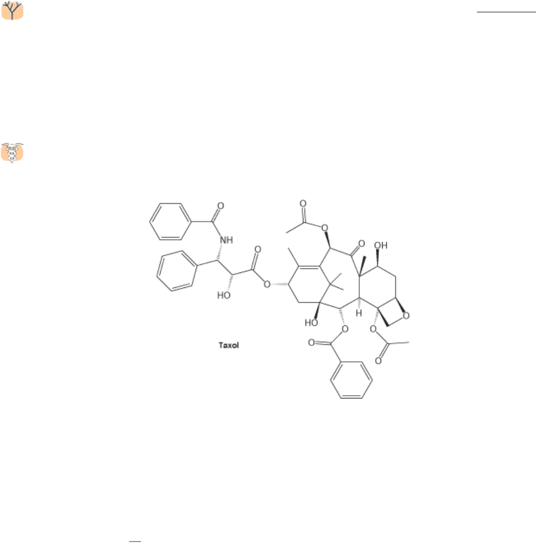
The continual lengthening and shortening of microtubules is essential to their role in cell division. Taxol, a
compound isolated from the bark of the Pacific yew tree, was discovered through its ability to interfere with cell
proliferation. Taxol binds to microtubules and stabilizes the polymerized form.
Taxol and its derivatives have been developed as anticancer agents because they preferentially affect rapidly dividing
cells, such as those in tumors.
34.3.2. Kinesin Motion Is Highly Processive
Kinesins are motor proteins that move along microtubules. We have seen that myosin moves along actin filaments by a
process in which actin is released in each cycle; a myosin head group acting independently dissociates from actin after
every power stroke. In contrast, when a kinesin molecule moves along a microtubule, the two head groups of the kinesin
molecule operate in tandem
one binds, and then the next one does. A kinesin molecule may take many steps before
both heads groups are dissociated at the same time. In other words, the motion of kinesin is highly processive. Single-
molecule measurements allow processive motion to be observed (Figure 34.24). A single kinesin molecule will typically
take 100 or more steps toward the plus end of a microtubule in a period of seconds before the molecule becomes
detached from the microtubule. These measurements also revealed that the average step size is approximately 80 Å, a
value that corresponds to the distance between consecutive α- or β-tubulin subunits along each protofilament.
An additional fact is crucial to the development of a mechanism for kinesin motion
namely, that the addition of ATP
strongly increases the affinity of kinesin for microtubules. This behavior stands in contrast with the behavior of myosin;
ATP binding to myosin promotes its dissociation from actin. Do these differences imply that kinesin and myosin operate
by completely different mechanisms? Indeed not. Kinesin-generated movement appears to proceed by a mechanism that
is quite similar to that used by myosin (Figure 34.25). Let us begin with a two-headed kinesin molecule in its ADP form,
dissociated from a microtubule. Recall that the neck linker binds the head domain when ATP is bound and is released
when ADP is bound. The initial interaction of one of the head domains with a tubulin dimer on a microtubule stimulates
the release of ADP from this head domain and the subsequent binding of ATP. The binding of ATP triggers a
conformational change in the head domain that leads to two important events. First, the affinity of the head domain for
the microtubule increases, essentially locking this head domain in place. Second, the neck linker binds to the head
domain. This change repositions the other head domain acting through the coiled-coil domain that connects the two
kinesin monomers. In its new position, the second head domain is close to a second tubulin dimer, 80 Å along the
microtubule in the direction of the plus end. Meanwhile, the intrinsic ATPase activity of the first head domain
hydrolyzes the ATP to ADP and P
i
. When the second head domain binds to the microtubule, the first head releases ADP
and binds ATP. Again, ATP binding favors a conformational change that pulls the first domain forward. This process
can continue for many cycles until, by chance, both head domains are in the ADP form simultaneously and kinesin
dissociates from the microtubule. Because of the relative rates of the component reactions, a simultaneous dissociation
occurs approximately every 100 cycles.
Kinesin hydrolyzes ATP at a rate of approximately 80 molecules per second. Thus, given the step size of 80 Å per
molecule of ATP, kinesin moves along a microtubule at a speed of 6400 Å per second. This rate is considerably slower
than the maximum rate for myosin, which moves relative to actin at 80,000 Å per second. Recall, however, that myosin
movement depends on the independent action of hundreds of different head domains working along the same actin
filament, whereas the movement of kinesin is driven by the processive action of kinesin head groups working in pairs.
Muscle myosin evolved to maximize the speed of the motion, whereas kinesin functions to achieve steady, but slower,
transport in one direction along a filament.
34.3.3. Small Structural Changes Can Reverse Motor Polarity
Most members of the kinesin family move toward the plus end of microtubules. However, a small number, including the
protein ncd (for nonclaret disjunctional, first identified in Drosophila), move toward the minus end. From an engineering
perspective, there are many ways to change the polarity of a motor. How is polarity changed in this case?
Determination of the core structure of ncd revealed great similarity to other kinesins in the mechanical parts of the motor
domain, including the structures of the switch regions, the relay helix, and the parts that bind microtubules. Significantly,
however, the motor domain of ncd lies near the carboxyl terminus of the protein, whereas it lies near the amino terminus
of conventional kinesin. Furthermore, when a larger fragment of ncd bound to ADP was analyzed, a short region just
before the motor domain was seen to form an α helix that docks against the motor domain in a position similar to that
occupied by the neck linker of conventional kinesin in the ATP form (Figure 34.26). This finding suggests that ncd
moves toward the minus end of a microtubule by a mechanism only slightly different from that used by conventional
kinesin to move in the opposite direction. Whereas ATP binding by conventional kinesin leads to the binding of the neck
linker, ATP binding by ncd releases the helical region. Its release allows the second motor domain of the ncd dimer to
bind to a site on the microtubule farther toward the minus end (Figure 34.27). We see once again the economical
refinement of a protein by evolution in this case, subtle adjustments have produced an opposite mechanical result in
the activity of the protein assembly.
IV. Responding to Environmental Changes 34. Molecular Motors 34.3. Kinesin and Dynein Move Along Microtubules
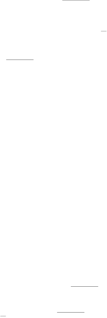
Figure 34.21. Microtubule Structure. Schematic views of the helical structure of a microtubule. α-tubulin is shown in
dark red and β-tubulin in light red. (A) Top view. (B) Side view.
IV. Responding to Environmental Changes 34. Molecular Motors 34.3. Kinesin and Dynein Move Along Microtubules
Figure 34.22. Microtubule Arrangement. Electron micrograph of a cross section of a flagellar axoneme shows nine
microtubule doublets surrounding two singlets. [Courtesy of Dr. Joel Rosenbaum.]
IV. Responding to Environmental Changes 34. Molecular Motors 34.3. Kinesin and Dynein Move Along Microtubules
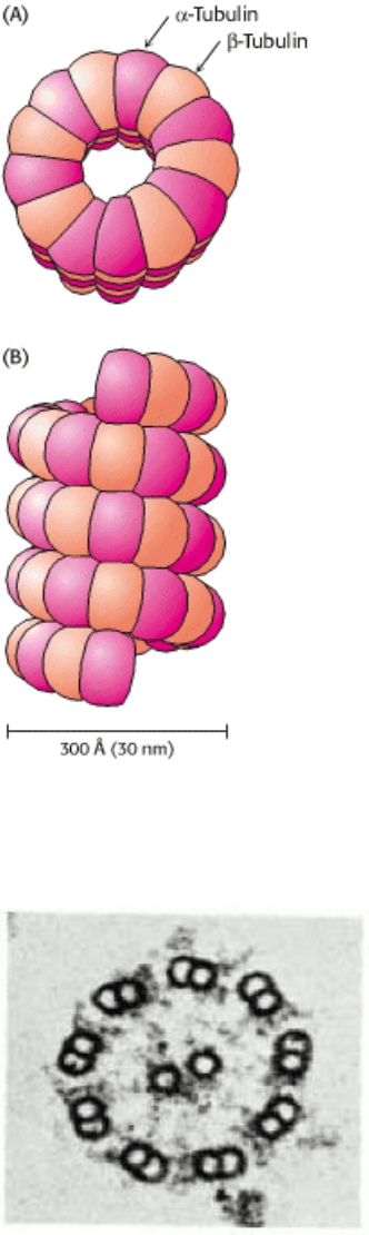
Figure 34.23. Tubulin.
Microtubules can be viewed as an assembly of α-tubulin - β-tubulin dimers. The structures of α-
tubulin and β-tubulin are quite similar; each includes a P-loop NTPase domain (purple shading) and a bound
guanine nucleotide.
IV. Responding to Environmental Changes 34. Molecular Motors 34.3. Kinesin and Dynein Move Along Microtubules
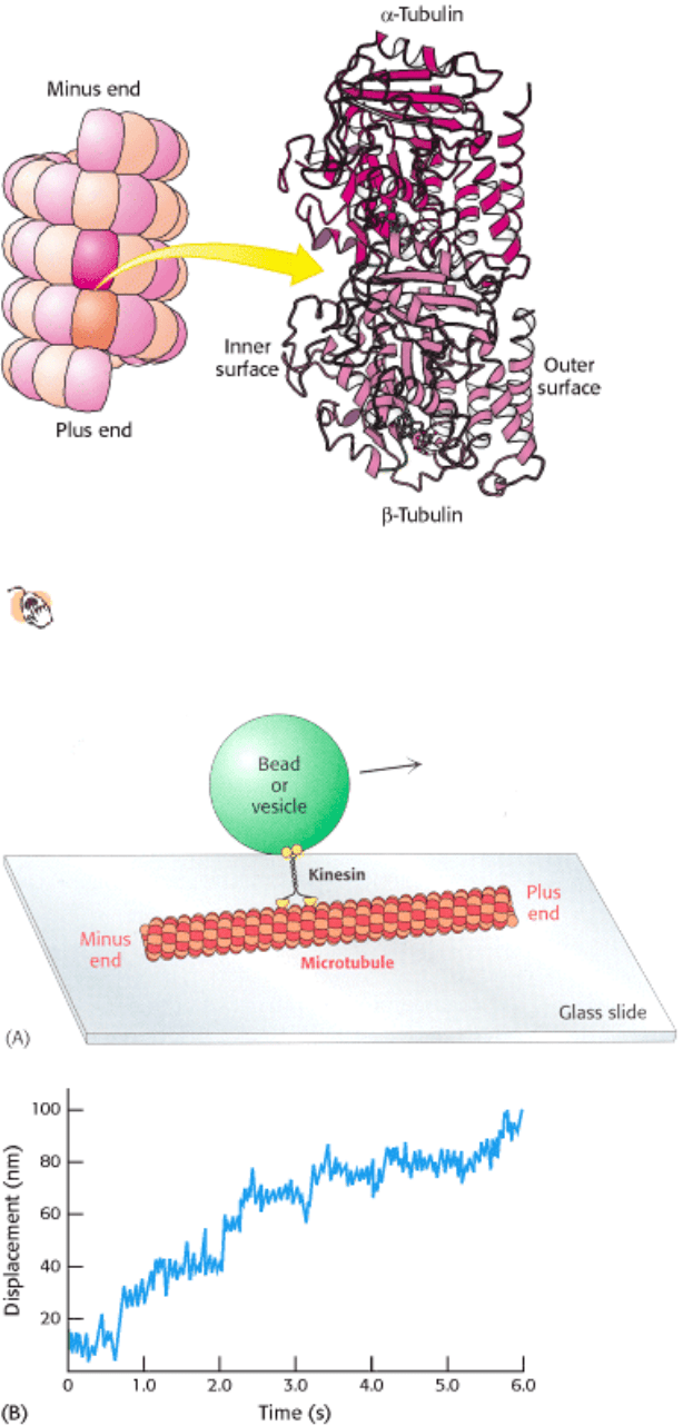
Figure 34.24. Monitoring Movements Mediated by Kinesin. (A) The movement of beads or vesicles, carried by
individual kinesin dimers along a microtubule, can be directly observed. (B) A trace shows the displacement of a bead
carried by a kinesin molecule. Multiple steps are taken in the 6-s interval. The average step size is about 8 nm (80 Å)
[Part B after K. Svoboda, C. F. Schmidt, B. J. Schnapp, and S. M. Block. Nature 365(1993):721.]
IV. Responding to Environmental Changes 34. Molecular Motors 34.3. Kinesin and Dynein Move Along Microtubules
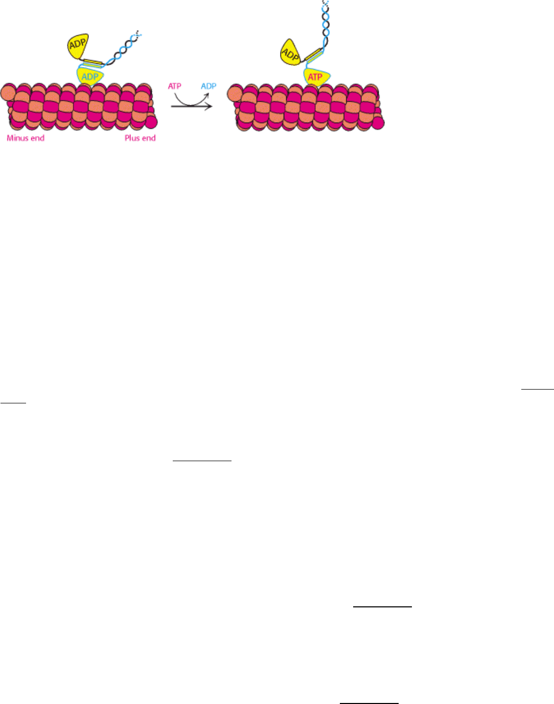
Figure 34.25. Kinesin Moving Along a Microtubule. (1) One head of a two-headed kinesin molecule, initially with
both heads in the ADP form, binds to a microtubule. (2) Release of ADP and binding of ATP results in a conformational
change that locks the head to the microtubule and pulls the neck linker (orange) to the head domain, throwing the second
domain toward the plus end of the microtubule. (3) ATP hydrolysis occurs while the second head interacts with the
microtubule. (4) The exchange of ATP for ADP in the second head pulls the first head off the microtubule, releasing Pi
and moving the first domain along the microtubule. (5) The cycle repeats, moving the kinesin dimer farther down the
microtubule.
IV. Responding to Environmental Changes 34. Molecular Motors 34.3. Kinesin and Dynein Move Along Microtubules
Figure 34.26. Structure of Ncd.
The head domain of ncd is quite similar to that of conventional kinesin, including the
presence of a P-loop NTPase domain (shaded in purple). In the ADP form of ncd (shown), the amino-terminal part
of this fragment forms an α-helix that docks into the site occupied by the neck linker in the ATP form of
conventional kinesin.
IV. Responding to Environmental Changes 34. Molecular Motors 34.3. Kinesin and Dynein Move Along Microtubules
Figure 34.27. Motion in Ncd. The replacement of ATP for ADP releases the α-helical region from the head domain,
moving the other head domain toward the minus end of the microtubule.
IV. Responding to Environmental Changes 34. Molecular Motors
34.4. A Rotary Motor Drives Bacterial Motion
In one second, a motile bacterium can move approximately 25 µm, or about 10 body lengths. A human being sprinting at
a proportional rate would complete the 100-meter dash in slightly more than 5 seconds. The motors that power this
impressive motion are strikingly different from the eukaryotic motors that we have seen so far. In the bacterial motor, an
element spins around a central axis rather than moving along a polymeric track. The direction of rotation can change
rapidly, a feature that is central to chemotaxis, the process by which bacteria swim preferentially toward an increasing
concentration of certain useful compounds and away from potentially harmful ones.
34.4.1. Bacteria Swim by Rotating Their Flagella
Bacteria such as Eschericia coli and Salmonella typhimurium swim by rotating flagella that lie on their surfaces (Figure
34.28). When the flagella rotate in a counterclockwise direction (viewed from outside the bacterium), the separate
flagella form a bundle that very efficiently propels the bacterium through solution.
Bacterial flagella are polymers approximately 15 nm in diameter and as much as 15 µm in length, composed of 53-kd
subunits of a protein called flagellin (Figure 34.29). These subunits associate into a helical structure that has 5.5 subunits
per turn, giving the appearance of 11 protofilaments. Each flagellum has a hollow core. Remarkably, flagella form not by
growing at the base adjacent to the cell body but, instead, by the addition of new subunits that pass through the hollow
core and add to the free end. Each flagellum is intrinsically twisted in a left-handed sense. At its base, each flagellum has
a rotory motor.
34.4.2. Proton Flow Drives Bacterial Flagellar Rotation
Early experiments by Julius Adler demonstrated that ATP is not required for flagellar motion. What powers these rotary
motors? The necessary free energy is derived from the proton gradient that exists across the plasma membrane. The
flagellar motor is quite complex, containing as many as 40 distinct proteins (Figure 34.30). Five components particularly
crucial to motor function have been identified through genetic studies. MotA is a membrane protein that appears to have
four transmembrane helices as well as a cytoplasmic domain. MotB is another membrane protein with a single
transmembrane helix and a large periplasmic domain. Approximately 11 MotA - MotB pairs form a ring around the base
of the flagellum. The proteins FliG, FliM, and FliN are part of a disc-like structure called the MS (membrane and
supramembrane) ring, with approximately 30 FliG subunits coming together to form the ring. The three-dimensional
structure of the carboxyl-terminal half of FliG reveals a wedge-shaped domain with a set of charged amino acids,
conserved among many species, lying along the thick edge of the wedge (Figure 34.31).