Berg J.M., Tymoczko J.L., Stryer L. Biochemistry
Подождите немного. Документ загружается.


Dynein has a rather different structure. As noted earlier, the dynein heavy chain includes six regions that are homologous
to the AAA subfamily of ATPase domains. Although no crystallographic data are yet available, the results of electron
microscopic studies and comparison with known structures of other AAA ATPases have formed the basis for the
construction of a model of the dynein head structure (Figure 34.9). The head domain is appended to a region of
approximately 1300 amino acids that forms an extended structure that links dynein units together to form oligomers and
interacts with other proteins.
34.1.2. ATP Binding and Hydrolysis Induce Changes in the Conformation and Binding
Affinity of Motor Proteins
A key feature of P-loop NTPases such as G proteins is that they undergo structural changes induced by NTP binding and
hydrolysis. Moreover, these structural changes alter their affinities for binding partners. Thus, it is not surprising that the
NTPase domains of motor proteins display analogous responses to nucleotide binding. The S1 fragment of myosin from
scallop muscle provides a striking example of the changes observed (Figure 34.10). The structure of the S1 fragment has
been determined for S1 bound to a complex formed of ADP and vanadate (VO
4
3-
), which is an analog of ATP, or, more
precisely, the ATP-hydrolysis transition state. In the presence of the ADP - VO
4
3-
complex, the long helix that binds the
light chains (hereafter referred to as the lever arm) protrudes outward from the head domain. In the presence of ADP
without VO
4
3-
, the lever arm has rotated by nearly 90 degrees relative to its position in the ADP - VO
4
3-
complex. How
does the identity of the species in the nucleotide-binding site cause this dramatic transition? Two regions around the
nucleotide-binding site, analogous to the switch regions of G proteins (Section 15.1.2), conform closely to the group in
the position of the γ-phosphate group of ATP and adopt a looser conformation when such a group is absent (Figure
34.11). This conformational change allows a long α helix (termed the relay helix) to adjust its position. The
carboxylterminal end of the relay helix interacts with structures at the base of the lever arm, and so a change in the
position of the relay helix leads to a reorientation of the lever arm.
The binding of ATP significantly decreases the affinity of the myosin head for actin filaments. No structures of myosin -
actin complexes have yet been determined at high resolution, so the mechanistic basis for this change remains to be
elucidated. However, the amino-terminal end of the relay helix interacts with the domains of myosin that bind to actin,
suggesting a clear pathway for the coupling of nucleotide binding to changes in actin affinity. The importance of the
changes in actin-binding affinity will be clear later when we examine the role of myosin in generating directed motion
(Section 34.2.4).
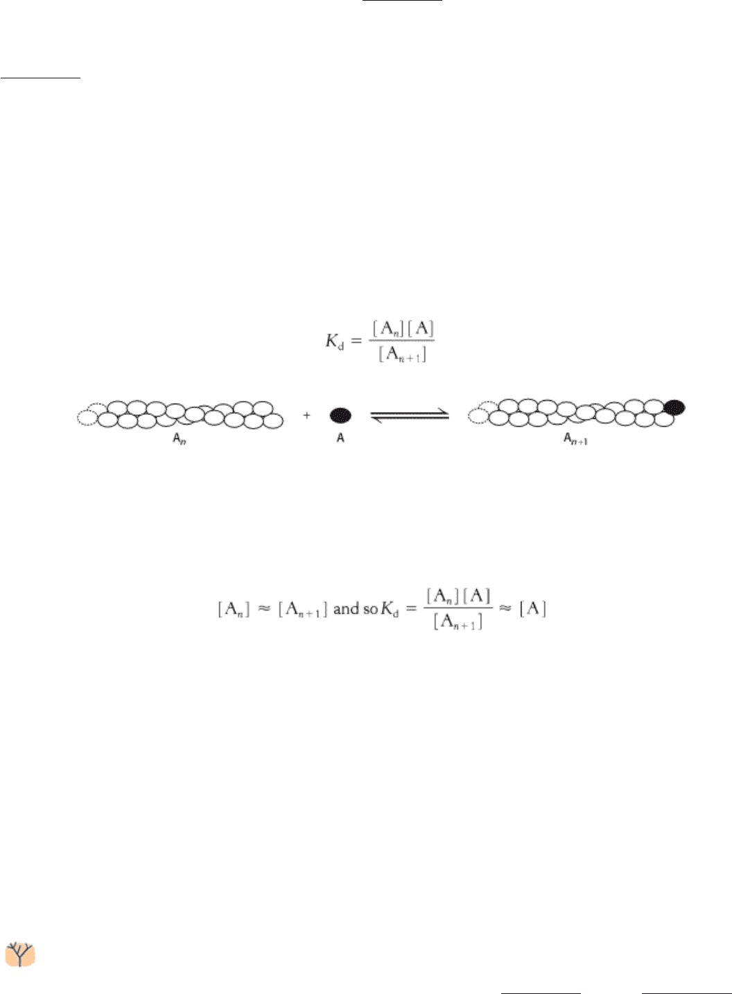
Analogous conformational changes take place in kinesin. The kinesins also have a relay helix that can adopt different
configurations when kinesin binds different nucleotides. Kinesin lacks an α-helical lever arm, however. Instead, a
relatively short segment termed the neck linker changes conformation in response to nucleotide binding (Figure 34.12).
The neck linker binds to the head domain of kinesin when ATP is bound but is released when the nucleotide-binding site
is vacant or occupied by ADP. Kinesin differs from myosin in that the binding of ATP to kinesin increases the affinity
between kinesin and its binding partner, microtubules. The properties of myosin, kinesin, and a heterotrimeric G protein
are compared in Table 34.1. Before turning to a discussion of how these properties are used to convert chemical energy
into motion, we must consider the properties of the tracks along which these motors move.
IV. Responding to Environmental Changes 34. Molecular Motors 34.1. Most Molecular-Motor Proteins Are Members of the P-Loop NTPase Superfamily
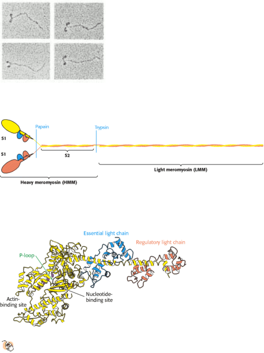
Figure 34.2. Myosin Structure at Low Resolution. Electron micrographs of myosin molecules reveal a two-headed
structure with a long, thin tail. [Courtesy of Dr. Paula Flicker, Dr. Theo Walliman, and Dr. Peter Vibert.]
IV. Responding to Environmental Changes 34. Molecular Motors 34.1. Most Molecular-Motor Proteins Are Members of the P-Loop NTPase Superfamily
Figure 34.3. Myosin Dissection. Treatment of muscle myosin with proteases forms stable fragments, including
subfragments S1 and S2 and light meromyosin. Each S1 fragment includes the head (shown in yellow and pink) from the
heavy chain and one copy of each light chain (shown in blue and orange).
IV. Responding to Environmental Changes 34. Molecular Motors 34.1. Most Molecular-Motor Proteins Are Members of the P-Loop NTPase Superfamily
Figure 34.4. Myosin Structure at High Resolution.
The structure of the S1 fragment from muscle myosin reveals the
presence of a P-loop NTPase domain (shaded in purple). An α helix that extends from this domain is the binding
site for the two light chains.
IV. Responding to Environmental Changes 34. Molecular Motors 34.1. Most Molecular-Motor Proteins Are Members of the P-Loop NTPase Superfamily
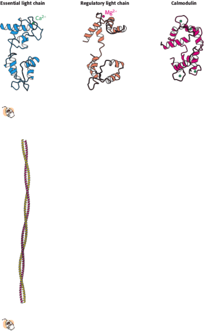
Figure 34.5. Myosin Light Chains.
The structures of the essential and regulatory light chains from muscle myosin are
compared with the structure of calmodulin. Each of these homologous proteins binds an α helix (not shown) by
wrapping around it.
IV. Responding to Environmental Changes 34. Molecular Motors 34.1. Most Molecular-Motor Proteins Are Members of the P-Loop NTPase Superfamily
Figure 34.6. Myosin Two-Stranded Coiled Coil.
The two α helices form left-handed supercoiled structures that spiral
around each other. Such structures are stabilized by hydrophobic residues at the contact points between the two
helices.
IV. Responding to Environmental Changes 34. Molecular Motors 34.1. Most Molecular-Motor Proteins Are Members of the P-Loop NTPase Superfamily
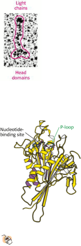
Figure 34.7. Kinesin at Low Resolution. An electron micrograph of conventional kinesin reveals an elongated structure
with two heads at one end. The position of the light chains was confirmed through the use of antibody labels. [After N.
Hirokawa, K. K. Pfister, H. Yorifuji, M. C. Wagner, S. T. Brady, and G. S. Broom. Cell 56 (1989):867.]
IV. Responding to Environmental Changes 34. Molecular Motors 34.1. Most Molecular-Motor Proteins Are Members of the P-Loop NTPase Superfamily
Figure 34.8. Structure of Head Domain of Kinesin at High Resolution.
The head domain of kinesin has the structure
of a P-loop NTPase core (indicated by purple shading).
IV. Responding to Environmental Changes 34. Molecular Motors 34.1. Most Molecular-Motor Proteins Are Members of the P-Loop NTPase Superfamily
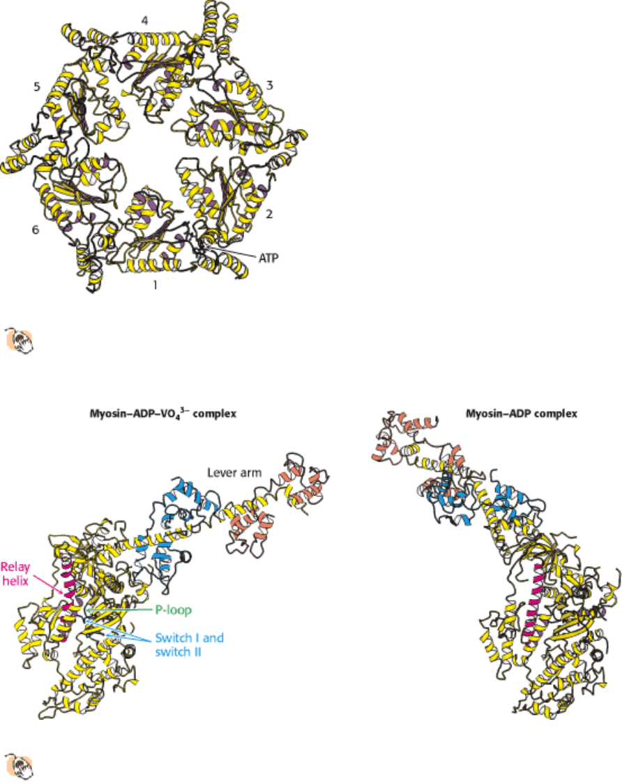
Figure 34.9. Dynein Head-Domain Model.
ATP is bound in the first of six P-loop NTPase domains (numbered) in this
model for the head domain of dynein. The model is based on electron micrographs and the structures of other
members of the AAA ATPase family.
IV. Responding to Environmental Changes 34. Molecular Motors 34.1. Most Molecular-Motor Proteins Are Members of the P-Loop NTPase Superfamily
Figure 34.10. Lever-Arm Motion.
Two forms of the S1 fragment of scallop muscle myosin. Dramatic conformational
changes are observed when the identity of the bound nucleotide changes from ADP-VO4
3-
to ADP or vice versa,
including a nearly 90-degree reorientation of the lever arm.
IV. Responding to Environmental Changes 34. Molecular Motors 34.1. Most Molecular-Motor Proteins Are Members of the P-Loop NTPase Superfamily
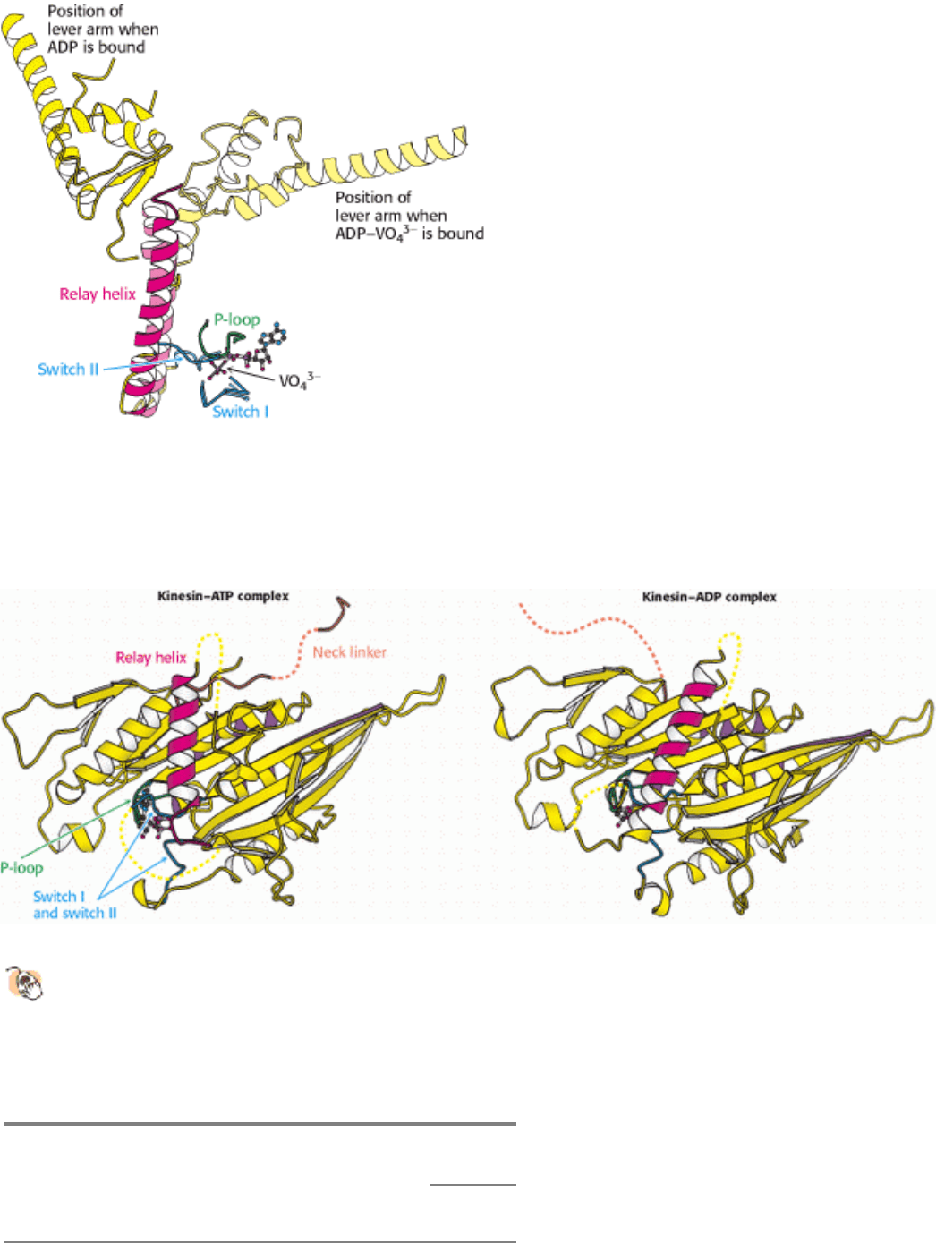
Figure 34.11. Relay Helix. A superposition of key elements in two forms of scallop myosin reveals the structural
changes that are transmitted by the relay helix from the switch I and switch II loops to the base of the lever arm. The
switch I and switch II loops interact with VO4
3-
in the position that would be occupied by the γ-phosphate group of ATP.
The structure of the ADP - VO4
3-
myosin complex is shown in lighter colors.
IV. Responding to Environmental Changes 34. Molecular Motors 34.1. Most Molecular-Motor Proteins Are Members of the P-Loop NTPase Superfamily
Figure 34.12. Neck Linker.
A comparison of the structures of a kinesin bound to ADP and bound to an ATP analog.
The neck linker (orange), which connects the head domain to the remainder of the kinesin molecule, is bound to
the head domain in the presence of the ATP analog but is free in the presence of ADP only.
IV. Responding to Environmental Changes 34. Molecular Motors 34.1. Most Molecular-Motor Proteins Are Members of the P-Loop NTPase Superfamily
Table 34.1. Effect of nucleotide binding on protein affinity
Bound to
Protein NTP NDP
Myosin (ATP or ADP)

Affinity for actin Low High
Kinesin (ATP or ADP)
Affinity for microtubules High Low
Heterotrimeric G protein (α subunit) (GTP or GDP)
Affinity for β γ dimer
Low High
Affinity for effectors High Low
IV. Responding to Environmental Changes 34. Molecular Motors
34.2. Myosins Move Along Actin Filaments
Myosins, kinesins, and dyneins move by cycling between states with different affinities for the long, polymeric
macromolecules that serve as their tracks. For myosin, the molecular track is a polymeric form of actin, a 42-kd protein
that is one of the most abundant proteins in eukaryotic cells, typically accounting for as much as 10% of the total protein.
Actin polymers are continually being assembled and disassembled in cells in a highly dynamic manner, accompanied by
the hydrolysis of ATP. On the microscopic scale, actin filaments participate in the dynamic reshaping of the cytoskeleton
and the cell itself and in other motility mechanisms that do not include myosin. In muscle, myosin and actin together are
the key components responsible for muscle contraction.
34.2.1. Muscle Is a Complex of Myosin and Actin
Vertebrate muscle that is under voluntary control has a banded (striated) appearance when examined under a light
microscope. It consists of multinucleated cells that are bounded by an electrically excitable plasma membrane. A muscle
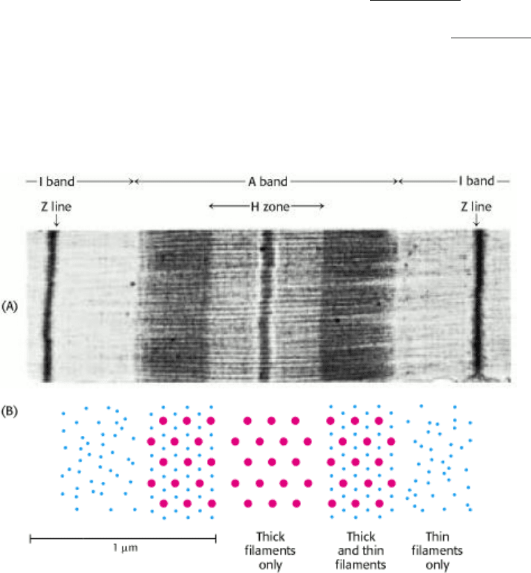
cell contains many parallel myofibrils, each about 1 µm in diameter. The functional unit, called a sarcomere, typically
repeats every 2.3 µm (23,000 Å) along the fibril axis in relaxed muscle (Figure 34.13). A dark A band and a light I band
alternate regularly. The central region of the A band, termed the H zone, is less dense that the rest of the band. The I
band is bisected by a very dense, narrow Z line.
The underlying molecular plan of a sarcomere is revealed by cross sections of a myofibril. These cross sections show the
presence of two kinds of interacting protein filaments. The thick filaments have diameters of about 15 nm (150 Å) and
consist primarily of myosin. The thin filaments have diameters of approximately 9 nm (90 Å) and consist of actin as well
as tropomyosin and the troponin complex. Muscle contraction is achieved through the sliding of the thin filaments along
the length of the thick filaments, driven by the hydrolysis of ATP (Figure 34.14). Tropomyosin and the troponin complex
regulate this sliding in response to nerve impulses. Under resting conditions, tropomyosin blocks the intimate interaction
between mysosin and actin. A nerve impulse leads to an increase in calcium ion concentration within the muscle cell. A
component of the troponin complex senses the increase in calcium and, in response, relieves the inhibition of myosin -
actin interactions by tropomyosin.
Although myosin was discovered through its role in muscle, other types of myosin play crucial roles in a number
of biological contexts. Some defects in hearing in both mice and human beings have been linked to mutations in
particular myosin homologs that are present in cells of the ear. For example, Usher syndrome in human beings and the
shaker mutation in mice have been linked to myosin VIIa, expressed in hair cells (Section 32.4.1). The mutation of this
myosin results in the formation of splayed stereocilia that do not function well. Myosin VIIa differs from muscle myosin
in that its tail region possesses a number of amino acid sequences that correspond to domains known to mediate specific
protein - protein interactions.
34.2.2. Actin Is a Polar, Self-Assembling, Dynamic Polymer
The structure of the actin monomer was determined to atomic resolution by x-ray crystallography and has been used to
interpret the structure of actin filaments, already somewhat understood through electron microscopy studies at lower
resolution. Each actin monomer comprises four domains (Figure 34.15). These domains come together to surround a
bound nucleotide, either ATP or ADP. The ATP form can be converted into the ADP form by hydrolysis.
Actin monomers (often called G-actin for globular) come together to form actin filaments (often called F-actin; see
Figure 34.15). F-actin has a helical structure; each monomer is related to the preceding one by a translation of 27.5 Å
and a rotation of 166 degrees around the helical axis. Because the rotation is nearly 180 degrees, F-actin resembles a two-
stranded cable. Note that each actin monomer is oriented in the same direction along the F-actin filament, and so the
structure is polar, with discernibly different ends. One end is called the barbed (plus) end, and the other is called the
pointed (minus) end. The names "barbed" and "pointed" refer to the appearance of an actin filament when myosin S1
fragments are bound to it.
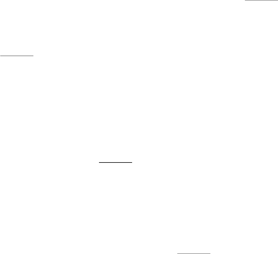
How are actin filaments formed? Like many biological structures, actin filaments self-assemble; that is, under
appropriate conditions, actin monomers will come together to form well-structured, polar filaments. The aggregation of
the first two or three monomers to form a filament is somewhat unfavorable. Once such a filament nucleus exists, the
addition of subunits is more favorable. Let us consider the polymerization reaction in more detail. We designate an actin
filament with n subunits A
n
. This filament can bind an additional actin monomer, A, to form A
n
+1
.
The dissociation constant for this reaction, K
d
, defines the monomer concentrations at which the polymerization reaction
will take place, because the concentration of polymers of length n + 1 will be essentially equal to that for polymers of
length n. Thus,
In other words, the polymerization reaction will proceed until the monomer concentration is reduced to the value of K
d
.
If the monomer concentration is below the value of K
d
, the polymerization reaction will not proceed at all; indeed,
existing filaments will depolymerize until the monomer concentration reaches the value of K
d
. Because of these
phenomena, K
d
is referred to as the critical concentration for the polymer. Recall that actin contains a nucleotide-
binding site that can contain either ATP or ADP. The critical concentration for the actin - ATP complex is approximately
20-fold lower than that for the actin - ADP complex; actin - ATP polymerizes more readily than does actin - ADP.
Actin filaments inside cells are highly dynamic structures that are continually gaining and losing monomers. The
concentration of free actin monomers is controlled by several mechanisms. For example, actinsequestering proteins such
as β-thymosin bind to actin monomers and inhibit polymerization. Furthermore, the concentration and properties of actin
filaments are closely regulated by proteins that sever an actin filament into two or that cap one of the ends of a filament.
Regulated actin polymerization is central to the changes in cell shape associated with cell motility in amebas as well as
in human cells such as macrophages.
A well-defined actin cytoskeleton is unique to eukaryotes; prokaryotes lack such structures. How did filamentous
actin evolve? Comparison of the three-dimensional structure of G-actin with other proteins revealed remarkable
similarity to several other proteins, including sugar kinases such as hexokinase (Figure 34.16; see also Section 16.1.1).
Notably, the nucleotide-binding site in actin corresponds to the ATP-binding site in hexokinase. Thus, actin evolved
from an enzyme that utilized ATP as a substrate.
More recently, a closer prokaryotic homolog of actin was characterized. This protein, called MreB, plays an important
role in determining cell shape in rod-shaped, filamentous, and helical bacteria. The internal structures formed by MreB
are suggestive of the actin cytoskeleton of eukaryotic cells, although they are far less extensive. Even though this protein
is only approximately 15% identical in sequence with actin, MreB folds into a very similar three-dimensional structure.
It also polymerizes into structures that are similar to F-actin in a number of ways, including the alignment of the
component monomers.
34.2.3. Motions of Single Motor Proteins Can Be Directly Observed
Muscle contraction is complex, requiring the action of many different myosin molecules. Studies of single myosin
molecules moving relative to actin filaments have been sources of deep insight into the mechanisms underlying muscle
contraction and other complex processes.
A powerful tool for these studies, called an optical trap, relies on highly focused laser beams (Figure 34.17). Small beads
can be caught in these traps and held in place in solution.
The position of the beads can be monitored with nanometer precision. James Spudich and coworkers designed an
experimental arrangement consisting of an actin filament that had a bead attached to each end. Each bead could be
caught in an optical trap (one at each end of the filament) and the actin filament pulled taut over a microscope slide
containing other beads that had been coated with fragments of myosin such as the heavy meromyosin fragment (see
Figure 34.17). On the addition of ATP, transient displacements of the actin filament were observed along its long axis.
The size of the displacement steps was fairly uniform with an average size of 11 nm.
The results of these studies, performed in the presence of varying concentrations of ATP, are interpreted as showing that
individual myosin heads bind the actin filament and undergo a conformational change (the power stroke) that pulls the
actin filament, leading to the displacement of the beads. After a period of time, the myosin head releases the actin, which
then snaps back into place.
34.2.4. Phosphate Release Triggers the Myosin Power Stroke
How does ATP hydrolysis drive the power stroke? A key observation is that the addition of ATP to a complex of myosin
and actin results in the dissociation of the complex. Thus, ATP binding and hydrolysis cannot be directly responsible for
the power stroke. We can combine this fact with the structural observations described earlier to construct a mechanism
for the motion of myosin along actin (Figure 34.18). Let us begin with myosin-ADP bound to actin. The release of ADP
and the binding of ATP to actin result in the dissociation of myosin from actin. As we saw earlier, the binding of ATP
with its γ-phosphate group to the myosin head leads to a significant conformational change, amplified by the lever arm.
This conformational change moves the myosin head along the actin filament by approximately 110 Å. The ATP in the
myosin is then hydrolyzed to ADP and P
i
, which remain bound to myosin. The myosin head can then bind to the surface
of actin, resulting in the dissociation of P
i
from the myosin. Phosphate release, in turn, leads to a conformational change
that increases the affinity of the myosin head for actin and allows the lever arm to move back to its initial position. The
conformational change associated with phosphate release corresponds to the power stroke. After the release of P
i
, the
myosin remains tightly bound to the actin and the cycle can begin again.
How does this cycle apply to muscle contraction? Myosin molecules self-assemble into thick bipolar structures with the
myosin heads protruding at both ends of a bare region in the center (Figure 34.19). Approximately 500 head domains
line the surface of each thick filament. These domains are paired in myosin dimers, but the two heads within each dimer
act independently. Actin filaments associate with each head-rich region, with the barbed ends of actin toward the Z-line.
In the presence of normal levels of ATP, most of the myosin heads are detached from actin. Each head can
independently hydrolyze ATP, bind to actin, release P
i
, and undergo its power stroke. Because few other heads are
attached, the actin filament is relatively free to slide. Each head cycles approximately five times per second with a
movement of 110 Å per cycle. However, because hundreds of heads are interacting with the same actin filament, the
overall rate of movement of myosin relative to the actin filament may reach 80,000 Å per second, allowing a sacromere
to contract from its fully relaxed to its fully contracted form rapidly. Having many myosin heads briefly and
independently attaching and moving an actin filament allows for much greater speed than could be achieved by a single
motor protein.
34.2.5. The Length of the Lever Arm Determines Motor Velocity
A key feature of myosin motors is the role of the lever arm as an amplifier. The lever arm amplifies small structural
changes at the nucleotide-binding site to achieve the 110-Å movement along the actin filament that takes place in each
ATP hydrolysis cycle. A strong prediction of the mechanism proposed for the movement of myosin along actin is that
the length traveled per cycle should depend on the length of this lever arm. Thus, the length of the lever arm should
influence the overall rate at which actin moves relative to a collection of myosin heads.
This prediction was tested with the use of mutated forms of myosin with lever arms of different lengths. The lever arm in
muscle myosin includes binding sites for two light chains (Section 34.1.2). Thus investigators shortened the lever arm by
deleting the sequences that correspond to one or both of these binding sites. They then examined the rates at which actin
filaments were transported along collections of these mutated myosins (Figure 34.20). As predicted, the rate decreased as
the lever arm was shortened. A mutated form of myosin with an unusually long lever arm was generated by inserting 23
amino acids corresponding to the binding site for an additional regulatory light chain. Remarkably, this form was found
to support actin movement that was faster than the wild-type protein. These results strongly support the proposed role of
the lever arm in contributing to myosin motor activity.
IV. Responding to Environmental Changes 34. Molecular Motors 34.2. Myosins Move Along Actin Filaments
Figure 34.13. Sarcomere. (A) Electron micrograph of a longitudinal section of a skeletal muscle myofibril, showing a
single sarcomere. (B) Schematic representations of cross sections correspond to the regions in the micrograph. [Courtesy
of Dr. Hugh Huxley.]