Berg J.M., Tymoczko J.L., Stryer L. Biochemistry
Подождите немного. Документ загружается.

IV. Responding to Environmental Changes 33. The Immune System 33.1. Antibodies Possess Distinct Antigen-Binding and Effector Units
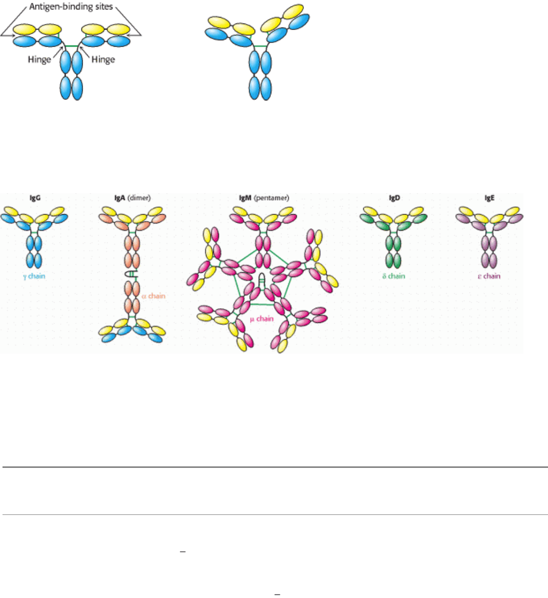
Figure 33.5. Segmental Flexibility. The linkages between the F
ab
and the F
c
regions of an IgG molecule are flexible,
allowing the two antigen-binding sites to adopt a range of orientations with respect to one another. This flexibility allows
effective interactions with a multivalent antigen without requiring that the epitopes on the target be a precise distance
apart.
IV. Responding to Environmental Changes 33. The Immune System 33.1. Antibodies Possess Distinct Antigen-Binding and Effector Units
Figure 33.6. Classes of Immuno-Globulin. Each of five classes of immuno-globulin has the same light chain (shown in
yellow) combined with a different heavy chain (γ, α, µ, δ, or ε). Disulfide bonds are indicated by green lines. The IgA
dimer and the IgM pentamer have a small polypeptide chain in addition to the light and heavy chains.
IV. Responding to Environmental Changes 33. The Immune System 33.1. Antibodies Possess Distinct Antigen-Binding and Effector Units
Table 33.1. Properties of immunoglobulin classes
Class Serum concentration (mg/
ml)
Mass (kd) Sedimentation coefficient(s) Light chains Heavy chains Chain structure
IgG 12 150 7
κ or λ γ κ
2
γ
2
or λ
2
γ
2
IgA 3 180
500 7, 10, 13
κ or λ α (κ
2
α
2
)
n
or (λ
2
α
2
)
n
IgM 1 950 18 20
κ or λ µ (κ
2
µ
2
)
5
or (λ
2
µ
2
)
5
IgD 0.1 175 7
κ or λ δ κ
2
δ
2
or λ
2
δ
2
IgE 0.001 200 8
κ or λ ε κ
2
ε
2
or λ
2
ε
2
Note: n = 1, 2, or 3. IgM and oligomers of IgA also contain J chains that connect immunoglobulin molecules. IgA in secretions
has an additional component.
IV. Responding to Environmental Changes 33. The Immune System 33.1. Antibodies Possess Distinct Antigen-Binding and Effector Units
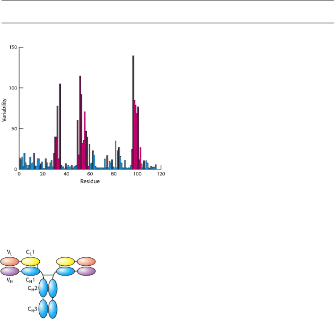
Figure 33.7. Immunoglobulin Sequence Diversity. A plot of sequence variability as a function of position along the
sequence of the amino-terminal immunglobulin domain of the H chain of human IgG molecules. Three regions (in red)
show remarkably high levels of variability. These hypervariable regions correspond to three loops in the
immunoglobulin domain structure. [After R. A. Goldsby, T. J. Kindt, and B. A. Osborne, Kuby Immunology, 4th ed. (W.
H. Freeman and Company, 2000), p. 91.]
IV. Responding to Environmental Changes 33. The Immune System 33.1. Antibodies Possess Distinct Antigen-Binding and Effector Units
Figure 33.8. Variable and Constant Regions. Each L and H chain includes one immunoglobulin domain at its amino
terminus that is quite variable from one antibody to another. These domains are referred to as V
L
and V
H
. The remaining
domains are more constant from one antibody to another and are referred to as constant domains (C
L
1, C
H
1, C
H
2, and
C
H
3).
IV. Responding to Environmental Changes 33. The Immune System
33.2. The Immunoglobulin Fold Consists of a Beta-Sandwich Framework with
Hypervariable Loops
An IgG molecule consists of a total of 12 immunoglobulin domains. These domains have many sequence features in
common and adopt a common structure, the immunoglobulin fold (Figure 33.9). Remarkably, this same structural
domain is found in many other proteins that play key roles in the immune system.
The immunoglobulin fold consists of a pair of β sheets, each built of antiparallel β strands, that surround a central
hydrophobic core. A single disulfide bond bridges the two sheets. Two aspects of this structure are particularly important
for its function. First, three loops present at one end of the structure form a potential binding surface. These loops contain
the hypervariable sequences present in antibodies and in T-cell receptors (see Sections 33.3 and 33.5.2). Variation of the
amino acid sequences of these loops provides the major mechanism for the generation of the vastly diverse set of
antibodies and T-cell receptors expressed by the immune system. These loops are referred to as hypervariable loops or
complementaritydetermining regions (CDRs). Second, the amino terminus and the carboxyl terminus are at opposite
ends of the structure, which allows structural domains to be strung together to form chains, as in the L and H chains of
antibodies. Such chains are present in several other key molecules in the immune system.
The immunoglobulin fold is one of the most prevalent domains encoded by the human genome more than 750
genes encode proteins with at least one immunoglobulin fold recognizable at the level of amino acid sequence.
Such domains are also common in other multicellular animals such as flies and nemotodes. However, from inspection of
amino acid sequence alone, immunoglobulin-fold domains do not appear to be present in yeast or plants. However,
structurally similar domains are present in these organisms, including the key photosynthetic electron-transport protein
plastocyanin in plants (Section 19.3.2). Thus, the immunglobulin-fold family appears to have expanded greatly along
evolutionary branches leading to animals particularly, vertebrates.
IV. Responding to Environmental Changes 33. The Immune System 33.2. The Immunoglobulin Fold Consists of a Beta-Sandwich Framework with Hypervariable Loops
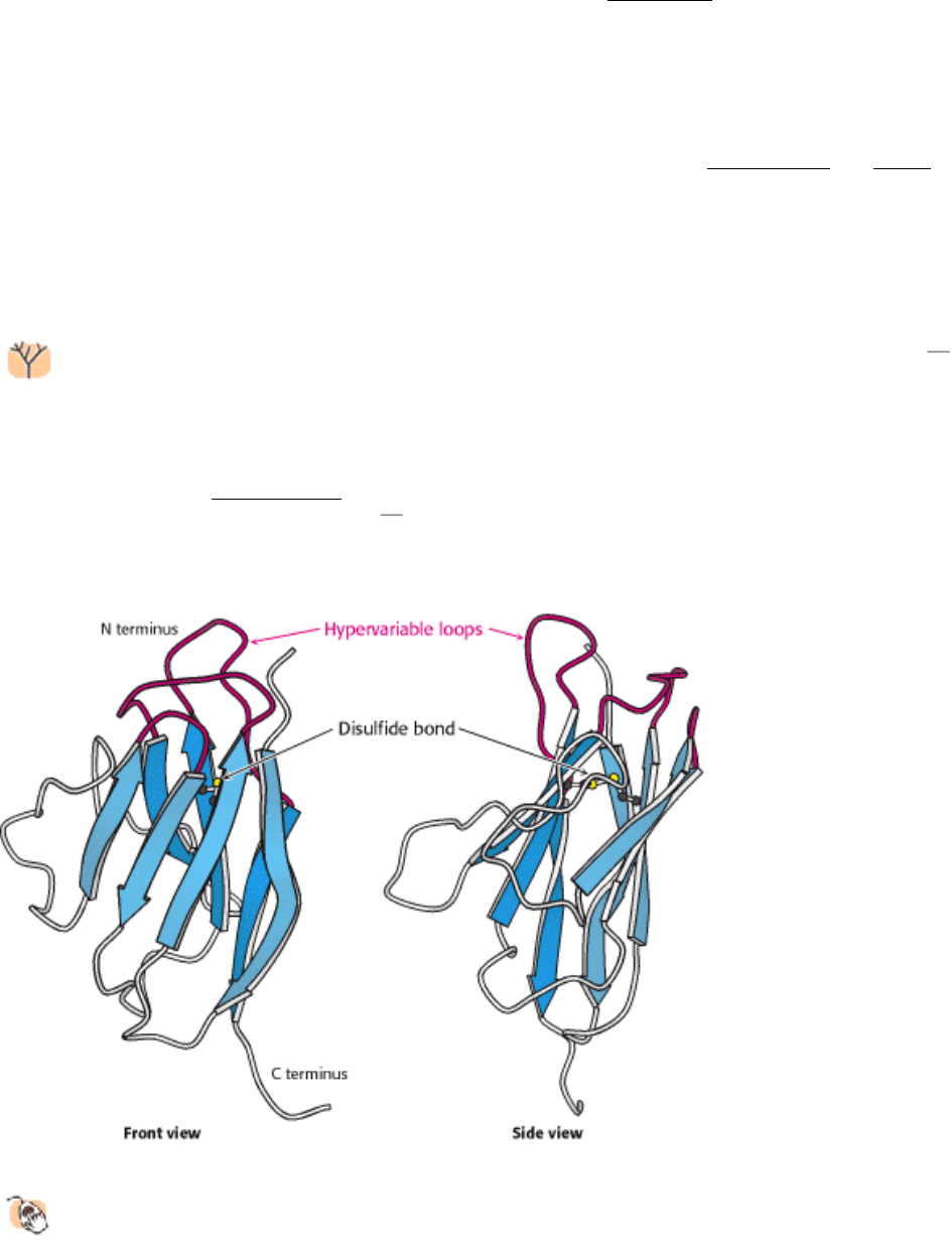
Figure 33.9. Immunoglobulin Fold.
An immunoglobulin domain consists of a pair of β-sheets linked by a disulfide
bond and hydrophobic interactions. Three hypervariable loops lie at one end of the structure.

IV. Responding to Environmental Changes 33. The Immune System
33.3. Antibodies Bind Specific Molecules Through Their Hypervariable Loops
For each class of antibody, the amino-terminal immunoglobin domains of the L and H chains (the variable domains,
designated V
L
and V
H
) come together at the ends of the arms extending from the structure. The positions of the
complementarity-determining regions are striking. These hypervariable sequences, present in three loops of each
domain, come together so that all six loops form a single surface at the end of each arm (Figure 33.10). Because virtually
any V
L
can pair with any V
H
, a very large number of different binding sites can be constructed by their combinatorial
association.
33.3.1. X-Ray Analyses Have Revealed How Antibodies Bind Antigens
The results of x-ray crystallographic studies of many large and small antigens bound to F
ab
molecules have been sources
of much insight into the structural basis of antibody specificity. The binding of antigens to antibodies is governed by the
same principles that govern the binding of substrates to enzymes. The apposition of complementary shapes results in
numerous contacts between amino acids at the binding surfaces of both molecules. Numerous hydrogen bonds,
electrostatic interactions, and van der Waals interactions, reinforced by hydrophobic interactions, combine to give
specific and strong binding.
A few aspects of antibody binding merit specific attention, inasmuch as they relate directly to the structure of
immunoglobulins. The binding site on the antibody has been found to incorporate some or all of the CDRs in the variable
domains of the antibody. Small molecules (e.g., octapeptides) are likely to make contact with fewer CDRs, with perhaps
15 residues of the antibody participating in the binding interaction. Macromolecules often make more extensive contact,
interacting with all six CDRs and 20 or more residues of the antibody. Small molecules often bind in a cleft of the
antigen-binding region. Macromolecules such as globular proteins tend to interact across larger, fairly flat apposed
surfaces bearing complementary protrusions and depressions.
A well-studied case of small-molecule binding is seen in an example of phosphorylcholine bound to F
ab
.
Crystallographic analysis revealed phosphorylcholine bound to a cavity lined by residues from five CDRs
two from
the L chain and three from the H chain (Figure 33.11). The positively charged trimethylammonium group of
phosphorylcholine is buried inside the wedge-shaped cavity, where it interacts electrostatically with two negatively
charged glutamate residues. The negatively charged phosphate group of phosphorylcholine binds to the positively
charged guanidinium group of an arginine residue at the mouth of the crevice and to a nearby lysine residue. The
phosphate group is also hydrogen bonded to the hydroxyl group of a tyrosine residue and to the guanidinium group of
the arginine side chain. Numerous van der Waals interactions, such as those made by a tryptophan side chain, also
stabilize this complex.
The binding of phosphorylcholine does not significantly change the structure of the antibody, yet induced fit plays a role
in the formation of many antibody-antigen complexes. A malleable binding site can accommodate many more kinds of
ligands than can a rigid one. Thus, induced fit increases the repertoire of antibody specificities.
33.3.2. Large Antigens Bind Antibodies with Numerous Interactions
How do large antigens interact with antibodies? A large collection of antibodies raised against hen egg-white lysozyme
has been structurally characterized in great detail (Figure 33.12). Each different antibody binds to a distinct surface of
lysozyme. Let us examine the interactions present in one of these complexes in detail. This antibody binds two
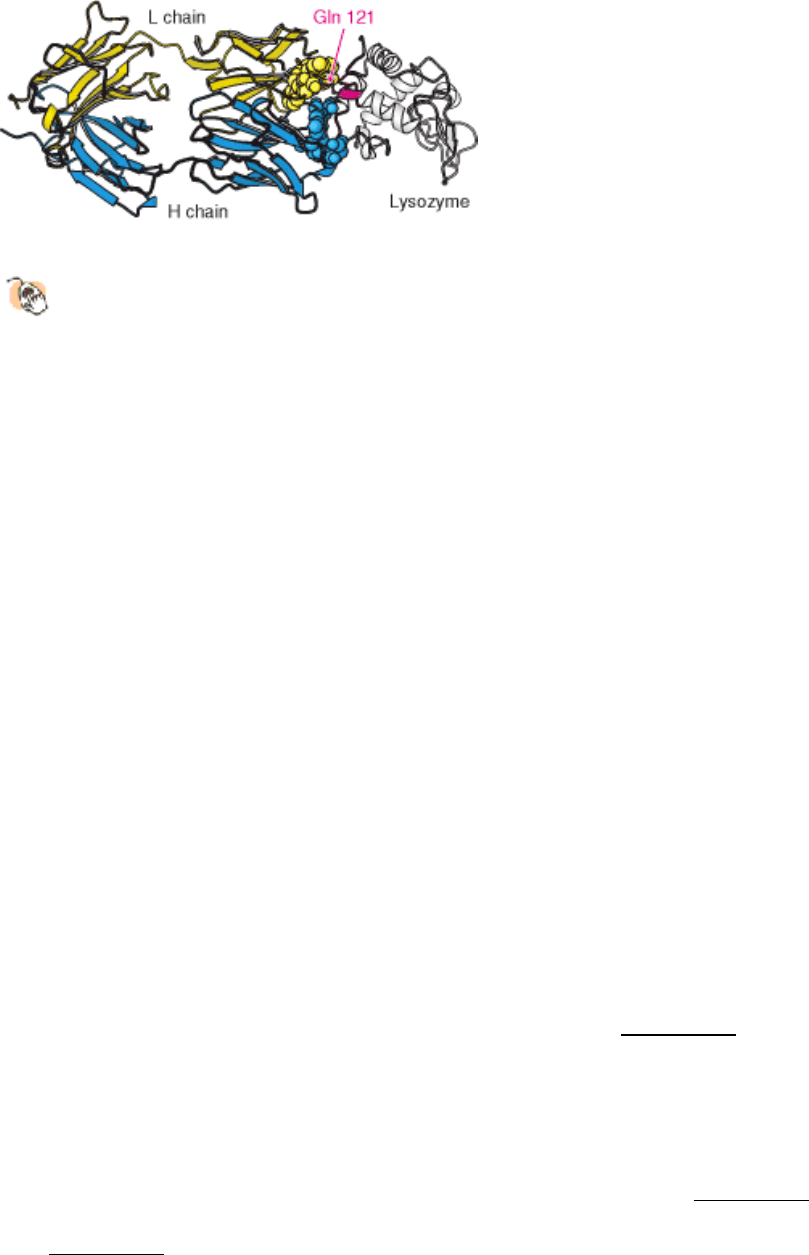
polypeptide segments that are widely separated in the primary structure, residues 18 to 27 and 116 to 129 (Figure 33.13).
All six CDRs of the antibody make contact with this epitope. The region of contact is quite extensive (about 30 × 20 Å).
The apposed surfaces are rather flat. The only exception is the side chain of glutamine 121 of lysozyme, which
penetrates deeply into the antibody binding site, where it forms a hydrogen bond with a main-chain carbonyl oxygen
atom and is surrounded by three aromatic side chains. The formation of 12 hydrogen bonds and numerous van der Waals
interactions contributes to the high affinity (K
d
= 20 nM) of this antibody-antigen interaction. Examination of the F
ab
molecule without bound protein reveals that the structures of the V
L
and V
H
domains change little on binding, although
they slide 1 Å apart to allow more intimate contact with lysozyme.
IV. Responding to Environmental Changes 33. The Immune System 33.3. Antibodies Bind Specific Molecules Through Their Hypervariable Loops
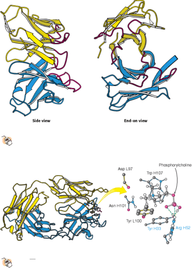
Figure 33.10. Variable Domains.
Two views of the variable domains of the L chain (yellow) and the H chain (blue); the
complementarity-determining regions (CDRs) are shown in red. The six CDRs come together to form a binding
surface. The specificity of the surface is determined by the sequences and structures of the CDRs.
IV. Responding to Environmental Changes 33. The Immune System 33.3. Antibodies Bind Specific Molecules Through Their Hypervariable Loops
Figure 33.11. Binding of a Small Antigen.
The structure of a complex between an F
ab
fragment of an antibody and its
target
in this case, phosphoryl-choline. Residues from the antibody interact with phosphorylcholine through
hydrogen bonding and electrostatic and van der Waals interactions.
IV. Responding to Environmental Changes 33. The Immune System 33.3. Antibodies Bind Specific Molecules Through Their Hypervariable Loops
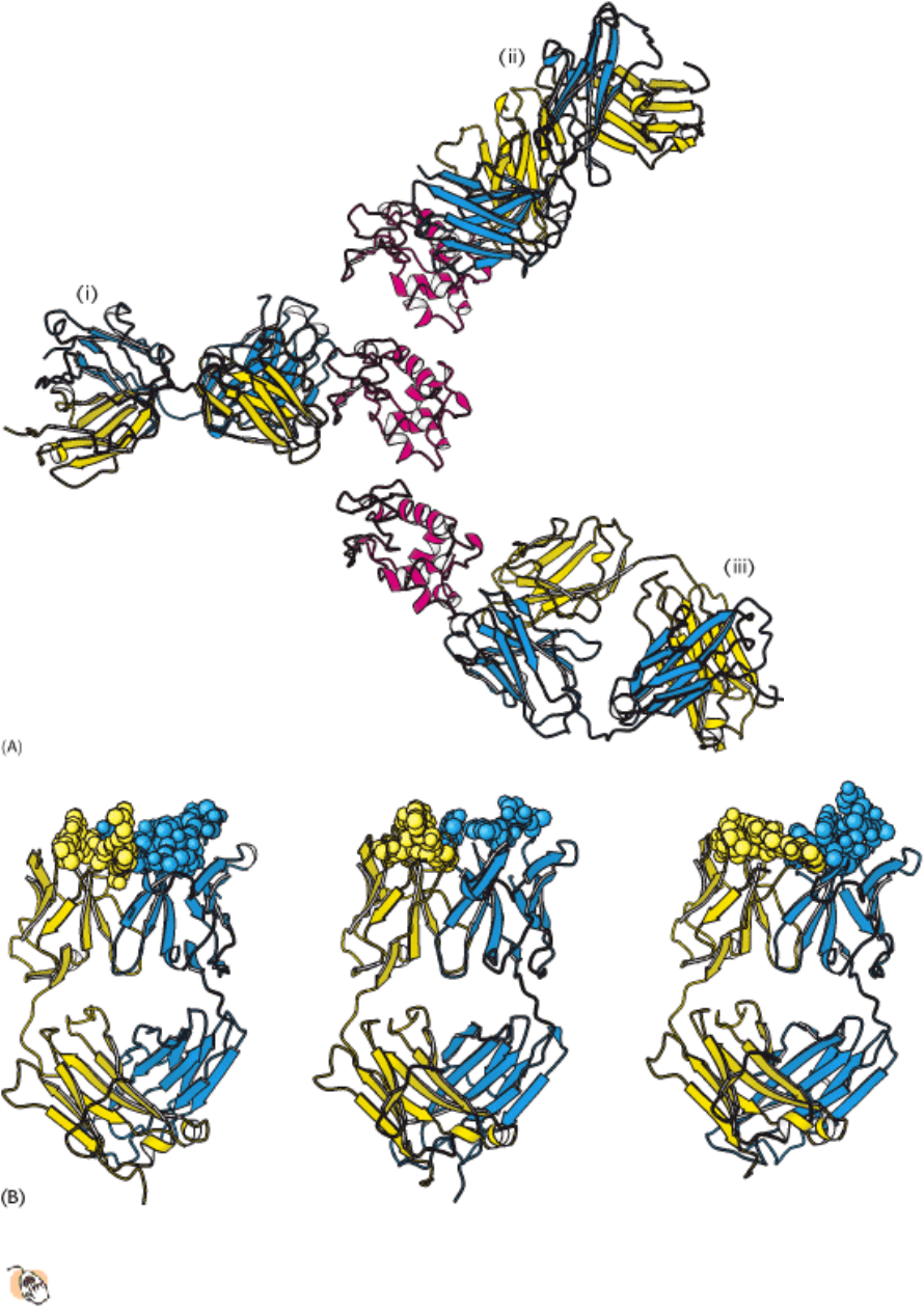
Figure 33.12. Antibodies Against Lysozyme.
(A) The structures of three complexes (i, ii, iii) between F
ab
fragments
(blue and yellow) and hen egg-white lysozyme (red) shown with lysozyme in the same orientation in each case.
The three antibodies recognize completely different epitopes on the lysozyme molecule. (B) The F
ab
fragments
from part A with points of contact highlighted as space-filling models, revealing the different shapes of the antigen-
binding sites.
IV. Responding to Environmental Changes 33. The Immune System 33.3. Antibodies Bind Specific Molecules Through Their Hypervariable Loops
Figure 33.13. Antibody - Protein Interactions.
The structure of a complex between an F
ab
fragment and lysozyme
reveals that the binding surfaces are complementary in shape over a large area. A single residue of lysozyme,
glutamine 121, penetrates more deeply into the antibody combining site.
IV. Responding to Environmental Changes 33. The Immune System
33.4. Diversity Is Generated by Gene Rearrangements
A mammal such as a mouse or a human being can synthesize large amounts of specific antibody against virtually any
foreign determinant within a matter of days of being exposed to it. We have seen that antibody specificity is determined
by the amino acid sequences of the variable regions of both light and heavy chains, which brings us to the key question:
How are different variable-region sequences generated?
The discovery of distinct variable and constant regions in the L and H chains raised the possibility that the genes that
encode immunoglobulins have an unusual architecture that facilitates the generation of a diverse set of polypeptide
products. In 1965, William Dreyer and Claude Bennett proposed that multiple V (variable) genes are separate from a
single C (constant) gene in embryonic (germ-line) DNA. According to their model, one of these V genes becomes joined
to the C gene in the course of differentiation of the antibody-producing cell. A critical test of this novel hypothesis had to
await the isolation of pure immunoglobulin mRNA and the development of techniques for analyzing mammalian
genomes. Twenty years later, Susumu Tonegawa found that V and C genes are indeed far apart in embryonic DNA but
are closely associated in the DNA of antibody-producing cells. Thus, immunoglobulin genes are rearranged in the
differentiation of lymphocytes.
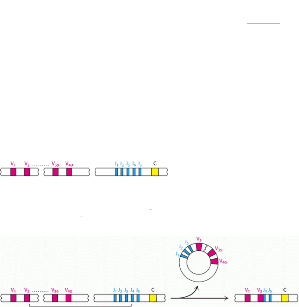
33.4.1. J (Joining) Genes and D (Diversity) Genes Increase Antibody Diversity
Sequencing studies carried out by Susumu Tonegawa, Philip Leder, and Leroy Hood revealed that V genes in embryonic
cells do not encode the entire variable region of L and H chains. Consider, for example, the region that encodes the κ
light-chain family. A tandem array of 40 segments, each of which encodes approximately the first 97 residues of the
variable domain of the L chain, is present on human chromosome 2 (Figure 33.14).
However, the variable region of the L chain extends to residue 110. Where is the DNA that encodes the last 13 residues
of the V region? For L chains in undifferentiated cells, this stretch of DNA is located in an unexpected place: near the C
gene. It is called the J gene because it joins the V and C genes in a differentiated cell. In fact, a tandem array of five J
genes is located near the C gene in embryonic cells. In the differentiation of an antibody-producing cell, a V gene
becomes spliced to a J gene to form a complete gene for the variable region (Figure 33.15). RNA splicing generates an
mRNA molecule for the complete L chain by linking the coding regions for the rearranged VJ unit with that for the C
unit (Figure 33.16).
J genes are important contributors to antibody diversity because they encode part of the last hypervariable segment
(CDR3). In forming a continuous variable-region gene, any of the 40 V genes can become linked to any of five J genes.
Thus, somatic recombination of these gene segments amplifies the diversity already present in the germ line. The linkage
between V and J is not precisely controlled. Recombination between these genes can take place at one of several bases

near the codon for residue 95, generating additional diversity. A similar array of V and J genes encoding the λ light chain
is present on human chromosome 22. This region includes 30 V
λ
gene segments and four J
λ
segments. In addition, this
region includes four distinct C genes, in contrast with the single C gene in the κ locus.
In human beings, the genes encoding the heavy chain are present on chromosome 14. Remarkably, the variable domain
of heavy chains is assembled from three rather than two segments. In addition to V
H
genes that encode residues 1 to 94
and J
H
segments that encode residues 98 to 113, this chromosomal region includes a distinct set of segments that encode
residues 95 to 97 (Figure 33.17). These gene segments are called D for diversity. Some 27 D segments lie between 51
V
H
and 6 J
H
segments. The recombination process first joins a D segment to a J
H
segment; a V
H
segment is then joined
to DJ
H
. A greater variety of antigen-binding patches and clefts can be formed by the H chain than by the L chain because
the H chain is encoded by three rather than two gene segments. Moreover, CDR3 of the H chain is diversified by the
action of terminal deoxyribonucleotidyl transferase, a special DNA polymerase that requires no template. This enzyme
inserts extra nucleotides between V
H
and D. The V(D)J recombination of both the L and the H chains is executed by
specific enzymes present in immune cells. These proteins, called RAG-1 and RAG-2, recognize specific DNA sequences
called recombination signal sequences (RSSs) adjacent to the V, D, and J segments and facilitate the cleavage and
religation of the DNA segments.
33.4.2. More Than 10
8
Antibodies Can Be Formed by Combinatorial Association and
Somatic Mutation
Let us recapitulate the sources of antibody diversity. The germ line contains a rather large repertoire of variable-region
genes. For κ light chains, there are about 40 V-segment genes and five J-segment genes. Hence, a total of 40 × 5 = 200
kinds of complete V
κ
genes can be formed by the combinations of V and J. A similar analysis suggests that at least 120
different λ light chains can be generated. A larger number of heavy-chain genes can be formed because of the role of the
D segments. For 51 V, 27 D, and 6 J gene segments, the number of complete V
H
genes that can be formed is 8262. The
association of 320 kinds of L chains with 8262 kinds of H chains would yield 2.6 × 10
6
different antibodies. Variability
in the exact points of segment joining and other mechanisms increases this value by at least two orders of magnitude.
Even more diversity is introduced into antibody chains by somatic mutation
that is, the introduction of mutations into
the recombined genes. In fact, a 1000-fold increase in binding affinity is seen in the course of a typical humoral immune
response, arising from somatic mutation, a process called affinity maturation. The generation of an expanded repertoire
leads to the selection of antibodies that more precisely fit the antigen. Thus, nature draws on each of three sources of
diversity a germ-line repertoire, somatic recombination, and somatic mutation to form the rich variety of
antibodies that protect an organism from foreign incursions.
33.4.3. The Oligomerization of Antibodies Expressed on the Surface of Immature B
Cells Triggers Antibody Secretion
The processes heretofore described generate a highly diverse set of antibody molecules
a key first step in the
generation of an immune response. The next stage is the selection of a particular set of antibodies directed against a
specific invader. How does this selection occur? Each immature B cell, produced in the bone marrow, expresses a
monomeric form of IgM attached to its surface (Figure 33.18). Each cell expresses approximately 10
5
IgM molecules,
but all of these molecules are identical in amino acid sequence and, hence, in antigen-binding specificity. Thus, the
selection of a particular immature B cell for growth will lead to the amplification of an antibody with a unique
specificity. The selection process begins with the binding of an antigen to the membrane-bound antibody.
Associated with each membrane-linked IgM molecule are two molecules of a heterodimeric membrane protein called Ig-
α-Ig-β (see Figure 33.18). Examination of the amino acid sequences of Ig-α and Ig-β is highly instructive. The amino
terminus of each protein lies outside the cell and corresponds to a single immunoglobulin, and the carboxyl terminus,
which lies inside the cell, includes a sequence of 18 amino acids called an immunoreceptor tyrosine-based activation
motif (ITAM) (see Figure 33.18). As its name suggests, each ITAM includes key tyrosine residues, which are subject to
phosphorylation by particular protein kinases present in immune-system cells.
A fundamental observation with regard to the mechanism by which the binding of antigen to membrane-bound antibody
triggers the subsequent steps of the immune response is that oligomerization or clustering of the antibody molecules is
required (Figure 33.19). The requirement for oligomerization is reminiscent of the dimerization of receptors triggered by
growth hormone and epidermal growth factor encountered in Sections 15.4 and 15.4.1; indeed, the associated signaling
mechanisms appear to be quite similar. The oligomerization of the membrane-bound antibodies results in the
phosphorylation of the tyrosine residues within the ITAMs by protein tyrosine kinases including Lyn, a homolog of Src
(Section 15.5). The phosphorylated ITAMs serve as docking sites for a protein kinase termed spleen tyrosine kinase
(Syk), which has two SH2 domains that interact with the pair of phosphorylated tyrosines in each ITAM. Syk, when
activated by phosphorylation, proceeds to phosphorylate other signal-transduction proteins including an inhibitory
subunit of a transcription factor called NF-κB and an isoform of phospholipase C. The signaling processes continue
downstream to activate gene expression, leading to the stimulation of cell growth and initiating further B-cell
differentiation.
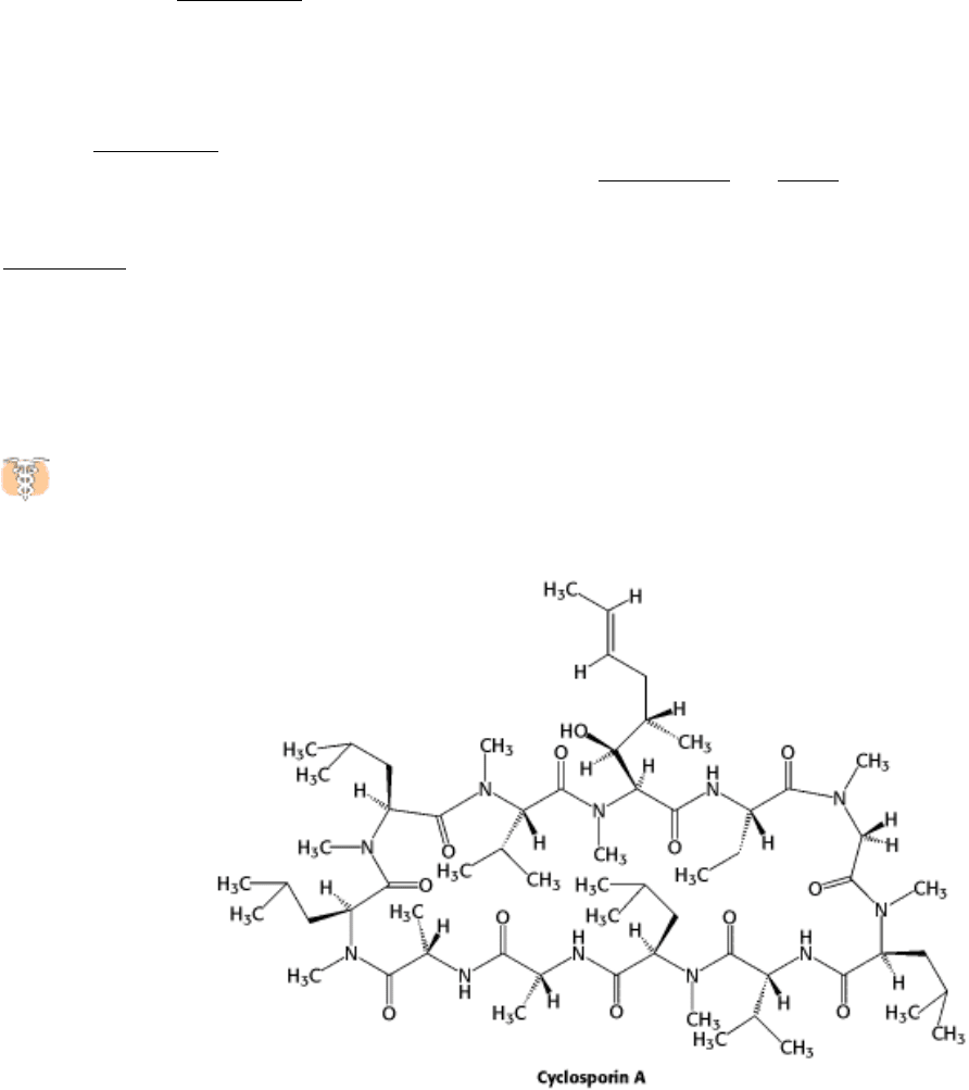
Drugs that modulate the immune system have served as sources, of insight into immune-system signaling
pathways. For example, cyclosporin, a powerful suppressor of the immune system, acts by blocking a phosphatase
called calcineurin, which normally activates a transcription factor called NF-AT by dephosphorylating it.
The potent immune supression that results reveals how crucial the activity of this transcription factor is to the
development of an immune response. Without drugs such as cyclosporin, organ transplantation would be extremely
difficult because transplanted tissue expresses a wide range of foreign antigens, which causes the immune system to
reject the new tissue.
The role of oligomerization in the B-cell signaling pathway is illuminated when we consider the nature of many antigens
presented by pathogens. The surfaces of many viruses, bacteria, and parasites are characterized by arrays of identical
membrane proteins or membrane-linked carbohydrates. Thus, most pathogens present multiple binding surfaces that will
naturally cause membrane-associated antibodies to oligomerize as they bind adjacent epitopes. In addition, the
mechanism accounts for the observation that most small molecules do not induce an immune response; however,
coupling multiple copies of the small molecule to a large oligomeric protein such as keyhole limpet hemocyanin (KLH),
which has a molecular mass of close to 1 million daltons or more, promotes antibody oligomerization and, hence, the
production of antibodies against the small-molecule epitope. The large protein is called the carrier of the attached
chemical group, which is called a haptenic determinant. The small foreign molecule by itself is called a hapten.
Antibodies elicited by attached haptens will bind unattached haptens as well.
33.4.4. Different Classes of Antibodies Are Formed by the Hopping of V
H
Genes
The development of an effective antibody-based immune response depends on the secretion into the blood of antibodies
that have appropriate effector functions. At the beginning of this response, an alternative mRNA splicing pathway is
activated so that the production of membrane-linked IgM is supplanted by the synthesis of secreted IgM. As noted in
Section 33.1, secreted IgM is pentameric and has a relatively high avidity for multivalent antigens. Later, the antibody-
producing cell makes either IgG, IgA, IgD, or IgE of the same specificity as the intially secreted IgM. In this switch, the
light chain is unchanged, as is the variable region of the heavy chain. Only the constant region of the heavy chain
changes. This step in the differentiation of an antibody-producing cell is called class switching (Figure 33.20). In
undifferentiated cells, the genes for the constant region of each class of heavy chain, called C
µ
, C
δ
, C
γ
, C
ε
, and C
α
,
are next to each other. There are eight in all, including four genes for the constant regions of γ chains. A complete gene
for the heavy chains of IgM antibody is formed by the translocation of a V
H
gene segment to a DJ
H
gene segment.
How are other heavy chains formed? Class switching is mediated by a gene-rearrangement process that moves a VDJ
gene from a site near one C gene to a site near another C gene. Importantly, the antigen-binding specificity is conserved
in class switching because the entire V
H
DJ
H
gene is translocated in an intact form. For example, the antigen-
combining specificity of IgA produced by a particular cell is the same as that of IgM synthesized at an earlier stage of its
development. The biological significance of C
H
switching is that a whole recognition domain (the variable domain) is
shifted from the early constant region (C
µ
) to one of several other constant regions that mediate different effector
functions.
IV. Responding to Environmental Changes 33. The Immune System 33.4. Diversity Is Generated by Gene Rearrangements
Figure 33.14. The κ Light-Chain Locus. This part of human chromosome 2 includes an array of 40 segments that
encode the variable (V) region (approximately residues 1
97) of the light chain, an array of 5 segments that encode the
joining (J) region (residues 98 110), and a single region that encodes the constant (C) region.
IV. Responding to Environmental Changes 33. The Immune System 33.4. Diversity Is Generated by Gene Rearrangements
Figure 33.15. VJ Recombination. A single V gene (in this case, V
2
) is linked to a J gene (here, J
4
) to form an intact VJ
region. The intervening DNA is released in a circular form. Because the V and J regions are selected at random and the
joint between them is not always in exactly the same place, many VJ combinations can be generated by this process.