Berg J.M., Tymoczko J.L., Stryer L. Biochemistry
Подождите немного. Документ загружается.

computer for potential 7TM receptor genes, and, indeed, one was detected and named T2R-1. Additional database
searches for sequences similar to this one detected 12 genes encoding full-length receptors as well as 7 pseudogenes
within the sequence of the human genome known at the time. The encoded proteins were between 30 and 70% identical
with T2R-1 (Figure 32.14). Further analysis suggests that there are from 50 to 100 members of this family of 7TM
receptors in the entire human genome. Similar sequences have been detected in the mouse and rat genomes.
Are these proteins, in fact, bitter receptors? Several lines of evidence suggest that they are. First, their genes are
expressed in taste-sensitive cells
in fact, in many of the same cells that express gustducin. Second, cells that express
individual members of this family respond to specific bitter compounds. For example, cells that express a specific mouse
receptor (mT2R-5) responded when exposed specifically to cycloheximide. Third, mice that had been found
unresponsive to cycloheximide were found to have point mutations in the gene encoding mT2R-5. Finally,
cycloheximide specifically stimulates the binding of GTP analogs to gustducin in the presence of the mT2R-5 protein
(Figure 32.15).
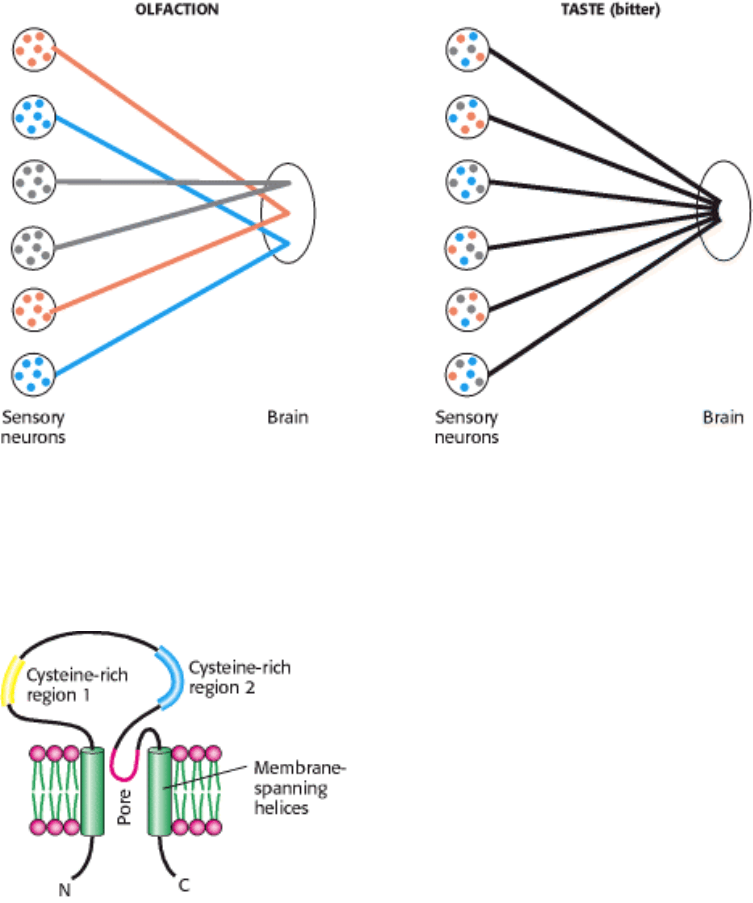
Importantly, each taste receptor cell expresses many different members of the T2R family. This pattern of expression
stands in sharp contrast to the pattern of one receptor type per cell that characterizes the olfactory system (Figure 32.16).
The difference in expression patterns accounts for the much greater specificity of our perceptions of smells compared
with tastes. We are able to distinguish among subtly different odors because each odorant stimulates a unique pattern of
neurons. In contrast, many tastants stimulate the same neurons. Thus, we perceive only "bitter" without the ability to
discriminate cycloheximide from quinine.
32.2.2. A Family of 7TM Receptors Almost Certainly Respond to Sweet Compounds
Most sweet compounds are carbohydrates, energy rich and easily digestible. Some noncarbohydrate compounds such as
saccharin and aspartame also taste sweet. The structural diversity among sweet-tasting compounds, though less than that
among bitter compounds, strongly suggested that a family of receptors detects these compounds. The observation that
mice in which the gene for gustducin was disrupted lost much of their ability to sense sweet, as well as bitter, compounds
strongly suggested that the sweet receptors would belong to the 7TM receptor superfamily. Recently, a small group of
7TM receptors that respond to sweet compounds has been identified. Interestingly, simultaneous expression of two
members of the family in the same cell is required for the cells to respond to sweet compounds. The biochemical
explanation for this observation remains to be elucidated.
32.2.3. Salty Tastes Are Detected Primarily by the Passage of Sodium Ions Through
Channels
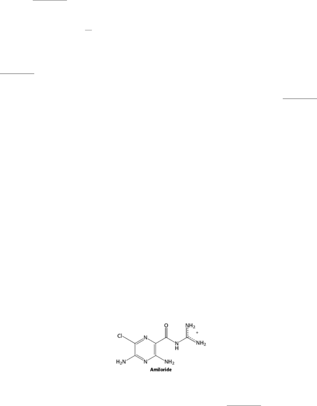
Salty tastants are not detected by 7TM receptors. Rather, they are detected directly by their passage through ion channels
expressed on the surface of cells in the tongue. Evidence for the role of these ion channels comes from examining known
properties of sodium channels characterized in other biological contexts. One class of channels, characterized first for
their role in salt reabsorption, are thought to be important in salt taste detection because they are sensitive to the
compound amiloride, which mutes the taste of salt and significantly lowers sensory neuron activation in response to
sodium.
An amiloride-sensitive sodium channel comprises four subunits that may be either identical or distinct but in any case
are homologous. An individual subunit ranges in length from 500 to 1000 amino acids and includes two presumed
membrane-spanning helices as well as a large extracellular domain in between them (Figure 32.17). The extracellular

region includes two (or, sometimes, three) distinct regions rich in cysteine residues (and, presumably, disulfide bonds). A
region just ahead of the second membrane-spanning helix appears to form part of the pore in a manner analogous to the
structurally characterized potassium channel (Section 13.5.6). The members of the amiloride-sensitive sodium-channel
family are numerous and diverse in their biological roles. We shall encounter them again in the context of the sense of
touch.
Sodium ions passing through these channels produce a significant transmembrane current. Amiloride blocks this current,
accounting for its effect on taste. However, about 20% of the response to sodium remains even in the presence of
amiloride, suggesting that other ion channels also contribute to salt detection.
32.2.4. Sour Tastes Arise from the Effects of Hydrogen Ions (Acids) on Channels
Like salty tastes, sour tastes are also detected by direct interactions with ion channels, but the incoming ions are
hydrogen ions (in high concentrations) rather than sodium ions. For example, in the absence of high concentrations of
sodium, hydrogen ion flow can induce substantial transmembrane currents through amiloride-sensitive sodium channels.
However, hydrogen ions are also sensed by mechanisms other than their direct passage through membranes. Binding by
hydrogen ions blocks some potassium channels and activates other types of channels. Together, these mechanisms lead
to changes in membrane polarization in sensory neurons that produce the sensation of sour taste.
32.2.5. Umami, the Taste of Glutamate, Is Detected by a Specialized Form of
Glutamate Receptor
Glutamate is an abundant amino acid that is present in protein-rich foods as well as in the widely used flavor enhancer
monosodium glutamate. This amino acid has a taste, termed umami, that is distinct from the other four basic tastes.
Adults can detect glutamate at a concentration of approximately 1 mM. Glutamate is also a widely used neurotransmitter,
and thus, not surprisingly, several classes of receptors for glutamate have been identified in the nervous system. One
class, called metabotrophic glutamate receptors, are 7TM receptors with large amino-terminal domains of approximately
600 amino acids. Sequence analysis reveals that the first half of the aminoterminal region is most likely a ligand-binding
domain, because it is homologous to such domains found in the Lac repressor (Section 31.1.3) and other bacterial ligand-
binding proteins.
One glutamate receptor gene, encoding a protein called the metabotrophic glutamate receptor 4 (mGluR4), has
been found to be expressed in taste buds. Further analysis of the mRNA that is expressed in taste buds reveals that
this mRNA lacks the region encoding the first 309 amino acids in brain mGluR4, which includes most of the high-
affinity glutamate-binding domain (Figure 32.18). The glutamate receptor found in taste buds shows a lowered affinity
for glutamate that is appropriate to glutamate levels in the diet. Thus, the receptor responsible for the perception of
glutamate taste appears to have evolved simply by changes in the expression of an existing glutamate-receptor gene. We
shall consider an additional receptor related to taste, that responsible for the "hot" taste of spicy food, when we deal with
mechanisms of touch perception.
IV. Responding to Environmental Changes 32. Sensory Systems 32.2. Taste Is a Combination of Senses that Function by Different Mechanisms
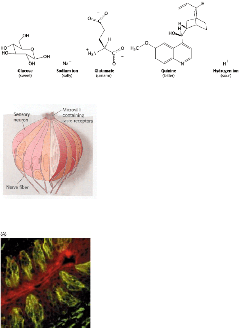
Figure 32.11. Examples of Tastant Molecules. Tastants fall into five groups: sweet, salty, umami, bitter, and sour.
IV. Responding to Environmental Changes 32. Sensory Systems 32.2. Taste Is a Combination of Senses that Function by Different Mechanisms
Figure 32.12. A Taste Bud. Each taste bud contains sensory neurons that extend microvilli to the surface of the tongue,
where they interact with tastants.
IV. Responding to Environmental Changes 32. Sensory Systems 32.2. Taste Is a Combination of Senses that Function by Different Mechanisms
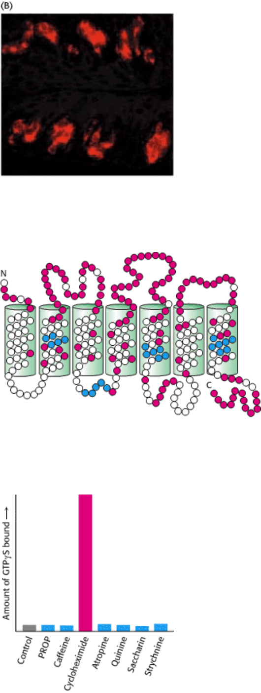
Figure 32.13. Expression of Gustducin in the Tongue. (A) A section of tongue stained with a fluorescent antibody
reveals the position of the taste buds. (B) The same region stained with a antibody directed against gustducin reveals that
this G protein is expressed in taste buds. [Courtesy of Charles S. Zuker.]
IV. Responding to Environmental Changes 32. Sensory Systems 32.2. Taste Is a Combination of Senses that Function by Different Mechanisms
Figure 32.14. Conserved and Variant Regions in Bitter Receptors. The bitter receptors are members of the 7TM
receptor family. Strongly conserved residues characteristic of this protein family are shown in blue, and highly variable
residues are shown in red.
IV. Responding to Environmental Changes 32. Sensory Systems 32.2. Taste Is a Combination of Senses that Function by Different Mechanisms
Figure 32.15. Evidence that T2R Proteins Are Bitter Taste Receptors. Cycloheximide uniquely stimulates the
binding of the GTP analog GTP γ S to gustducin in the presence of the mT2R protein. [Adapted from J. Chandrashekar,
K. L. Mueller, M. A. Hoon, E. Adler, L. Feng, W. Guo, C. S. Zuker, and N. J. Ryba. Cell 100(2000):703.]
IV. Responding to Environmental Changes 32. Sensory Systems 32.2. Taste Is a Combination of Senses that Function by Different Mechanisms
Figure 32.16. Differing Gene Expression and Connection Patterns in Olfactory and Bitter Taste Receptors. In
olfaction, each neuron expresses a single OR gene, and the neurons expressing the same OR converge to specific sites in
the brain, enabling specific perception of different odorants. In gustation, each neuron expresses many bitter receptor
genes, so the identity of the tastant is lost in transmission.
IV. Responding to Environmental Changes 32. Sensory Systems 32.2. Taste Is a Combination of Senses that Function by Different Mechanisms
Figure 32.17. Schematic Structure of the Amiloride-Sensitive Sodium Channel. Only one of the four subunits that
constitute the functional channel is illustrated. The amiloride-sensitive sodium channel belongs to a superfamily having
common structural features, including two hydrophobic membrane-spanning regions, intracellular amino and carboxyl
termini; and a large, extracellular region with conserved cysteine-rich domains.
IV. Responding to Environmental Changes 32. Sensory Systems 32.2. Taste Is a Combination of Senses that Function by Different Mechanisms
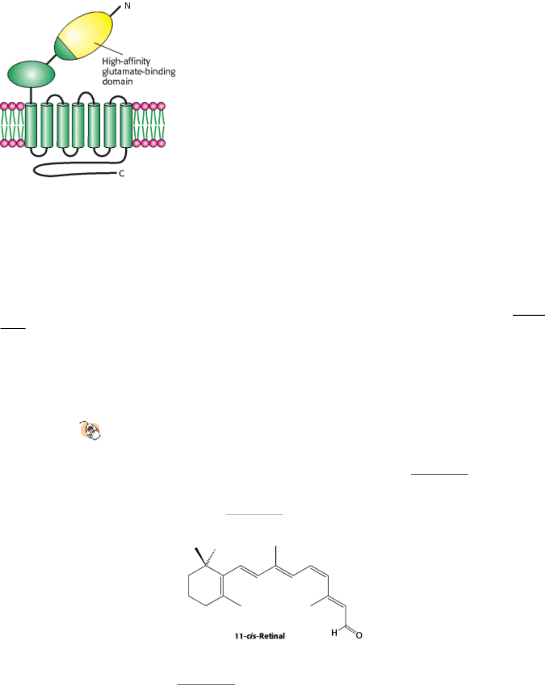
Figure 32.18. Schematic Structure of a Metabotrophic Glutamate Receptor. The umami receptor is a variant of a
brain glutamate receptor. A substantial part of the high-affinity glutamate-binding domain (shown in yellow) is missing
in the form expressed in the tongue.
IV. Responding to Environmental Changes 32. Sensory Systems
32.3. Photoreceptor Molecules in the Eye Detect Visible Light
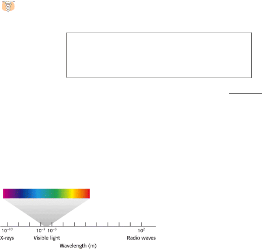
Vision is based on the absorption of light by photoreceptor cells in the eye. These cells are sensitive to light in a
relatively narrow region of the electromagnetic spectrum, the region with wavelengths between 300 and 850 nm (Figure
32.19). Vertebrates have two kinds of photoreceptor cells, called rods and cones because of their distinctive shapes.
Cones function in bright light and are responsible for color vision, whereas rods function in dim light but do not perceive
color. A human retina contains about 3 million cones and 100 million rods. Remarkably, a rod cell can respond to a
single photon, and the brain requires fewer than 10 such responses to register the sensation of a flash of light.
32.3.1. Rhodopsin, a Specialized 7TM Receptor, Absorbs Visible Light
Structural Insights, Rhodopsin: A G Protein Coupled 7TM Receptor offers
a more detailed look at rhodopsin structure and function (Figure 32.5).
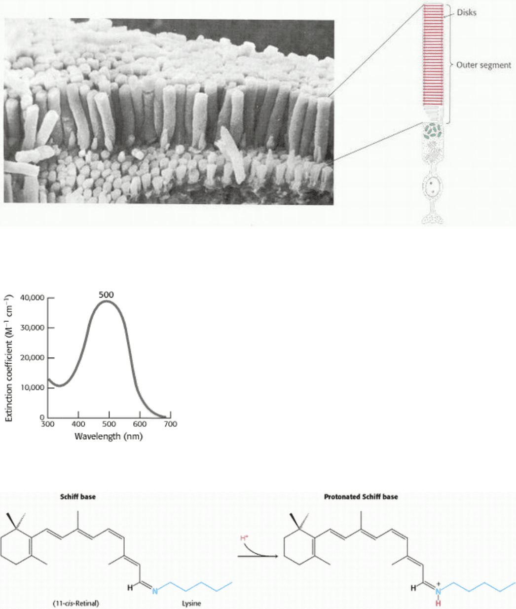
Rods are slender elongated structures; the outer segment is specialized for photoreception (Figure 32.20). It contains a
stack of about 1000 discs, which are membrane-enclosed sacs densely packed with photoreceptor molecules. The
photosensitive molecule is often called a visual pigment because it is highly colored owing to its ability to absorb light.
The photoreceptor molecule in rods is rhodopsin (Section 15.1), which consists of the protein opsin linked to 11-cis-
retinal, a prosthetic group.
Rhodopsin absorbs light very efficiently in the middle of the visible spectrum, its absorption being centered on 500 nm,
which nicely matches the solar output (Figure 32.21). A rhodopsin molecule will absorb a high percentage of the photons
of the correct wavelength that strike it, as indicated by the extinction coefficient of 40,000 M
-1
cm
-1
at 500 nm. The
extinction coefficient for rhodopsin is more than an order of magnitude greater than that for tryptophan, the most
efficient absorber in proteins that lack prosthetic groups.

Opsin, the protein component of rhodopsin, is a member of the 7TM receptor family. Indeed, rhodopsin was the first
member of this family to be purified, its gene was the first to be cloned and sequenced, and its three-dimensional
structure was the first to be determined. The color of rhodopsin and its responsiveness to light depend on the presence of
the light-absorbing group (chromophore) 11-cis-retinal. This compound is a powerful absorber of light because it is a
polyene; its six alternating single and double bonds constitute a long, unsaturated electron network. Recall that
alternating single and double bonds account for the chromophoric properties of chlorophyll (Section 19.2). The aldehyde
group of 11-cis-retinal forms a Schiff base (Figure 32.22) with the ε-amino group of lysine residue 296, which lies in the
center of the seventh transmembrane helix. Free retinal absorbs maximally at 370 nm, and its unprotonated Schiff-base
adduct absorbs at 380 nm, whereas the protonated Schiff base absorbs at 440 nm or longer wavelengths. Thus, the 500-
nm absorption maximum for rhodopsin strongly suggests that the Schiff base is protonated; additional interactions with
opsin shift the absorption maximum farther toward the red. The positive charge of the protonated Schiff base is
compensated by the negative charge of glutamate 113 located in helix 2; the glutamate residue closely approaches the
lysine-retinal linkage in the three-dimensional structure of rhodopsin.
32.3.2. Light Absorption Induces a Specific Isomerization of Bound 11-cis-Retinal
How does the absorption of light by the retinal Schiff base generate a signal? George Wald and his coworkers discovered
that light absorption results in the isomerization of the 11-cis-retinal group of rhodopsin to its all-trans form (Figure
32.23). This isomerization causes the Schiff-base nitrogen atom to move approximately 5 Å, assuming that the
cyclohexane ring of the retinal group remains fixed. In essence, the light energy of a photon is converted into atomic
motion. The change in atomic positions, like the binding of a ligand to other 7TM receptors, sets in train a series of
events that lead to the closing of ion channels and the generation of a nerve impulse.
The isomerization of the retinal Schiff base takes place within a few picoseconds of a photon being absorbed. The initial
product, termed bathorhodopsin, contains a strained all-trans-retinal group. Within approximately 1 millisecond, this
intermediate is converted through several additional intermediates into metarhodopsin II. In metarhodopsin II, the Schiff
base is deprotonated and the opsin protein has undergone significant reorganization.
Metarhodopsin II (also referred to as R*) is analogous to the ligand-bound state of 7TM receptors such as the β
2
-
adrenergic receptor (Section 15.1) and the odorant and tastant receptors heretofore discussed (Figure 32.24). Like these
receptors, this form of rhodopsin activates a heterotrimeric G protein that propagates the signal. The G protein associated
with rhodopsin is called transducin. Metarhodopsin II triggers the exchange of GDP for GTP by the α subunit of
transducin (Figure 32.25). On the binding of GTP, the β γ subunits of transducin are released and the α subunit switches
on a cGMP phosphodiesterase by binding to and removing an inhibitory subunit. The activated phosphodiesterase is a
potent enzyme that rapidly hydrolyzes cGMP to GMP. The reduction in cGMP concentration causes cGMP-gated ion
channels to close, leading to hyperpolarization of the membrane and neuronal signaling. At each step in this process, the
initial signal the absorption of a single photon is amplified so that it leads to sufficient membrane hyperpolarization
to result in signaling.
Conceptual Insights, Signaling Pathways: Response and Recovery presents
an animated version of Figure 32.25 and a comparison to olfactory signal
transduction (Figure 32.5).
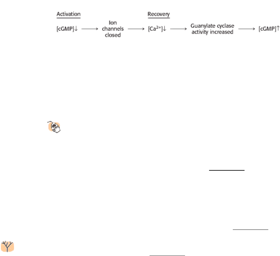
32.3.3. Light-Induced Lowering of the Calcium Level Coordinates Recovery
As we have seen, the visual system responds to changes in light and color within a few milliseconds, quickly enough that
we are able to perceive continuous motion at nearly 1000 frames per second. To achieve a rapid response, the signal
must also be terminated rapidly and the system must be returned to its initial state. First, activated rhodopsin must be
blocked from continuing to activate transducin. Rhodopsin kinase catalyzes the phosphorylation of the carboxyl terminus
of R* at multiple serine and threonine residues. Arrestin, an inhibitory protein (Section 15.1.4), then binds
phosphorylated R* and prevents additional interaction with transducin.
Second, the α subunit of transducin must be returned to its inactive state to prevent further signaling. Like other G
proteins, the α subunit possesses built-in GTPase activity that hydrolyzes bound GTP to GDP. Hydrolysis takes place in
less than a second when transducin is bound to the phosphodiesterase. The GDP form of transducin then leaves the
phosphodiesterase and reassociates with the β γ subunits, and the phosphodiesterase returns to its inactive state. Third,
the level of cGMP must be raised to reopen the cGMP-gated ion channels. The action of guanylate cyclase accomplishes
this third step by synthesizing cGMP from GTP.
Calcium ion plays an essential role in controlling guanylate cyclase because it markedly inhibits the activity of the
enzyme. In the dark, Ca
2+
as well as Na
+
enter the rod outer segment through the cGMP-gated channels. Calcium ion
influx is balanced by its efflux through an exchanger, a transport system that uses the thermodynamically favorable flow
of four Na
+
ions into the cell and one K
+
ion out of the cell to extrude one Ca
2+
ion. After illumination, the entry of Ca
2+
through the cGMP-gated channels stops, but its export through the exchanger continues. Thus, the cytosolic Ca
2+
level
drops from 500 nM to 50 nM after illumination. This drop markedly stimulates guanylate cyclase, rapidly restoring the
concentration of cGMP to reopen the cGMP-gated channels.
By controlling the rate of cGMP synthesis, Ca
2
+
levels govern the speed with which the system is restored to its initial
state.
32.3.4. Color Vision Is Mediated by Three Cone Receptors That Are Homologs of
Rhodopsin
Structural Insights, Rhodopsin: A G Protein Coupled 7TM Receptor
explores the structural basis of color vision and night blindness in more detail.
Cone cells, like rod cells, contain visual pigments. Like rhodopsin, these photoreceptor proteins are members of the 7TM
receptor family and utilize 11-cis-retinal as their chromophore. In human cone cells, there are three distinct
photoreceptor proteins with absorption maxima at 426, 530, and ~ 560 nm (Figure 32.26). These absorbances
correspond to (in fact, define) the blue, green, and red regions of the spectrum. Recall that the absorption maximum for
rhodopsin is 500 nm.
The amino acid sequences of the cone photoreceptors have been compared with each other and with rhodopsin. The
result is striking. Each of the cone photoreceptors is approximately 40% identical in sequence with rhodopsin. Similarly,
the blue photoreceptor is 40% identical with each of the green and red photoreceptors. The green and red photoreceptors,
however, are > 95% identical with each other, differing in only 15 of 364 positions (Figure 32.27).
These observations are sources of insight into photoreceptor evolution. First, the green and red photoreceptors are
clearly products of a recent evolutionary event (Figure 32.28). The green and red pigments appear to have
diverged in the primate lineage approximately 35 million years ago. Mammals, such as dogs and mice, that diverged
from primates earlier have only two cone photoreceptors, blue and green. They are not sensitive to light as far toward the
infrared region as we are, and they do not discriminate colors as well. In contrast, birds such as chickens have a total of
six pigments: rhodopsin, four cone pigments, and a pineal visual pigment called pinopsin. Birds have highly acute color
perception.
Second, the high level of similarity between the green and red pigments has made it possible to identify the specific
amino acid residues that are responsible for spectral tuning. Three residues (at positions 180, 277, and 285) are
responsible for most of the difference between the green and red pigments. In the green pigment, these residues are
alanine, phenylalanine, and alanine, respectively; in the red pigment, they are serine, tyrosine, and threonine. A hydroxyl
group has been added to each amino acid in the red pigment. The hydroxyl groups can interact with the photoexcited
state of retinal and lower its energy, leading to a shift toward the lower-energy (red) region of the spectrum.
32.3.5. Rearrangements in the Genes for the Green and Red Pigments Lead to "Color
Blindness"
The genes for the green and red pigments lie adjacent to each other on the human X chromosome. These genes are
more than 98% identical in nucleotide sequence, including introns and untranslated regions as well as the protein-
coding region. Regions with such high similarity are very susceptible to unequal homologous recombination.
Homologous recombination
The exchange of DNA segments at equivalent positions between
chromosomes with substantial sequence similarity.
Recombination can take place either between or within transcribed regions of the gene (Figure 32.29). If recombination
takes place between transcribed regions, the product chromosomes will differ in the number of pigment genes that they
carry. One chromosome will lose a gene and thus may lack the gene for, say, the green pigment; the other chromosome
will gain a gene. Consistent with this scenario, approximately 2% of human X chromosomes carry only a single color
pigment gene, approximately 20% carry two, 50% carry three, 20% carry four, and 5% carry five or more. A person
lacking the gene for the green pigment will have trouble distinguishing red and green color, characteristic of the most
common form of color blindness. Approximately 5% of males have this form of color blindness. Recombination can also
take place within the transcription units, resulting in genes that encode hybrids of the green and red photoreceptors. The
absorption maximum of such a hybrid lies between that of the red and green pigments. A person with such hybrid genes
who also lacks either a functional red or a functional green pigment gene does not discriminate color well.
IV. Responding to Environmental Changes 32. Sensory Systems 32.3. Photoreceptor Molecules in the Eye Detect Visible Light
Figure 32.19. The Electromagnetic Spectrum. Visible light has wavelengths between 300 and 850 nanometers.
IV. Responding to Environmental Changes 32. Sensory Systems 32.3. Photoreceptor Molecules in the Eye Detect Visible Light
Figure 32.20. The Rod Cell. (Left) Scanning electron micrograph of retinal rod cells. (Right) Schematic representation
of a rod cell. [Photograph courtesy of Dr. Deric Bownds.]
IV. Responding to Environmental Changes 32. Sensory Systems 32.3. Photoreceptor Molecules in the Eye Detect Visible Light
Figure 32.21. Rhodopsin Absorption Spectrum.
IV. Responding to Environmental Changes 32. Sensory Systems 32.3. Photoreceptor Molecules in the Eye Detect Visible Light
Figure 32.22. Retinal-Lysine Linkage. Retinal is linked to lysine 296 in opsin by a Schiff-base linkage. In the resting
state of rhodopsin, this Schiff base is protonated.