Berg J.M., Tymoczko J.L., Stryer L. Biochemistry
Подождите немного. Документ загружается.

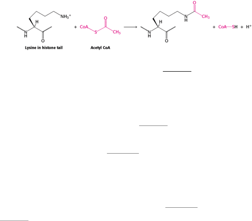
Enzymes that catalyze such reactions are called histone acetyltransferases (HATs). The histone tails are readily
extended; so they can fit into the HAT active site and become acetylated (Figure 31.28).
What are the consequences of histone acetylation? Lysine bears a positively charged ammonium group at neutral pH.
The addition of an acetyl group generates an uncharged amide group. This change dramatically reduces the affinity of
the tail for DNA and modestly decreases the affinity of the entire histone complex for DNA. The loosening of the histone
complex from the DNA exposes additional DNA regions to the transcription machinery. In addition, the acetylated
lysine residues interact with a specific acetyllysine-binding domain that is present in many proteins that regulate
eukaryotic transcription. This domain, termed a bromodomain, comprises approximately 110 amino acids that form a
four-helix bundle containing a peptide-binding site at one end (Figure 31.29).
Bromodomain-containing proteins are components of two large complexes essential for transcription. One is a complex
of more than 10 polypeptides that binds to the TATA-box-binding protein. Recall that the TATA-box-binding protein is
an essential transcription factor for many genes (Section 28.2.4). Proteins that bind to the TATA-box-binding protein are
called TAFs (for TATA-box-binding protein associated factors). In particular, TAFII250 (named for its participation in
RNA polymerase II transcription and its apparent molecular weight of 250 kd) contains a pair of bromodomains near its
carboxyl terminus. The two domains are oriented so that each can bind one of two acetyllysine residues at positions 5
and 12 in the histone H4 tail. Thus, acetylation of the histone tails provides a mechanism for recruiting other
components of the transcriptional machinery.
Bromodomains are also present in some components of large complexes known as chromatin-remodeling engines. These
complexes, which also contain domains homologous to those of helicases (Section 27.2.5), utilize the free energy of ATP
hydrolysis to shift the positions of nucleosomes along the DNA and to induce other conformational changes in chromatin
(Figure 31.30). Histone acetylation can lead to reorganization of the chromatin structure, potentially exposing binding
sites for other factors. Thus, histone acetylation can activate transcription through a combination of three mechanisms:
by reducing the affinity of the histones for DNA, by recruiting other components of the transcriptional machinery, and by
initiating the active remodeling of the chromatin structure.
31.3.5. Histone Deacetylases Contribute to Transcriptional Repression
Just as in prokaryotes, some changes in a cell's environment lead to the repression of genes that had been active. The
modification of histone tails again plays an important role. However, in repression, a key reaction appears to be the
deacetylation of acetylated lysine, catalyzed by specific histone deacetylase enzymes.
In many ways, the acetylation and deacetylation of lysine residues in histone tails (and, likely, in other proteins) is
analogous to the phosphorylation and dephosphorylation of serine, threonine, and tyrosine residues in other stages of
signaling processes. Like the addition of phosphoryl groups, the addition of acetyl groups can induce conformational
changes and generate novel binding sites. Without a means of removing these groups, however, these signaling switches
will become stuck in one position and lose their effectiveness. Like phosphatases, deacetylases help reset the switches.
31.3.6. Ligand Binding to Membrane Receptors Can Regulate Transcription Through
Phosphorylation Cascades
In Chapter 15, we examined several signaling pathways that begin with the binding of molecules to receptors in the cell
membrane. Some of these pathways lead to the regulation of gene expression. Let us review the pathway initiated by
epinephrine. The binding of epinephrine to a 7TM receptor results in the activation of a G protein. The activated G
protein, in turn, binds to and activates adenylate cyclase, increasing the intracellular concentration of cAMP. This cAMP
binds to the regulatory subunit of protein kinase A (PKA), activating the enzyme. We have previously examined the role
of phosphorylation by PKA of a variety of enzymes for example, those controlling glycogen metabolism. PKA also
phosphorylates the cyclic AMP-response element binding protein (CREB), a transcription factor that binds specific DNA
sequences as a dimer. Each monomer contributes a long α helix; together, the two helices grab the DNA in the manner
of a pair of chopsticks (Figure 31.31).
How does the phosphorylation of CREB affect its ability to activate transcription? Phosphorylation does not appear to
alter the DNA-binding properties of this protein. Instead, phosphorylated CREB binds a coactivator protein termed CBP,
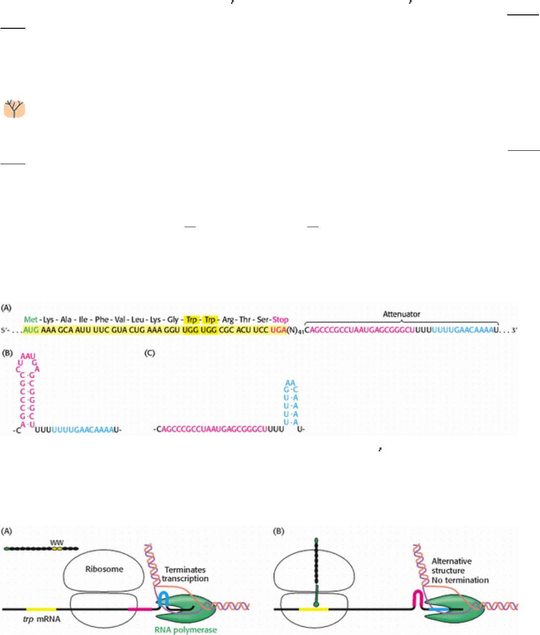
for CREB-binding protein. CBP possesses a highly revealing domain structure (Figure 31.32).
Its domains include a KIX domain (for kinase-inducible interaction) that binds to the phosphorylated region of CREB
(Figure 31.33); a bromodomain that binds acetylated histone tails, and two TAZ domains, zinc-binding domains that
facilitate the binding of CBP to a variety of proteins through a remarkable triangular structure (see Figure 31.32). Thus,
the pathway initiated by epinephrine binding induces the phosphorylation of a transcription factor, the recruitment of a
coactivator, and the assembly of complexes that participate in chromatin remodeling and transcription initiation.
31.3.7. Chromatin Structure Effectively Decreases the Size of the Genome
The transcriptional regulatory mechanisms utilized by prokaryotes and eukaryotes have some significant differences,
many of which are related to the significant difference in genome sizes between these classes of organisms. However,
much of the DNA in a eukaryotic cell is stably assembled into chromatin. The packaging of DNA with chromatin
renders many potential binding sites for transcription factors inaccessible
in effect, reducing the size of the genome.
Thus, rather than scanning through the entire genome, a eukaryotic DNA-binding protein scans a set of accessible
binding sites that is close in size to the genome of a prokaryote. The cell type is determined by the genes that are
accessible.
III. Synthesizing the Molecules of Life 31. The Control of Gene Expression 31.3. Transcriptional Activation and Repression Are Mediated by Protein-Protein Interactions
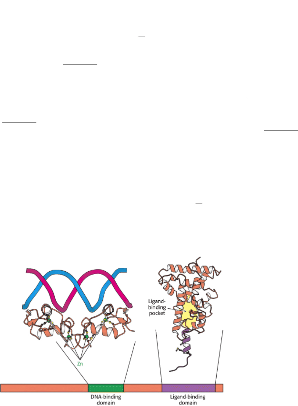
Figure 31.22. Structure of Two Nuclear Hormone Receptor Domains. Nuclear hormone receptors contain two crucial
conserved domains: (1) a DNA-binding domain toward the center of the sequence and (2) a ligand-binding domain
toward the carboxyl terminus. The structure of a dimer of the DNA-binding domain bound to DNA is shown, as is
one monomer of the normally dimeric ligand-binding domain.
III. Synthesizing the Molecules of Life 31. The Control of Gene Expression 31.3. Transcriptional Activation and Repression Are Mediated by Protein-Protein Interactions
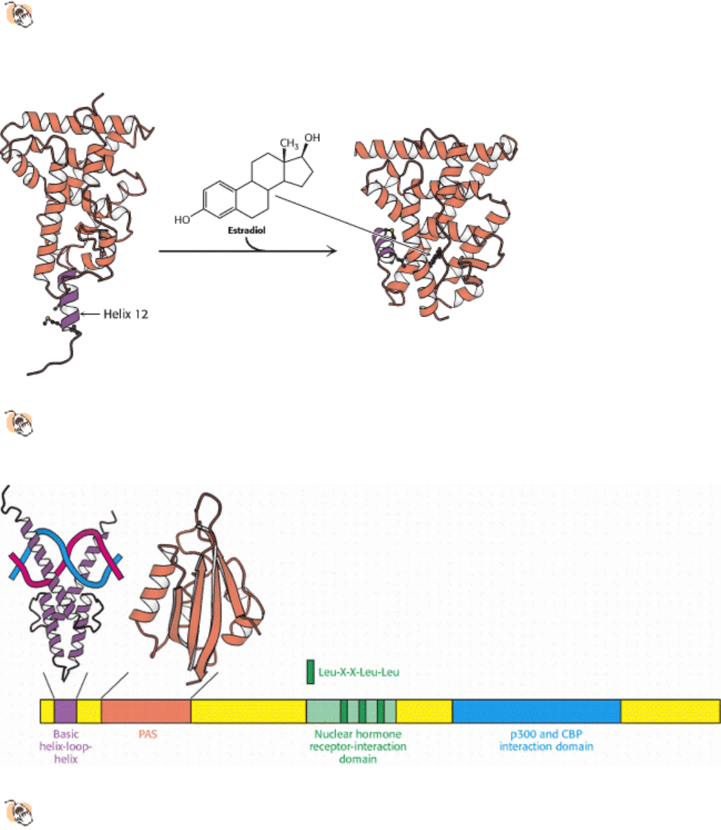
Figure 31.23. Ligand Binding to Nuclear Hormone Receptor.
The ligand lies completely surrounded within a pocket
in the ligand-binding domain. The last α helix, helix 12 (shown in purple), folds into a groove on the side of the
structure on ligand binding.
III. Synthesizing the Molecules of Life 31. The Control of Gene Expression 31.3. Transcriptional Activation and Repression Are Mediated by Protein-Protein Interactions
Figure 31.24. Coactivator Structure.
The p160 family of coactivators includes a number of domains that can be
recognized at the amino acid sequence level, including a basic helix-loop-helix domain that takes part in DNA
binding, a PAS domain that participates in protein-protein interactions, a central domain that contacts the ligand-
binding domain of the nuclear hormone receptors, and a domain that interacts with additional coactivators such as p300
and CREB-binding protein (CBP). (CREB stands for cyclic AMP-response element binding protein.) The nuclear
hormone receptor interaction domain includes three Leu-X-X-Leu-Leu sequences.
III. Synthesizing the Molecules of Life 31. The Control of Gene Expression 31.3. Transcriptional Activation and Repression Are Mediated by Protein-Protein Interactions
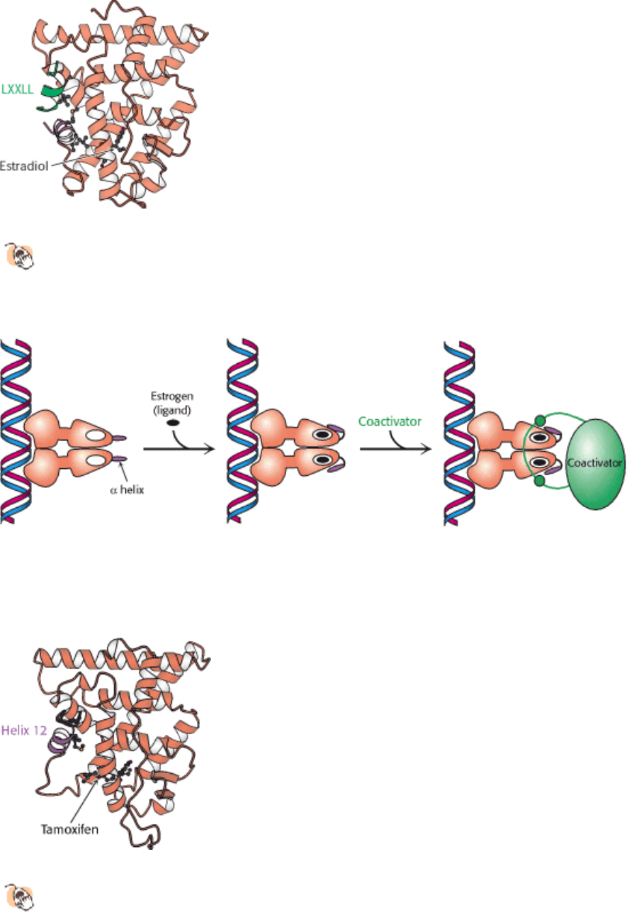
Figure 31.25. Coactivator-Nuclear Hormone Receptor Interactions.
The structure of a complex between the ligand-
binding domain of the estrogen receptor with estradiol bound and a peptide from a coactivator reveals that the Leu-
X-X-Leu-Leu (LXXLL) sequence forms a helix that binds in a groove on the surface of the ligand-binding
domain.
III. Synthesizing the Molecules of Life 31. The Control of Gene Expression 31.3. Transcriptional Activation and Repression Are Mediated by Protein-Protein Interactions
Figure 31.26. Coactivator Recruitment. The binding of ligand to a nuclear hormone receptor induces a conformational
change in the ligand-binding domain. This change in conformation generates favorable sites for the binding of a
coactivator.
III. Synthesizing the Molecules of Life 31. The Control of Gene Expression 31.3. Transcriptional Activation and Repression Are Mediated by Protein-Protein Interactions
Figure 31.27. Estrogen Receptor-Tamoxifen Complex.
Tamoxifen binds in the pocket normally occupied by estrogen.
However, part of the tamoxifen structure extends from this pocket, and so helix 12 cannot pack in its usual
position. Instead, this helix blocks the coactivator-binding site.
III. Synthesizing the Molecules of Life 31. The Control of Gene Expression 31.3. Transcriptional Activation and Repression Are Mediated by Protein-Protein Interactions
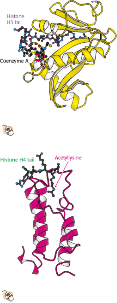
Figure 31.28. Structure of Histone Acetyltransferase.
The amino-terminal tail of histone H3 extends into a pocket in
which a lysine side chain can accept an acetyl group from acetyl CoA bound in an adjacent site.
III. Synthesizing the Molecules of Life 31. The Control of Gene Expression 31.3. Transcriptional Activation and Repression Are Mediated by Protein-Protein Interactions
Figure 31.29. Structure of a Bromodomain.
This four-helix-bundle domain binds peptides containing acetyllysine. An
acetylated peptide of histone H4 is bound in the structure shown.
III. Synthesizing the Molecules of Life 31. The Control of Gene Expression 31.3. Transcriptional Activation and Repression Are Mediated by Protein-Protein Interactions
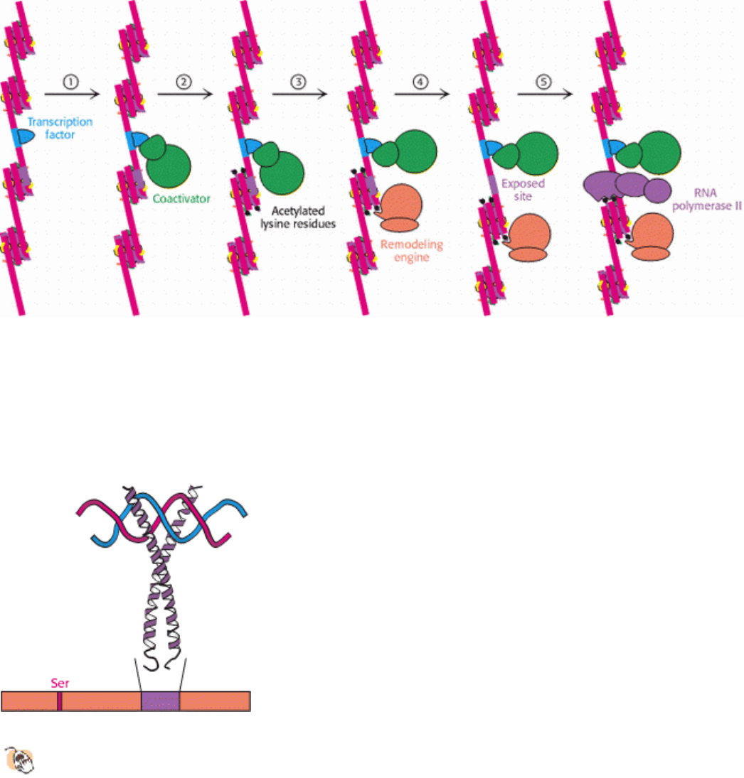
Figure 31.30. Chromatin Remodeling. Eukaryotic gene regulation begins with an activated transcription factor bound
to a specific site on DNA. One scheme for the initiation of transcription by RNA polymerase II requires five steps: (1)
recruitment of a coactivator, (2) acetylation of lysine residues in the histone tails, (3) binding of a remodeling engine
complex to the acetylated lysine residues, (4) ATP-dependent remodeling of the chromatin structure to expose a binding
site for RNA polymerase or for other factors, and (5) recruitment of RNA polymerase. Only two subunits are shown for
each complex, although the actual complexes are much larger. Other schemes are possible.
III. Synthesizing the Molecules of Life 31. The Control of Gene Expression 31.3. Transcriptional Activation and Repression Are Mediated by Protein-Protein Interactions
Figure 31.31. Cyclic AMP-Response Element Binding Protein (CREB).
Each of two CREB subunits contributes a
long α helix. The two helices coil around each other to form a dimeric DNA-binding unit. CREB is
phosphorylated on a specific serine residue by protein kinase A.
III. Synthesizing the Molecules of Life 31. The Control of Gene Expression 31.3. Transcriptional Activation and Repression Are Mediated by Protein-Protein Interactions
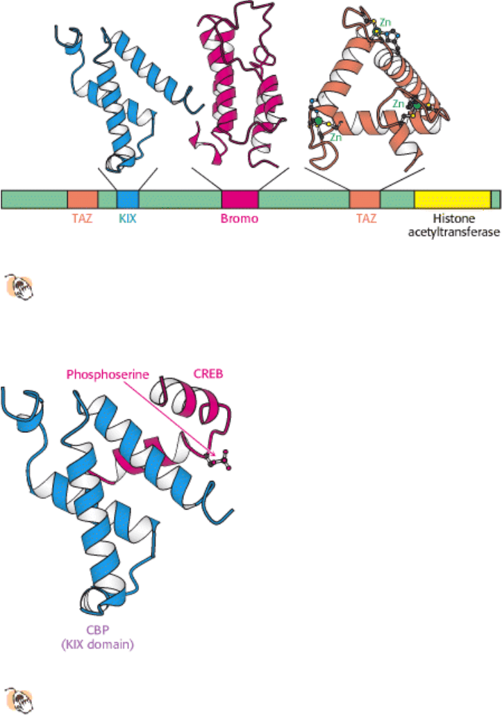
Figure 31.32. Domain Structure of CREB-Binding Protein (CBP).
The CREB-binding protein includes at least three
types of protein-protein interaction domains in addition to a histone acetyltransferase domain that lies near the
carboxyl terminus. The kinase-inducible interaction (KIX) domain interacts specifically with a region of CREB in
its phosphorylated form.
III. Synthesizing the Molecules of Life 31. The Control of Gene Expression 31.3. Transcriptional Activation and Repression Are Mediated by Protein-Protein Interactions
Figure 31.33. Interaction between CBP and CREB.
The KIX domain of CBP binds a region of CREB in its
phosphorylated form.
III. Synthesizing the Molecules of Life 31. The Control of Gene Expression
31.4. Gene Expression Can Be Controlled at Posttranscriptional Levels
Modulation of the rate of transcriptional initiation is the most common mechanism of gene regulation. However, other
stages of transcription also are targets for regulation in some cases. In addition, the process of translation provides other
points of intervention for regulating the level of a protein produced in a cell. These mechanisms are quite distinct in
prokaryotic and eukaryotic cells because prokaryotes and eukaryotes differ greatly in how transcription and translation
are coupled and in how translation is initiated. We shall consider two important examples of posttranscriptional
regulation: one from prokaryotes and the other from eukaryotes. In both examples, regulation depends on the formation
of distinct secondary structures in mRNA.
31.4.1. Attenuation Is a Prokaryotic Mechanism for Regulating Transcription Through
Modulation of Nascent RNA Secondary Structure

A novel mechanism for regulating transcription in bacteria was discovered by Charles Yanofsky and his colleagues as a
result of their studies of the tryptophan operon. The 7-kb mRNA transcript from this operon encodes five enzymes that
convert chorismate into tryptophan (Section 24.2.10). The mode of regulation of this operon is called attenuation, and it
depends on features at the 5
end of the mRNA product (Figure 31.34). Upstream of the coding regions for the enzymes
responsible for tryptophan biosynthesis lies a short open reading frame encoding a 14-amino-acid leader peptide.
Following this open reading frame is a region of RNA, called an attenuator, that is capable of forming several alternative
structures. Recall that transcription and translation are tightly coupled in bacteria. Thus, the translation of the trp mRNA
begins soon after the ribosome-binding site has been synthesized.
A ribosome is able to translate the leader region of the mRNA product only in the presence of adequate concentrations of
tryptophan. When enough tryptophan is present, a stem-loop structure forms in the attenuator region, which leads to the
release of RNA polymerase from the DNA (Figure 31.35). However, when tryptophan is scarce, transcription is
terminated less frequently. How does the level of tryptophan alter transcription of the trp operon? An important clue was
the finding that the 14-amino-acid leader peptide includes two adjacent tryptophan residues. When tryptophan is scarce,
little tryptophanyl-tRNA is present. Thus, the ribosome stalls at the tandem UGG codons encoding tryptophan. This
delay leaves the adjacent region of the mRNA exposed as transcription continues. An alternative RNA structure that
does not function as a terminator is formed and transcription continues into and through the coding regions for the
enzymes. Thus, attenuation provides an elegant means of sensing the supply of tryptophan required for protein synthesis.
Several other operons for the biosynthesis of amino acids in E. coli also are regulated by attenuator sites. The leader
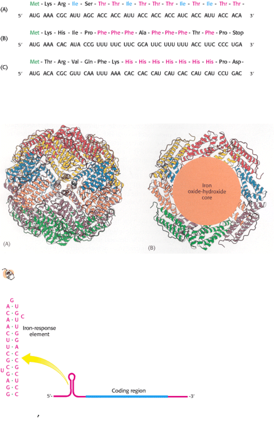
peptide of each contains an abundance of the amino acid residues of the type controlled by the operon (Figure 31.36).
For example, the leader peptide for the phenylalanine operon includes 7 phenylalanine residues among 15 residues. The
threonine operon encodes enzymes required for the synthesis of both threonine and isoleucine; the leader peptide
contains 8 threonine and 4 isoleucine residues in a 16-residue sequence. The leader peptide for the histidine operon
includes 7 histidine residues in a row. In each case, low levels of the corresponding charged tRNA causes the ribosome
to stall, trapping the nascent mRNA in a state that can form a structure that allows RNA polymerase to read through the
attenuator site.
31.4.2. Genes Associated with Iron Metabolism Are Translationally Regulated in
Animals
RNA secondary structure plays a role in the regulation of iron metabolism in eukaryotes. Iron is an essential nutrient,
required for the synthesis of hemoglobin, cytochromes, and many other proteins. However, excess iron can be quite
harmful because, untamed by a suitable protein environment, iron can initiate a range of free-radical reactions that
damage proteins, lipids, and nucleic acids. Animals have evolved sophisticated systems for the accumulation of iron in
times of scarcity and for the safe storage of excess iron for later use. Key proteins include transferrin, a transport protein
that carries iron in the serum, transferrin receptor, a membrane protein that binds iron-loaded transferrin and initiates its
entry into cells, and ferritin, an impressively efficient iron-storage protein found primarily in the liver and kidneys.
Twenty-four ferritin polypeptides form a nearly spherical shell that encloses as many as 2400 iron atoms, a ratio of one
iron atom per amino acid (Figure 31.37).
Ferritin and transferrin receptor expression levels are reciprocally related in their responses to changes in iron levels.
When iron is scarce, the amount of transferrin receptor increases and little or no new ferritin is synthesized. Interestingly,
the extent of mRNA synthesis for these proteins does not change correspondingly. Instead, regulation takes place at the
level of translation.
Consider ferritin first. Ferritin mRNA includes a stem-loop structure termed an iron-response element (IRE) in its 5
untranslated region (Figure 31.38). This stem-loop binds a 90-kd protein, called an IRE-binding protein (IRE-BP), that
blocks the initiation of translation. When the iron level increases, the IRE-BP binds iron as a 4Fe-4S cluster. The IRE-BP
bound to iron cannot bind RNA, because the binding sites for iron and RNA substantially overlap. Thus, in the presence
of iron, ferritin mRNA is released from the IRE-BP and translated to produce ferritin, which sequesters the excess iron.
An examination of the nucleotide sequence of transferrin-receptor mRNA reveals the presence of several IRE-like
regions. However, these regions are located in the 3
untranslated region rather than in the 5 untranslated region (Figure
31.39). Under low-iron conditions, IRE-BP binds to these IREs. However, given the location of these binding sites, the
transferrin-receptor mRNA can still be translated. What happens when the iron level increases and the IRE-BP no longer
binds transferrin-receptor mRNA? Freed from the IRE-BP, transferrin-receptor mRNA is rapidly degraded. Thus, an
increase in the cellular iron level leads to the destruction of transferrin-receptor mRNA and, hence, a reduction in the
production of transferrin-receptor protein.
The purification of the IRE-BP and the cloning of its cDNA were sources of truly remarkable insight into
evolution. The IRE-BP was found to be approximately 30% identical in amino acid sequence with the citric acid
cycle enzyme aconitase from mitochondria. Further analysis revealed that the IRE-BP is, in fact, an active aconitase
enzyme; it is a cytosolic aconitase that had been known for a long time, but its function was not well understood (Figure
31.40). The iron-sulfur center at the active site of the IRE-BP is rather unstable, and loss of the iron triggers significant
changes in protein conformation. Thus, this protein can serve as an iron-sensing factor.
Other mRNAs, including those taking part in heme synthesis, have been found to contain IREs. Thus, genes encoding
proteins required for iron metabolism acquired sequences that, when transcribed, provided binding sites for the iron-
sensing protein. An environmental signal
the concentration of iron controls the translation of proteins required for
the metabolism of this metal. The IREs have evolved appropriately in the untranslated regions of mRNAs to lead to
beneficial regulation by iron levels.
III. Synthesizing the Molecules of Life 31. The Control of Gene Expression 31.4. Gene Expression Can Be Controlled at Posttranscriptional Levels
Figure 31.34. Leader Region of TRP mRNA. (A) The nucleotide sequence of the 5
end of trp mRNA includes a short
open reading frame that encodes a peptide comprising 14 amino acids; the leader encodes two tryptophan residues and
has an untranslated attenuator region (blue and red nucleotides). (B and C) The attenuator region can adopt two distinct
stem-loop structures.
III. Synthesizing the Molecules of Life 31. The Control of Gene Expression 31.4. Gene Expression Can Be Controlled at Posttranscriptional Levels
Figure 31.35. Attenuation. (A) In the presence of adequate concentrations of tryptophan (and, hence, Trp-tRNA),
translation proceeds rapidly and an RNA structure forms that terminates transcription. (B) At low concentrations of
tryptophan, translation stalls awaiting Trp-tRNA, giving time for an alternative RNA structure to form that does not
terminate transcription efficiently.
III. Synthesizing the Molecules of Life 31. The Control of Gene Expression 31.4. Gene Expression Can Be Controlled at Posttranscriptional Levels
Figure 31.36. Leader Peptide Sequences. Amino acid sequences and the corresponding mRNA nucleotide sequences of
the (A) threonine operon, (B) phenylalanine operon, and (C) histidine operon. In each case, an abundance of one amino
acid in the leader peptide sequence leads to attenuation.
III. Synthesizing the Molecules of Life 31. The Control of Gene Expression 31.4. Gene Expression Can Be Controlled at Posttranscriptional Levels
Figure 31.37. Structure of Ferritin.
(A) Twenty-four ferritin polypeptides form a nearly spherical shell. (B) A cut-
away view reveals the core that stores iron as an iron oxide-hydroxide complex.
III. Synthesizing the Molecules of Life 31. The Control of Gene Expression 31.4. Gene Expression Can Be Controlled at Posttranscriptional Levels
Figure 31.38. Iron-Response Element. Ferritin mRNA includes a stem-loop structure, termed an iron-response element
(IRE), in its 5
untranslated region. The IRE binds a specific protein that blocks the translation of this mRNA under low