Berg J.M., Tymoczko J.L., Stryer L. Biochemistry
Подождите немного. Документ загружается.


Megabase (Mb)
A length of DNA consisting of 10
6
base pairs (if double stranded) or
10
6
bases (if single stranded).
Another source of complexity in eukaryotic gene regulation is the many different cell types present in most eukaryotes.
Liver and pancreatic cells, for example, differ dramatically in the genes that are highly expressed (see Table 31.1).
Moreover, eukaryotic genes are not generally organized into operons. Instead, genes that encode proteins for steps within
a given pathway are often spread widely across the genome. Finally, transcription and translation are uncoupled in
eukaryotes, eliminating some potential gene-regulatory mechanisms.
31.2.1. Nucleosomes Are Complexes of DNA and Histones
The DNA in eukaryotic chromosomes is not bare. Rather eukaryotic DNA is tightly bound to a group of small basic
proteins called histones. In fact, histones constitute half the mass of a eukaryotic chromosome. The entire complex of a
cell's DNA and associated protein is called chromatin. Five major histones are present in chromatin: four histones, called
H2A, H2B, H3, and H4, associate with one another; the other histone is called H1. Histones have strikingly basic
properties because a quarter of the residues in each histone is either arginine or lysine.
In 1974, Roger Kornberg proposed that chromatin is made up of repeating units, each containing 200 bp of DNA and
two copies each of H2A, H2B, H3, and H4, called the histone octomer. These repeating units are known as nucleosomes.
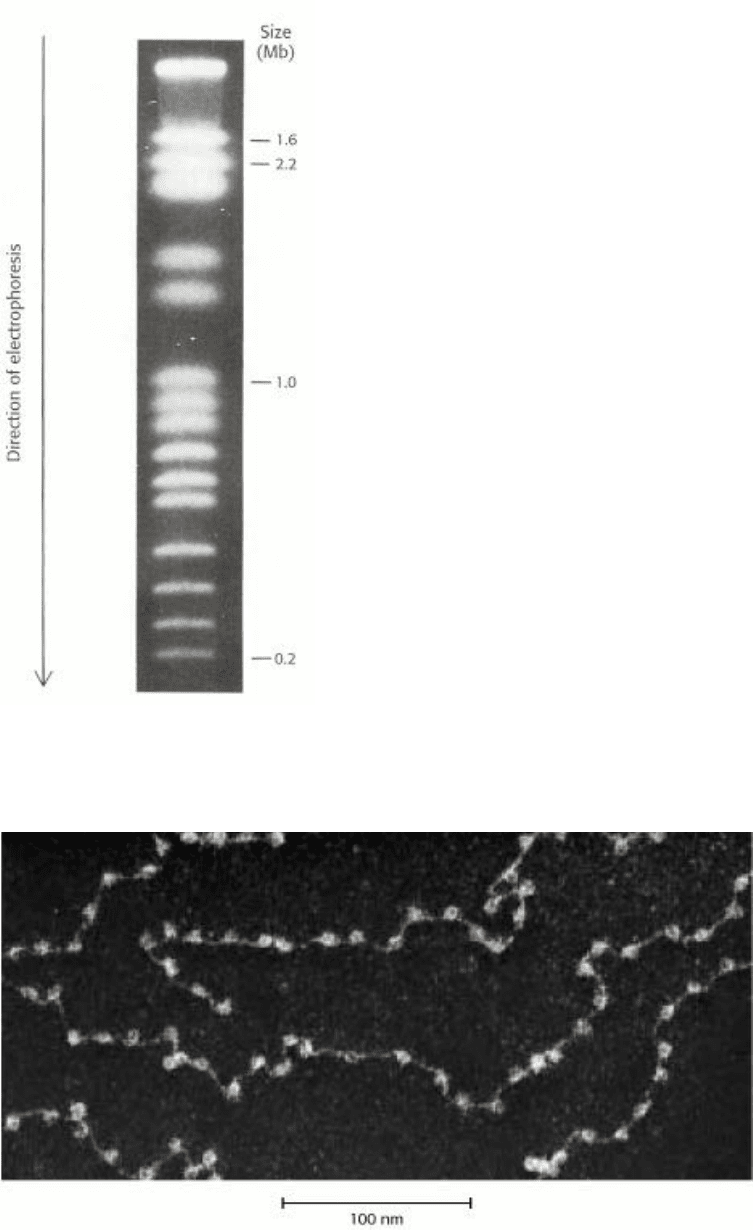
Strong support for this model comes from the results of a variety of experiments, including observations of appropriately
prepared samples of chromatin viewed by electron microscopy (Figure 31.15). Chromatin viewed with the electron
microscope has the appearance of beads on a string; each bead has a diameter of approximately 100 Å. Partial digestion
of chromatin with DNAse yields the isolated beads. These particles consist of fragments of DNA 200 bp in length
bound to the eight histones. More extensive digestion yields a reduced DNA fragment of 145 bp bound to the histone
octamer. This smaller complex of the histone octamer and the 145-bp DNA fragment is the nucleosome core particle.
The DNA connecting core particles in undigested chromatin is called linker DNA. Histone H1 binds, in part, to the linker
DNA.
31.2.2. Eukaryotic DNA Is Wrapped Around Histones to Form Nucleosomes
The overall structure of the nucleosome was revealed through electron microscopic and x-ray crystallographic studies
pioneered by Aaron Klug and his colleagues. More recently, the three-dimensional structure of a reconstituted
nucleosome core (Figure 31.16) was determined to relatively high resolution by x-ray diffraction methods. As was
shown by Evangelos Moudrianakis, the four types of histone that make up the protein core are homologous and similar
in structure (Figure 31.17). The eight histones in the core are arranged into a (H3)
2
(H4)
2
tetramer and a pair of H2A-
H2B dimers. The tetramer and dimers come together to form a left-handed superhelical ramp around which the DNA
wraps. In addition, each histone has an amino-terminal tail that extends out from the core structure. These tails are
flexible and contain a number of lysine and arginine residues. As we shall see, covalent modifications of these tails play
an essential role in modulating the affinity of the histones for DNA and other properties.
The DNA forms a left-handed superhelix as it wraps around the outside of the histone octamer. The protein core forms
contacts with the inner surface of the superhelix at many points, particularly along the phosphodiester backbone and the
minor groove of the DNA. Nucleosomes will form on almost all DNA sites, although some sequences are preferred
because the dinucleotide steps are properly spaced to favor bending around the histone core. Histone H1, which has a
different structure from the other histones, seals off the nucleosome at the location at which the linker DNA enters and
leaves the nucleosome. The amino acid sequences of histones, including their amino- terminal tails, are remarkably
conserved from yeast through human beings.
The winding of DNA around the nucleosome core contributes to DNA's packing by decreasing its linear extent. An
extended 200-bp stretch of DNA would have a length of about 680 Å. Wrapping this DNA around the histone octamer
reduces the length to approximately 100 Å along the long dimension of the nucleosome. Thus the DNA is compacted by
a factor of seven. However, human chromosomes in metaphase, which are highly condensed, are compacted by a factor
of 10
4
. Clearly, the nucleosome is just the first step in DNA compaction. What is the next step? The nucleosomes
themselves are arranged in a helical array approximately 360 Å across, forming a series of stacked layers approximately
110 Å apart (Figure 31.18). The folding of these fibers of nucleosomes into loops further compacts DNA.
The writhing of DNA around the histone core in a left-handed helical manner also stores negative supercoils; if the DNA
in a nucleosome is straightened out, it will be underwound (Section 23.3.2). This underwinding is exactly what is needed
to separate the two DNA strands during replication and transcription (Sections 27.5 and 28.1.5).
31.2.3. The Control of Gene Expression Requires Chromatin Remodeling
Does chromatin structure play a role in the control of gene expression? Early observations suggested that it does indeed.
The treatment of cell nuclei with the nonspecific DNA-cleaving enzyme DNAse I revealed that regions adjacent to genes
that are being actively transcribed are more sensitive to cleavage than are other sites in the genome, suggesting that the
DNA in these regions is less compacted than it is elsewhere in the genome and more accessible to proteins. In addition,
some sites, usually within 1 kb of the start site of an active gene, are exquisitely sensitive to DNAse I and other
nucleases. These hypersensitive sites correspond to regions that have few nucleosomes or have nucleosomes in an altered
conformational state. Hyper-sensitive sites are cell-type specific and developmentally regulated. For example, globin
genes in the precursors of erythroid cells from 20-hour-old chicken embryos are insensitive to DNAse I. However, when
hemoglobin synthesis begins at 35 hours, regions adjacent to these genes become highly susceptible to digestion. In
tissues such as the brain that produce no hemoglobin, the globin genes remain resistant to DNAse I throughout
development and into adulthood. The results of these studies suggest that a prerequisite for gene expression is a relaxing
of the chromatin structure.
Recent experiments even more clearly revealed the role of chromatin structure in regulating access to DNA binding sites.
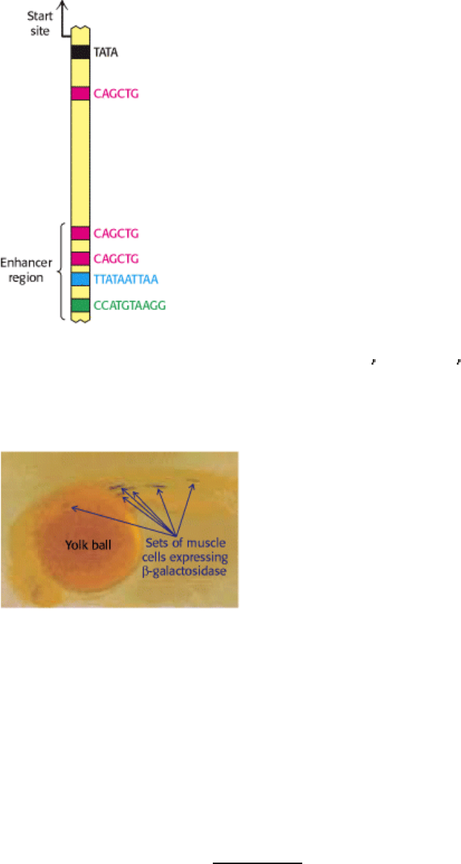
Genes required for galactose utilization in yeast are activated by a DNA-binding protein called GAL4, which recognizes
DNA binding sites with two 5
-CGG-3 sequences separated by 11 base pairs (Figure 31.19). Approximately 4000
potential GAL4 binding sites of the form 5
-CGG(N)
11
CCG-3 are present in the yeast genome, but only 10 of them
regulate genes necessary for galactose metabolism. What fraction of the potential binding sites are actually bound by
GAL4? This question is addressed through the use of a technique called chromatin immunoprecipitation (ChIP). GAL4
is first cross-linked to the DNA to which it is bound in chromatin. The DNA is then fragmented into small pieces, and
antibodies to GAL4 are used to isolate the chromatin fragments containing GAL4. The cross-linking is reversed, and the
DNA is isolated and characterized. The results of these studies reveal that only approximately 10 of the 4000 potential
GAL4 sites are occupied by GAL4 when the cells are growing on galactose; more than 99% of the sites appear to be
blocked. Thus, whereas in prokaryotes all sites appear to be equally accessible, chromatin structure shields a large
number of the potential binding sites in eukaryotic cells. GAL4 is thereby prevented from binding to sites that are
unimportant in galactose metabolism.
These lines of evidence and others reveal that chromatin structure is altered in active genes compared with inactive ones.
How is chromatin structure modified? As we shall see in Section 31.3.4, specific covalent modifications of histone
proteins are crucial. In addition, the binding of specific proteins to DNA sequences called enhancers at specific sites in
the genome plays a role.
31.2.4. Enhancers Can Stimulate Transcription by Perturbing Chromatin Structure
We can now understand the action of enhancers, already introduced in Section 28.2.6. Recall that these DNA sequences,
although they have no promoter activity of their own, greatly increase the activities of many promoters in eukaryotes,
even when the enhancers are located at a distance of several thousand base pairs from the gene being expressed.
Enhancers function by serving as binding sites for specific regulatory proteins. (Figure 31.20). An enhancer is effective
only in the specific cell types in which appropriate regulatory proteins are expressed. In many cases, these DNA-binding
proteins influence transcription initiation by perturbing the local chromatin structure to expose a gene or its regulatory
sites rather than by direct interactions with RNA polymerase. This mechanism accounts for the ability of enhancers to act
at a distance.
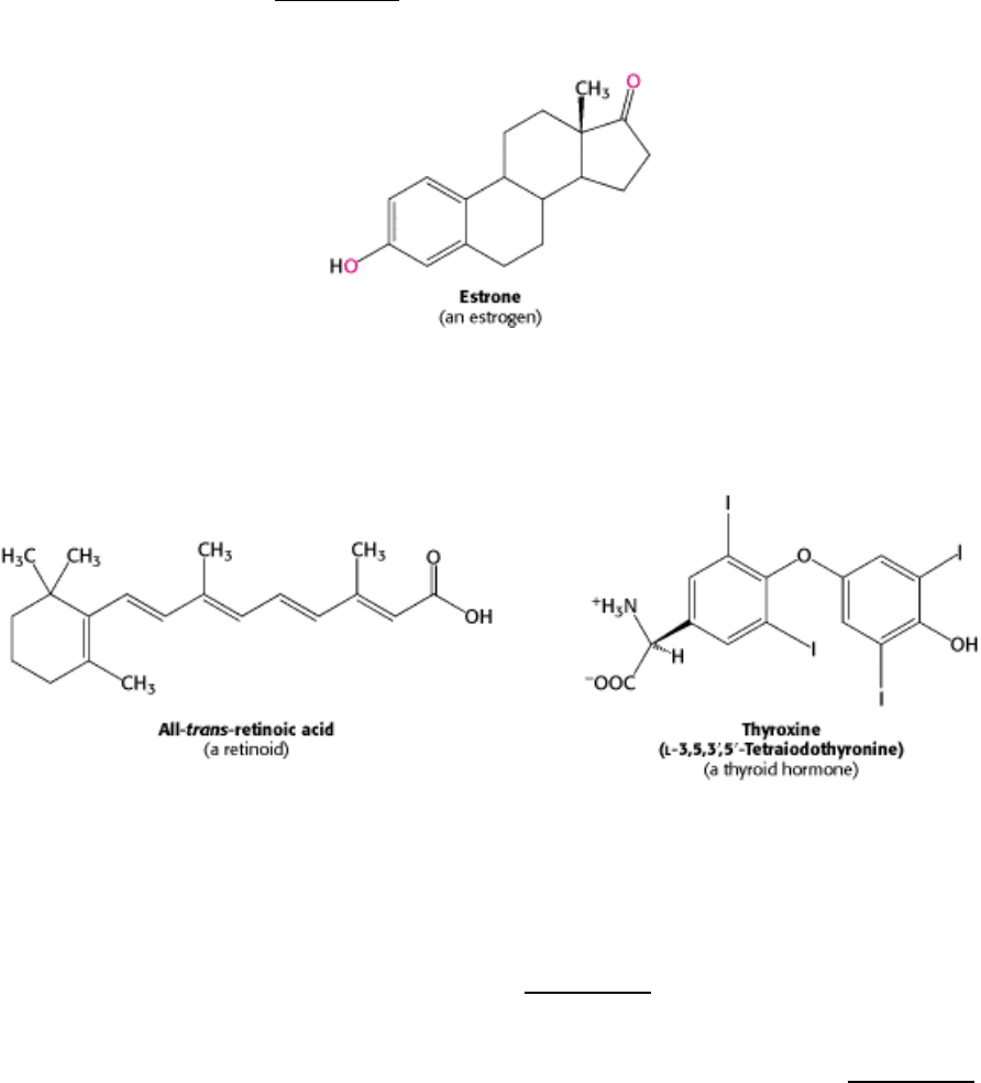
The properties of enhancers are illustrated by studies of the enhancer controlling the muscle isoform of creatine kinase
(Section 14.1.5). The results of mutagenesis and other studies revealed the presence of an enhancer located between
1350 and 1050 base pairs upstream of the start site of the gene for this enzyme. Experimentally inserting this enhancer
near a gene not normally expressed in muscle cells is sufficient to cause the gene to be expressed at high levels in muscle
cells, but not other cells (Figure 31.21).
31.2.5. The Modification of DNA Can Alter Patterns of Gene Expression
The modification of DNA provides another mechanism, in addition to packaging with histones, for inhibiting
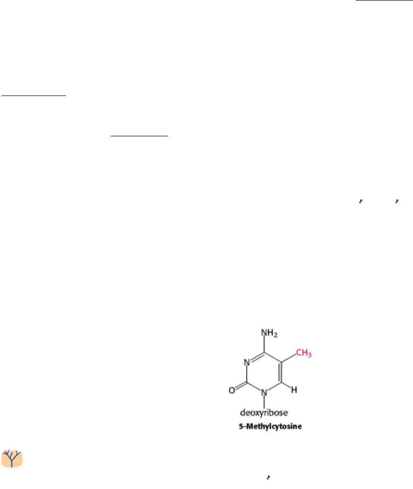
inappropriate gene expression in specific cell types. Approximately 70% of the 5
-CpG-3 sequences in mammalian
genomes are methylated at the C-5 position of cytosine by specific methyltransferases. However, the distribution of these
methylated cytosines varies, depending on the cell type. Consider, again, the globin genes. In cells that are actively
expressing hemoglobin, the region from approximately 1 kb upstream of the start site of the β-globin gene to
approximately 100 bp downstream of the start site contains fewer 5-methylcytosine residues than does the corresponding
region in cells that do not express these genes. The relative absence of 5-methylcytosines near the start site is referred to
as hypomethylation. The methyl group of 5-methylcytosine protrudes into the major groove where it could easily
interfere with the binding of proteins that stimulate transcription.
The distribution of CpG sequences in mammalian genomes is not uniform. The deamination of 5-methylcytosine
produces thymine; so CpG sequences are subject to mutation to TpG. Many CpG sequences have been converted
into TpG through this mechanism. However, sites near the 5
ends of genes have been maintained because of their role in
gene expression. Thus, most genes are found in CpG islands, regions of the genome that contain approximately four
times as many CpG sequences as does the remainder of the genome. Note that methylation is not a universal regulatory
device, even in multicellular eukaryotes. For example, Drosophila DNA is not methylated at all.
III. Synthesizing the Molecules of Life 31. The Control of Gene Expression 31.2. The Greater Complexity of Eukaryotic Genomes Requires Elaborate Mechanisms for Gene Regulation
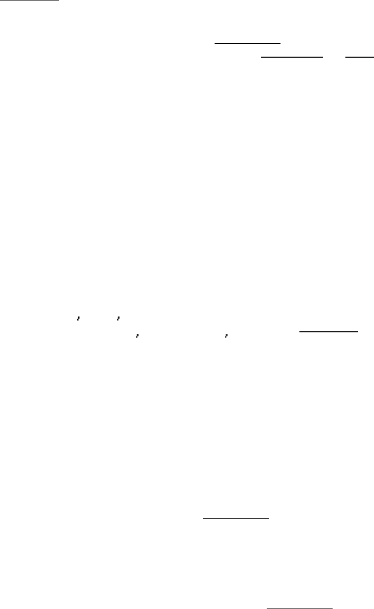
Figure 31.14. Yeast Chromosomes. Pulsed-field electrophoresis allows the separation of 16 yeast chromosomes. [From
G. Chu, D. Wollrath, and R. W. Davis. Science 234(1986):1583.]
III. Synthesizing the Molecules of Life 31. The Control of Gene Expression 31.2. The Greater Complexity of Eukaryotic Genomes Requires Elaborate Mechanisms for Gene Regulation
Figure 31.15. Chromatin Structure. An electron micrograph of chromatin showing its "beads on a string" character.
[Courtesy of Dr. Ada Olins and Dr. Donald Olins.]
III. Synthesizing the Molecules of Life 31. The Control of Gene Expression 31.2. The Greater Complexity of Eukaryotic Genomes Requires Elaborate Mechanisms for Gene Regulation
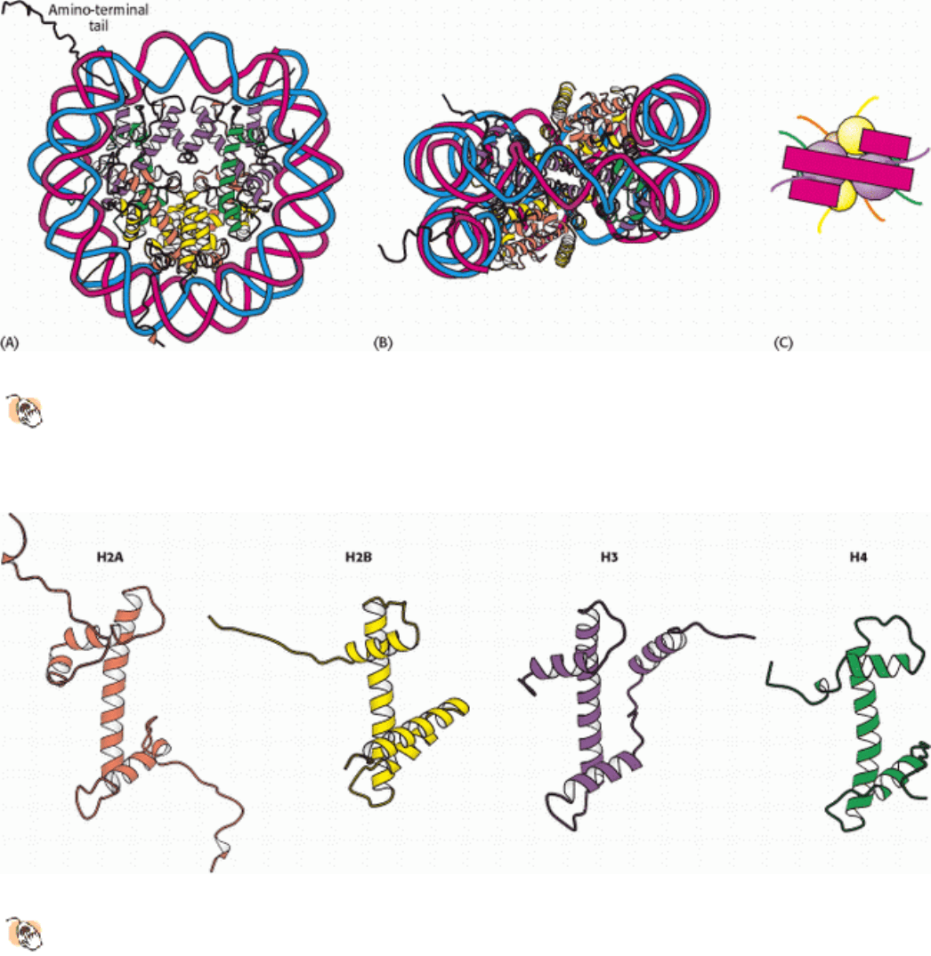
Figure 31.16. Nucleosome Core Particle.
The structure consists of a core of eight histone proteins surrounded by DNA.
(A) A view showing the DNA wrapping around the histone core. (B) A view related to that in part A by a 90-
degree rotation shows that the DNA forms a left-handed superhelix as it wraps around the core. (C) A schematic
view.
III. Synthesizing the Molecules of Life 31. The Control of Gene Expression 31.2. The Greater Complexity of Eukaryotic Genomes Requires Elaborate Mechanisms for Gene Regulation
Figure 31.17. Homologous Histones.
Histones H2A, H2B, H3, and H4 each adopt a similar three-dimensional structure
as a consequence of common ancestry. Some parts of the tails present at the termini of the proteins are not shown.
III. Synthesizing the Molecules of Life 31. The Control of Gene Expression 31.2. The Greater Complexity of Eukaryotic Genomes Requires Elaborate Mechanisms for Gene Regulation
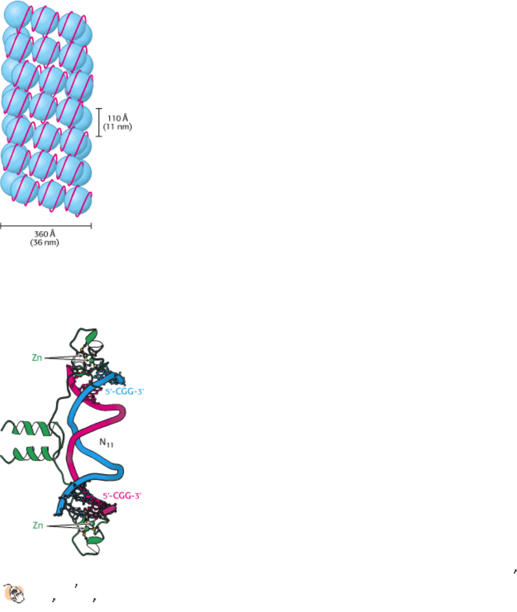
Figure 31.18. Higher-Order Chromatin Structure. A proposed model for chromatin arranged in a helical array
consisting of six nucleosomes per turn of helix. The DNA double helix (shown in red) is wound around each histone
octamer (shown in blue). [After J. T. Finch and A. Klug. Proc. Natl. Acad. Sci. USA 73(1976):1900.]
III. Synthesizing the Molecules of Life 31. The Control of Gene Expression 31.2. The Greater Complexity of Eukaryotic Genomes Requires Elaborate Mechanisms for Gene Regulation
Figure 31.19. Gal4 Binding Sites.
The yeast transcription factor GAL4 binds to DNA sequences of the form 5 -CGG(N)
11CCG-3
. Two zinc-based domains are present in the DNA-binding region of this protein. These domains contact
the 5
-CGG-3 sequences, leaving the center of the site uncontacted.
III. Synthesizing the Molecules of Life 31. The Control of Gene Expression 31.2. The Greater Complexity of Eukaryotic Genomes Requires Elaborate Mechanisms for Gene Regulation
Figure 31.20. Enhancer Binding Sites. A schematic structure for the region 1 kb upstream of the start site for the
muscle creatine kinase gene. One binding site of the form 5
-CAGCTG-3 is present near the TATA box. The enhancer
region farther upstream contains two binding sites for the same protein and two additional binding sites for other
proteins.
III. Synthesizing the Molecules of Life 31. The Control of Gene Expression 31.2. The Greater Complexity of Eukaryotic Genomes Requires Elaborate Mechanisms for Gene Regulation
Figure 31.21. An Experimental Demonstration of Enhancer Function. A promoter for muscle creatine kinase
artificially drives the transcription of β-galactosidase in a zebrafish embryo. Only specific sets of muscle cells produce β-
galactosidase, as visualized by the formation of the blue product on treatment of the embryo with X-Gal. [From F.
Müller, D. W. Williamson, J. Kobolák, L. Gauvry, G. Goldspink, L. Orbán, and N. MacLean. Molecular Reproduction
and Development 47(1997): 404.]
III. Synthesizing the Molecules of Life 31. The Control of Gene Expression
31.3. Transcriptional Activation and Repression Are Mediated by Protein-Protein
Interactions
We have seen how interactions between DNA-binding proteins such as CAP and RNA polymerase can activate
transcription in prokaryotic cells (Section 31.1.6). Such protein-protein interactions play a dominant role in eukaryotic
gene regulation. In contrast with those of prokaryotic transcription, few eukaryotic transcription factors have any effect
on transcription on their own. Instead, each factor recruits other proteins to build up large complexes that interact with
the transcriptional machinery to activate or repress transcription.
A major advantage of this mode of regulation is that a given regulatory protein can have different effects, depending on
what other proteins are present in the same cell. This phenomenon, called combinatorial control, is crucial to
multicellular organisms that have many different cell types. Even in unicellular eukaryotes such as yeast, combinatorial
control allows the generation of distinct cell types.
31.3.1. Steroids and Related Hydrophobic Molecules Pass Through Membranes and
Bind to DNA-Binding Receptors
Just as prokaryotes can adjust their patterns of gene expression in response to chemicals in their environment, eukaryotes
have many systems for responding to specific molecules with which they come in contact. We first consider a system
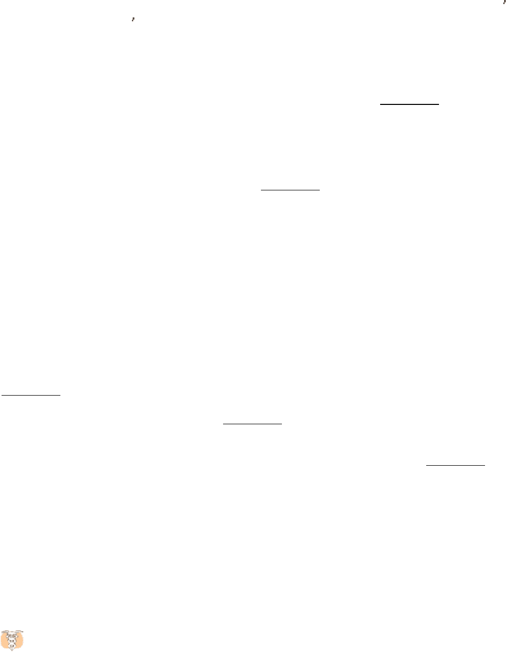
that detects and responds to estrogens. Synthesized and released by the ovaries, estrogens, such as estrone, are
cholesterol-derived, steroid hormones (Section 26.4). They are required for the development of female secondary sex
characteristics and, along with progesterone, participate in the ovarian cycle.
Because they are hydrophobic molecules, estrogens easily diffuse across cell membranes. When inside a cell, estrogens
bind to highly specific, soluble receptor proteins. Estrogen receptors are members of a large family of proteins that act as
receptors for a wide range of hydrophobic molecules, including other steroid hormones, thyroid hormones, and retinoids.
These receptors all have a similar mode of action. On binding of the signal molecule (called, generically, a ligand), the
ligand-receptor complex modifies the expression of specific genes by binding to control elements in the DNA. The
human genome encodes approximately 50 members of this family, often referred to as nuclear hormone receptors. The
genomes of other multicellular eukaryotes encode similar numbers of nuclear hormone receptors, although they are
absent in yeast. A comparison of the amino acid sequences of members of this family reveals two highly conserved
domains: a DNA-binding domain and a ligand-binding domain (Figure 31.22). The DNA-binding domain lies toward the
center of the molecule and includes nine conserved cysteine residues. This domain provides these receptors with
sequence-specific DNA activity. Eight of the cysteine residues are conserved because of their role in binding zinc ions:
the first four cysteine residues bind one zinc ion, and the second four bind a second zinc ion (see Figure 31.22). The zinc
ions stabilize the structure of this small domain; without the bound zinc ions, the domains unfold. Such domains are
often referred to as zinc finger domains.
The structure of the zinc-binding region of a steroid receptor includes an α helix that begins at the end of the first zinc
finger domain. This helix lies in the major groove in the specific DNA complexes formed by estrogen receptors and
binds with specific DNA sequences, analogously to prokaryotic DNA-binding proteins. Estrogen receptors bind to
specific DNA sites (referred to as estrogen response elements or EREs) that contain the consensus sequence 5 -
AGGTCANNNTGACCT-3
. As expected from the symmetry of this sequence, an estrogen receptor binds to such sites
as a dimer.
The second highly conserved region of the nuclear receptor proteins lies near the carboxyl terminus and is the ligand-
binding site. This domain folds into a structure that consists almost entirely of α helices, arranged in three layers. The
ligand binds in a hydrophobic pocket that lies in the center of this array of helices (Figure 31.23). The ligand-binding
domain also participates in receptor dimerization.
A comparison of the structures of the ligand-binding domains with and without bound ligand reveals that ligand binding
leads to substantial structural rearrangement. In particular, the last α helix (referred to as helix 12), which has
hydrophobic residues lining one face but extends out from the receptor in the ligand-free form, folds into a shallow
groove on the side of the receptor on ligand binding (see Figure 31.23). How does ligand binding lead to changes in gene
expression? The simplest model would have the binding of ligand alter the DNA-binding properties of the receptor,
analogously to the lac repressor in prokaryotes. However, the results of experiments with purified nuclear hormone
receptors revealed that ligand binding does not significantly alter DNA-binding affinity and specificity. Another
mechanism must be operative.
31.3.2. Nuclear Hormone Receptors Regulate Transcription by Recruiting Coactivators
and Corepressors to the Transcription Complex
Because ligand binding does not alter the ability of nuclear hormone receptors to bind DNA, investigators sought to
determine whether specific proteins might bind to the nuclear hormone receptors only in the presence of ligand. Such
searches led to the identification of several related proteins called coactivators, such as SRC-1 (steroid receptor
coactivator-1), GRIP-1 (glucocorticoid receptor interacting protein-1), and NcoA-1 (nuclear hormone receptor
coactivator-1). These coactivators, referred to as the p160 family because of their size, have a common modular structure
(Figure 31.24). Each coactivator protein contains three sequences of the form Leu-X-X-Leu-Leu within a central region
of 200 amino acids. These sequences form short α helices that bind to a hydrophobic patch on the surface of the ligand-
binding domains of a nuclear hormone receptor (Figure 31.25). The binding site for the coactivator is fully formed only
when ligand is bound, inasmuch as it is adjacent to helix 12. It is likely that a coactivator molecule binds to the ligand-
binding domains of a receptor dimer through two of its three Leu-X-X-Leu-Leu sequences. Thus, the binding of ligand
to the receptor induces a conformational change that allows the recruitment of a coactivator (Figure 31.26).
Some members of the nuclear hormone receptor family, such as the receptors for thyroid hormone and retinoic acid,
repress transcription in the absence of ligand. This repression also is mediated by the ligand-binding domain. In their
unbound forms, the ligand-binding domains of these receptors bind to corepressor proteins. Members of this family of
proteins include SMRT (Silencing mediator for retinoid and thyroid hormone receptors) and N-Cor (nuclear hormone
receptor corepressor). Such a corepressor binds to a site in the ligand-binding domain that overlaps the coactivator
binding site; ligand binding triggers the release of the corepressor and frees the ligand-binding domain for binding to a
coactivator.
31.3.3. Steroid-Hormone Receptors Are Targets for Drugs
Molecules such as estradiol that bind to a receptor and trigger signaling pathways are called agonists. Athletes
sometimes take natural and synthetic agonists of the androgen receptor, a member of the nuclear hormone receptor
family, because their binding to the androgen receptor stimulates the expression of genes that enhance the development
of lean muscle mass.
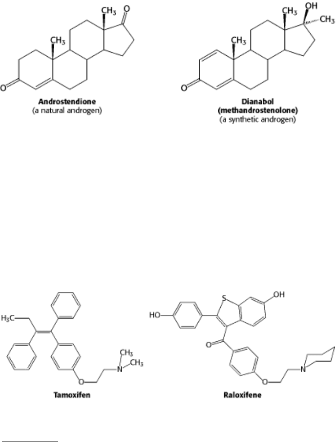
Referred to as anabolic steroids, such compounds used in excess are not without side effects. In men, excessive use leads
to a decrease in the secretion of testosterone, to testicular atrophy, and sometimes to breast enlargement (gynecomastia)
if some of the excess androgen is converted into estrogen. In women, excess testosterone causes a decrease in ovulation
and estrogen secretion; it also causes breast regression and growth of facial hair.
Other molecules bind to nuclear hormone receptors but do not effectively trigger signaling pathways. Such compounds
are called antagonists and are, in many ways, like competitive inhibitors of enzymes. Some important drugs are
antagonists that target the estrogen receptor. For example, tamoxifen and raloxifene are used in the treatment and
prevention of breast cancer, because some breast tumors rely on estrogen-mediated pathways for growth. These
compounds are sometimes called selective estrogen receptor modulators (SERMs).
The determination of the structures of complexes between the estrogen receptor and these drugs revealed the basis for
their antagonist effect (Figure 31.27). Tamoxifen binds to the same site as estradiol. However, tamoxifen (and other
antagonists) have groups that extend out of the normal ligand-binding pocket. These groups prevent helix 12 from
binding in its usual position; instead, this helix binds to the site normally occupied by the coactivator. Tamoxifen blocks
the binding of coactivators and thus inhibits the activation of gene expression.
31.3.4. Chromatin Structure Is Modulated Through Covalent Modifications of Histone
Tails
We have seen that nuclear receptors respond to signal molecules by recruiting coactivators and corepressors to the
chromatin. Now we can ask, How do coactivators and corepressors modulate transcriptional activity? Much of their
effectiveness appears to result from their ability to covalently modify the amino-terminal tails of histones and perhaps
other proteins. Some of the p160 coactivators and, in addition, the proteins that they recruit catalyze the transfer of acetyl
groups from acetyl CoA to specific lysine residues in the amino-terminal tails of histones.