Berg J.M., Tymoczko J.L., Stryer L. Biochemistry
Подождите немного. Документ загружается.


IV. Responding to Environmental Changes 32. Sensory Systems 32.3. Photoreceptor Molecules in the Eye Detect Visible Light
Figure 32.23. Atomic Motion in Retinal. The Schiff-base nitrogen atom moves 5 Å as a consequence of the light-
induced isomerization of 11-cis-retinal to all-trans-retinal by rotation about the bond shown in red.
IV. Responding to Environmental Changes 32. Sensory Systems 32.3. Photoreceptor Molecules in the Eye Detect Visible Light
Figure 32.24. Analogous 7TM Receptors. The conversion of rhodopsin into metarhodopsin II activates a signal-
transduction pathway analogously to the activation induced by the binding of other 7TM receptors to appropriate ligands.
IV. Responding to Environmental Changes 32. Sensory Systems 32.3. Photoreceptor Molecules in the Eye Detect Visible Light
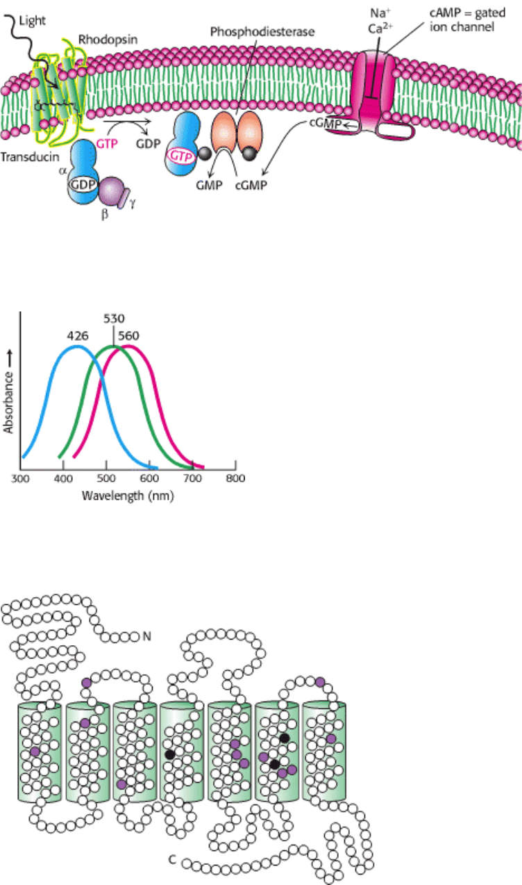
Figure 32.25. Visual Signal Transduction. The light-induced activation of rhodopsin leads to the hydrolysis of cGMP,
which in turn leads to ion channel closing and the initiation of an action potential.
IV. Responding to Environmental Changes 32. Sensory Systems 32.3. Photoreceptor Molecules in the Eye Detect Visible Light
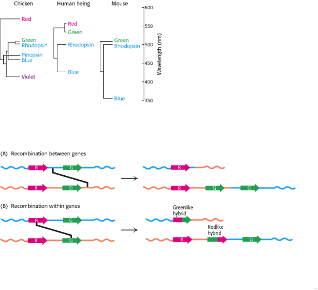
Figure 32.26. Cone-Pigment Absorption Spectra. The absorption spectra of the cone visual pigment responsible for
color vision.
IV. Responding to Environmental Changes 32. Sensory Systems 32.3. Photoreceptor Molecules in the Eye Detect Visible Light
Figure 32.27. Comparison of the Amino Acid Sequences of the Green and Red Photoreceptors. Open circles
correspond to identical residues, whereas colored circles mark residues that are different. The differences in the three
black positions are responsible for most of the difference in their absorption spectra.
IV. Responding to Environmental Changes 32. Sensory Systems 32.3. Photoreceptor Molecules in the Eye Detect Visible Light
Figure 32.28. Evolutionary Relationships among Visual Pigments. Visual pigments have evolved by gene duplication
along different branches of the animal evolutionary tree. The branch lengths of the "trees" correspond to the percentage
of amino acid divergence. [Adapted from Nathans, J. Neuron 24(1999):299; by permission of Cell Press.]
IV. Responding to Environmental Changes 32. Sensory Systems 32.3. Photoreceptor Molecules in the Eye Detect Visible Light
Figure 32.29. Recombination Pathways Leading to Color Blindness. Rearrangements in the course of DNA
replication may lead to (A) the loss of visual pigment genes or (B) the formation of hybrid pigment genes that encode
photoreceptors with anomolous absorption spectra. Because the amino acids most important for determining absorption
spectra are in the carboxyl-terminal half of each photoreceptor protein, the part of the gene that encodes this region most
strongly affects the absorption characteristics of hybrid receptors. [Adapted from J. Nathans. Neuron 24(1999):299
312;
by permission of Cell Press.]
IV. Responding to Environmental Changes 32. Sensory Systems
32.4. Hearing Depends on the Speedy Detection of Mechanical Stimuli
Hearing and touch are based on the detection of mechanical stimuli. Although the proteins of these senses have not been
as well characterized as those of the senses already discussed, anatomical, physiological, and biophysical studies have
elucidated the fundamental processes. A major clue to the mechanism of hearing is its speed. We hear frequencies
ranging from 200 to 20,000 Hz (cycles per second), corresponding to times of 5 to 0.05 ms. Furthermore, our ability to
locate sound sources, one of the most important functions of hearing, depends on the ability to detect the time delay
between the arrival of a sound at one ear and its arrival at the other. Given the separation of our ears and the speed of
sound, we must be able to accurately sense time differences of 0.7 ms. In fact, human beings can locate sound sources
associated with temporal delays as short as 0.02 ms. This high time resolution implies that hearing must employ direct
transduction mechanisms that do not depend on second messengers. Recall that, in vision, for which speed also is
important, the signal-transduction processes take place in milliseconds.

32.4.1. Hair Cells Use a Connected Bundle of Stereocilia to Detect Tiny Motions
Sound waves are detected inside the cochlea of the inner ear. The cochlea is a fluid-filled, membranous sac that is coiled
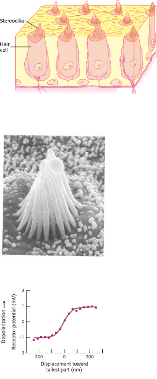
like a snail shell. The primary detection is accomplished by specialized neurons inside the cochlea called hair cells
(Figure 32.30). Each cochlea contains approximately 16,000 hair cells, and each hair cell contains a hexagonally shaped
bundle of 20 to 300 hairlike projections called stereocilia (Figure 32.31). These stereocilia are graded in length across
the bundle. Mechanical deflection of the hair bundle, as occurs when a sound wave arrives at the ear, creates a change in
the membrane potential of the hair cell.
Micromanipulation experiments have directly probed the connection between mechanical stimulation and membrane
potential. Displacement toward the direction of the tallest part of the hair bundle results in depolarization of the hair cell,
whereas displacement in the opposite direction results in hyperpolarization (Figure 32.32). Motion perpendicular to the
hair-length gradient does not produce any change in resting potential. Remarkably, displacement of the hair bundle by as
little as 3 Å (0.3 nm) results in a measurable (and functionally important) change in membrane potential. This motion of
0.003 degree corresponds to a 1-inch movement of the top of the Empire State Building.
How does the motion of the hair bundle create a change in membrane potential? The rapid response, within
microseconds, suggests that the movement of the hair bundle acts on ion channels directly. An important observation is
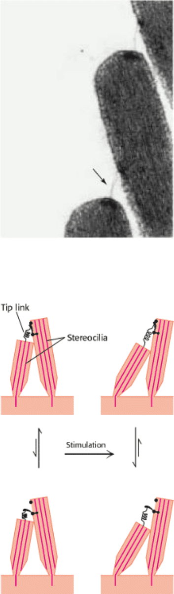
that adjacent stereocilia are linked by individual filaments called tip links (Figure 32.33).
The presence of these tip links suggests a simple mechanical model for transduction by hair cells (Figure 32.34). The tip
links are coupled to ion channels in the membranes of the stereocilia that are gated by mechanical stress. In the absence
of a stimulus, approximately 15% of these channels are open. When the hair bundle is displaced toward its tallest part,
the stereocilia slide across one another and the tension on the tip links increases, causing additional channels to open.
The flow of ions through the newly opened channels depolarizes the membrane. Conversely, if the displacement is in the
opposite direction, the tension on the tip links decreases, the open channels close, and the membrane hyperpolarizes.
Thus, the mechanical motion of the hair bundle is directly converted into current flow across the hair-cell membrane.
32.4.2. Mechanosensory Channels Have Been Identified in Drosophila and Bacteria
Although the ion channel that functions in human hearing has not been identified, other mechanosensory channels in
other organisms have been. Drosophila have sensory bristles used for detecting small air currents. These bristles respond
to mechanical displacement in ways similar to those of hair cells; displacement of a bristle in one direction leads to
substantial transmembrane current. Strains of mutant fruit flies that show uncoordinated motion and clumsiness have
been examined for their electrophysiological responses to displacement of the sensory bristles. In one set of strains,
transmembrane currents were dramatically reduced. The mutated gene in these strains was found to encode a protein of
1619 amino acids, called NompC for no mechanoreceptor potential.
The carboxyl-terminal 469 amino acids of NompC resemble a class of ion channel proteins called TRP (transient
receptor potential) channels. This region includes six putative transmembrane helices with a porelike region between the
fifth and sixth helices. The amino-terminal 1150 amino acids consist almost exclusively of 29 ankyrin repeats (Figure
32.35). Ankyrin repeats are structural motifs formed by 33 amino acids folded into a hairpin loop followed by a helix-
turn-helix. Importantly, in other proteins, regions with tandem arrays of these motifs mediate protein-protein
interactions, suggesting that these arrays couple the motions of other proteins to the activity of the NompC channel.
Prokaryotes such as E. coli have ion channels in their membranes that open in response to mechanical changes. These
channels play a role in regulating the osmotic pressure within the bacteria. The three-dimensional structure of one such
channel, that from Mycobaterium tuberculosis, has been determined. The channel is constructed of five identical
subunits arranged such that an alpha helix from each subunit lines the inner surface of the pore. Further studies should
reveal whether the transduction channel in hearing is homologous to either of these clases of mechanosensory channels
or represents a novel class.
IV. Responding to Environmental Changes 32. Sensory Systems 32.4. Hearing Depends on the Speedy Detection of Mechanical Stimuli
Figure 32.30. Hair Cells, the Sensory Neurons Crucial for Hearing. [Adapted from Hudspeth, A. J. Nature 341
(1989):397.]
IV. Responding to Environmental Changes 32. Sensory Systems 32.4. Hearing Depends on the Speedy Detection of Mechanical Stimuli
Figure 32.31. An Electron Micrograph of a Hair Bundle. [Courtesy of A. Jacobs and A. J. Hudspeth.]
IV. Responding to Environmental Changes 32. Sensory Systems 32.4. Hearing Depends on the Speedy Detection of Mechanical Stimuli
Figure 32.32. Micromanipulation of a Hair Cell. Movement toward the tallest part of the bundle depolarizes the cell as
measured by the microelectrode. Movement toward the shortest part hyperpolarizes the cell. Lateral movement has no
effect. [Adapted from Hudspeth, A. J. Nature 341(1989):397.]
IV. Responding to Environmental Changes 32. Sensory Systems 32.4. Hearing Depends on the Speedy Detection of Mechanical Stimuli
Figure 32.33. Electron Micrograph of Tip Links. The tip link between two hair fibers is marked by an arrow.
[Courtesy of A. Jacobs and A. J. Hudspeth.]
IV. Responding to Environmental Changes 32. Sensory Systems 32.4. Hearing Depends on the Speedy Detection of Mechanical Stimuli
Figure 32.34. Model for Hair-Cell Transduction. When the hair bundle is tipped toward the tallest part, the tip link
pulls on and opens an ion channel. Movement in the opposite direction relaxes the tension in the tip link, increasing the
probability that any open channels will close. [Adapted from A. J. Hudspeth. Nature 341 (1989):397.]
IV. Responding to Environmental Changes 32. Sensory Systems 32.4. Hearing Depends on the Speedy Detection of Mechanical Stimuli
Figure 32.35. Ankyrin Repeat Structure.
Four ankyrin repeats are shown with one shown in red. These domains
interact with other proteins, primarily through their loops.
IV. Responding to Environmental Changes 32. Sensory Systems
32.5. Touch Includes the Sensing of Pressure, Temperature, and Other Factors
Like taste, touch is a combination of sensory systems that are expressed in a common organ
in this case, the skin. The
detection of pressure and the detection of temperature are two key components. Amiloride-sensitive sodium channels,
homologous to those of taste, appear to play a role. Other systems are responsible for detecting painful stimuli such as
high temperature, acid, or certain specific chemicals. Although our understanding of this sensory system is not as
advanced as that of the other sensory systems, recent work has revealed a fascinating relation between pain and taste
sensation, a relation well known to anyone who has eaten "spicy" food.
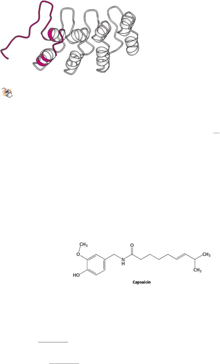
32.5.1. Studies of Capsaicin, the Active Ingredient in "Hot" Peppers, Reveal a Receptor
for Sensing High Temperatures and Other Painful Stimuli
Our sense of touch is intimately connected with the sensation of pain. Specialized neurons, termed nociceptors, transmit
signals to pain-processing centers in the spinal cord and brain in response to the onset of tissue damage. What is the
molecular basis for the sensation of pain? An intriguing clue came from the realization that capsaicin, the chemical
responsible for the "hot" taste of spicy food, activates nociceptors.
Early research suggested that capsaicin would act by opening ion channels that are expressed in nociceptors. Thus, a cell
that expresses the capsaicin receptor should take up calcium on treatment with the molecule. This insight led to the
isolation of the capsaicin receptor with the use of cDNA from cells expressing this receptor. Such cells had been detected
by their fluorescence when loaded with the calcium-sensitive compound Fura-2 and then treated with capsaicin or related
molecules. Cells expressing the capsaicin receptor, which is called VR1 (for vanilloid receptor 1), respond to capsaicin
below a concentration of 1 µM. The deduced 838-residue sequence of VR1 revealed it to be a member of the TRP
channel family (Figure 32.36). The amino-terminal region of VR1 includes three ankyrin repeats.
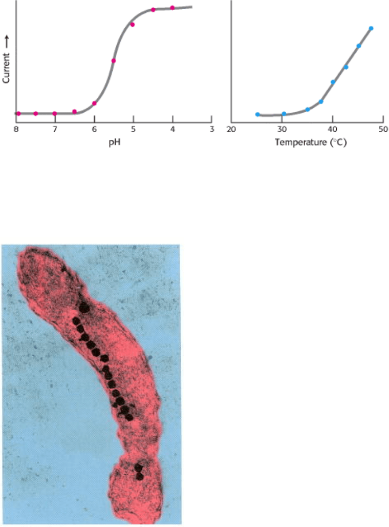
Currents through VR1 are also induced by temperatures above 40°C and by exposure to dilute acid, with a midpoint for
activation at pH 5.4 (Figure 32.37). Temperatures and acidity in these ranges are associated with infection and cell
injury. The responses to capsaicin, temperature, and acidity are not independent. The response to heat is greater at lower
pH, for example. Thus, VR1 acts to integrate several noxious stimuli. We feel these responses as pain and act to avoid
the potentially destructive conditions that caused the unpleasant sensation. Mice that do not express VR1 suggest that
this is the case; such mice do not mind food containing high concentrations of capsaicin and are, indeed, less responsive
than control mice to normally noxious heat. Plants such as chili peppers presumably gained the ability to synthesize
capsaicin and other "hot" compounds to protect themselves from being consumed by mammals. Birds, which play the
beneficial role of spreading pepper seeds into new territory, do not appear to respond to capsaicin.
Because of its ability to simulate VR1, capsaicin is used in pain management for arthritis, neuralgia, and other
neuropathies. How can a compound that induces pain assist in its alleviation? Chronic exposure to capsaicin
overstimulates pain-transmitting neurons, leading to their desensitization.
32.5.2. Subtle Sensory Systems Detect Other Environmental Factors Such as Earth's
Magnetic Field
In addition to the five primary senses, human beings may have counterparts to less-familiar sensory systems
characterized in other organisms. These sensory systems respond to environmental factors other than light, molecular
shape, or air motion. For example, some species of bacteria are magnetotactic; that is, they move in directions dictated
by Earth's magnetic field (Figure 32.38). In the Northern Hemisphere, Earth's magnetic field points northward but also
has a component directed downward, toward Earth's center. Magnetotactic bacteria not only swim northward but also
swim downward, away from the surface and the presence of high levels of oxygen, toxic to these bacteria. Remarkably,
these bacteria synthesize intracellular chains of small particles containing a magnetic ore called magnetite (Fe
3
O
4
) that
run through the center of each bacterium. Such chains are called magnetosomes. The magnetic force exerted by these
particles is sufficiently strong in relation to the size of the bacterium that it causes the bacterium to become passively
aligned with Earth's magnetic field. Intriguingly, similar magnetite particles have been detected in the brains of birds,
fish, and even human beings, although their role in sensing magnetic fields has not yet been established.
There may exist other subtle senses that are able to detect environmental signals that then influence our behavior. The
biochemical basis of these senses is now under investigation. One such sense is our ability to respond, often without our
awareness, to chemical signals called pheromones, released by other persons. Another is our sense of time, manifested in
our daily (circadian) rhythms of activity and restfulness. Daily changes in light exposure strongly influence these
rhythms. The foundations for these senses have been uncovered in other organisms; future studies should reveal to what
extent these mechanisms apply to human beings as well.
IV. Responding to Environmental Changes 32. Sensory Systems 32.5. Touch Includes the Sensing of Pressure, Temperature, and Other Factors
Figure 32.36. The Membrane Topology Deduced for VR1, the Capsaicin Receptor. The proposed site of the
membrane pore is indicated in red, and the three ankyrin (A) repeats are shown in orange. The active receptor comprises
four of these subunits. [Adapted from Caterina, M. J., Schumacher, M. A., Tominaga, M., Rosen, T. A., Levine, J. D.,
and Julius, D. Nature 389 (1997):816.]
IV. Responding to Environmental Changes 32. Sensory Systems 32.5. Touch Includes the Sensing of Pressure, Temperature, and Other Factors
Figure 32.37. Response of the Capsaicin Receptor to pH and Temperature. [Adapted from Tominaga, M., Caterina,
M. J., Malmberg, A. B., Rosen, T. A., Gilbert, H., Skinner, K., Raumann, B. E., Basbaum, A. I., and Julius, D. Neuron 21
(1998):531.]
IV. Responding to Environmental Changes 32. Sensory Systems 32.5. Touch Includes the Sensing of Pressure, Temperature, and Other Factors
Figure 32.38. Magnetotactic Bacterium. The magnetosome, visible as a chain of opaque membrane-bound magnetite
crystals, acts as a compass to orient the bacteria with the earth's magnetic field. The bacterium is artificially colored.
[Courtesy of Richard B. Frankel, California Polytechnic State University, San Luis Obispo, California.]

IV. Responding to Environmental Changes 32. Sensory Systems
Summary
Smell, Taste, Vision, Hearing, and Touch Are Based on Signal-Transduction Pathways
Activated by Signals from the Environment.
These sensory systems function similarly to the signal-transduction pathways for many hormones. These intercellular
signaling pathways appear to have been appropriated and modified to process environmental information.
A Wide Variety of Organic Compounds Are Detected by Olfaction
The sense of smell, or olfaction, is remarkable in its specificity
it can, for example, discern stereoisomers of small
organic compounds as distinct aromas. The 7TM receptors that detect these odorants operate in conjunction with G
(olf)
, a
G protein that activates a cAMP cascade resulting in the opening of an ion channel and the generation of a nerve
impulse. An outstanding feature of the olfactory system is its ability to detect a vast array of odorants. Each olfactory
neuron expresses only one type of receptor and connects to a particular region of the olfactory bulb. Odors are decoded
by a combinatorial mechanism
each odorant activates a number of receptors, each to a different extent, and most
receptors are activated by more than one odorant.
Taste Is a Combination of Senses That Function by Different Mechanisms
We can detect only five tastes: bitter, sweet, salt, sour, and umami. The transduction pathways that detect taste are,
however, diverse. Bitter and sweet tastants are experienced through 7TM receptors acting through a special G protein
called gustducin. Salty and sour tastants act directly through membrane channels. Salt tastants are detected by passage
though sodium channels, whereas sour taste results from the effects of hydrogen ions on a number of types of channels.
The end point is the same in all cases
membrane polarization that results in the transmission of a nerve impulse.
Umami, the taste of glutamate, is detected by a receptor that is a modified form of a brain receptor that responds to
glutamate as a neurotransmitter rather than as a tastant.
Photoreceptor Molecules in the Eye Detect Visible Light
Vision is perhaps the best understood of the senses. Two classes of photoreceptor cells exist: cones, which respond to
bright lights and colors, and rods, which respond only to dim light. The photoreceptor in rods is rhodopsin, a 7TM
receptor that is a complex of the protein opsin and the chromophore 11-cis-retinal. Absorption of light by 11-cis-retinal
changes its structure into that of all-trans-retinal, setting in motion a signal-transduction pathway that leads to the
breakdown of cGMP, to membrane hyperpolarization, and to a subsequent nerve impulse. Color vision is mediated by
three distinct 7TM photoreceptors that employ 11-cis-retinal as a chromophore and absorb light in the blue, green, and
red parts of the spectrum.
Hearing Depends on the Speedy Detection of Mechanical Stimuli
The immediate receptors for hearing are found in the hair cells of the cochleae, which contain bundles of stereocilia.
When the stereocilia move in response to sound waves, cation channels will open or close, depending on the direction of
movement. The mechanical motion of the cilia is converted into current flow and then into a nerve impulse.
Touch Includes the Sensing of Pressure, Temperature, and Other Factors
Touch, detected by the skin, senses pressure, temperature, and pain. Specialized nerve cells called nociceptors transmit
signals that are interpreted in the brain as pain. A receptor responsible for the perception of pain has been isolated on the