ASM Metals HandBook Vol. 17 - Nondestructive Evaluation and Quality Control
Подождите немного. Документ загружается.


object and the detector decreases the fraction of the scatter radiation that is detected. The use of focused collimators or
directionally sensitive detectors also reduces the amount of detected scatter radiation. Because the scatter radiation varies
slowly with distance, special reference detectors outside of the primary radiation beam can be used to measure the level of
the scatter signal.
Other Systemic Errors. A number of other systemic errors in data measurement can produce inconsistent data and can
result in artifacts or degraded image quality. These inconsistencies may be in the measured signal or may be the result of
poor characterization of the spatial position of the measured ray paths.
In medical CT imaging, patient motion is a major concern. If the object being scanned moves or changes configuration
during data acquisition, the measured data set does not correspond to a specific object. In addition to image blurring,
streaking from boundaries and high-density structures occurs. This is less of a problem with rigid structures in industrial
imaging. However, objects undergoing dynamic changes relative to the scan time, as in a fluid flow study, or poorly
fixtured components that wobble or vibrate during data acquisition can degrade the image quality.
Data are acquired along many ray paths, and the precise combination of this data produces the reconstructed image.
Imprecise manipulator motions, geometric misalignment, or insufficiently characterized positioning can also cause
blurring and image artifacts. An example is a series of tangential streaks forming a star pattern on translate-rotate systems
that can be caused by misalignments between traverses.
Inconsistencies in the data measurements may be due to variations in x-ray source output, the detector, or the detector
electronics. The x-ray tube output is normally measured by reference detectors, and the data are scaled appropriately. This
is effective for tube current changes that do not alter the effective energy of the x-ray beam. Changes in the tube voltage,
however, produce changes in both x-ray intensity and effective energy and are more difficult to correct. A highly stable x-
ray generator with feedback control of the voltage is normally required.
Measurement errors may also be the result of nonlinear detector performance over its entire dynamic range, detector
overranging, or differences between detector elements in a multidetector array. Detector overranging for a limited number
of measurements typically produces well-defined streaks through the image. Third-generation, rotate-only CT systems in
particular require a highly consistent detector array because uncharacterized variations between detectors cause concentric
ring artifacts. Oscillating changes in the detected signal can also occur because of mechanical vibration of the detector or
components within the detector or because of the pickup of electronic interference, especially at the line voltage
frequency.
References cited in this section
31.
S.M. Blumenfeld and G. Glover, Spatial Resolution in Computed Tomography, in Radiology of
the Skull
and Brain, Vol 5, Technical Aspects of Computed Tomography,
T.H. Newton and D.G. Potts, Ed., C.V.
Mosby Company, 1981
32.
M.W. Yester and G.T. Barnes, Geometrical Limitations of Computed Tomography (CT) Scanner
Resolution, Appl. Opt. Instr. Med. VI, Proc. SPIE, Vol 127, 1977, p 296-303
33.
P.M. Joseph, Image Noise and Smoothing in Computed Tomography (CT) Scanners, Opt. Eng.,
Vol 17,
1978, p 396-399
34.
H.H. Barrett and W. Swindell, Radiological Imaging: The Theory of Image Formation, Detectio
n, and
Processing, Academic Press, 1981
35.
G. Cohen and F.A. DiBianca, The Use of Contrast-Detail-
Dose Evaluation of Image Quality in Computed
Tomography Scanners, J. Comput. Asst. Tomogr., Vol 3, 1979, p 189
36.
Standard Guide for Computed Tomography (
CT) Imaging, American Society for Testing and Materials,
1989
37.
K.M. Hanson, Detectability in the Presence of Computed Tomography Reconstruction Noise,
Appl. Opt.
Instr. Med. VI, Proc. SPIE, Vol 127, 1977, p 304
38.
E. Chew, G.H. Weiss, R.H. Brooks, an
d G. DiChiro, Effect of CT Noise on the Detectability of Test
Objects, Am. J. Roentgenol., Vol 131, 1978, p 681

39.
P.M. Joseph, Artifacts in Computed Tomography, Phys. Med. Biol., Vol 23, 1978, p 1176-1182
40.
R.H. Brooks, et al., Aliasing: A Source of Streaks in Computed Tomograms, J. Comput. Asst. Tomgr.,
Vol
3 (No. 4), 1979, p 511-518
41.
D.A. Chesler, et al., Noise Due to Photon Counting Statistics in Computed X-Ray Tomography,
J. Comput.
Asst. Tomgr., Vol 1 (No. 1), 1977, p 64-74
42.
K.M. Hanson, Detectability in Computed Tomographic Images, Med. Phys., Vol 6 (No. 5), 1979
43.
W.D. McDavid, R.G. Waggener, W.H. Payne, and M.J. Dennis, Spectral Effects on Three-
Dimensional
Reconstruction From X-Rays, Med. Phys., Vol 2 (No. 6), 1975, p 321-324
44.
P
.M. Joseph and R.D. Spital, A Method for Correcting Bone Induced Artifacts in Computed Tomography
Scanners, J. Comput. Asst. Tomogr., Vol 2 (No. 3), 1978, p 100-108
Industrial Computed Tomography
Michael J. Dennis, General Electric Company, NDE Systems and Services
Special Features
The components that constitute a CT system often allow for additional flexibility and capabilities beyond providing the
cross-sectional CT images. The x-ray source, detector, and manipulation system provide the ability to acquire
conventional radiographic images. The acquisition of digital images along with the computer system facilitates the use of
image processing and automated analysis. In addition, the availability of the cross-sectional data permits three-
dimensional data processing, image generation, and analysis.
Digital Radiography. One of the limitations of CT inspection is that the CT image provides detailed information only
over the limited volume of the cross-sectional slice. Full inspection of the entire volume of a component with computed
tomography requires many slices, limiting the inspection throughput of the system. Therefore, CT equipment is often used
in a DR mode during production operations, with the CT imaging mode used for specific critical areas or to obtain more
detailed information on indications found in the DR image. Digital radiography capabilities and throughput can be
significant operational considerations for the overall system usage.
Computed tomography systems generally provide a DR imaging mode, producing a two-dimensional radiographic image
of the overall testpiece. The high-resolution detector of the rotate-only systems normally requires a single z-axis
translation (Fig. 25) to produce a high-quality DR image. For even higher resolution, interleaved data can be obtained by
repeating with a shift of a fraction of the detector spacing. For large objects, a scan-shift-scan approach can be used. The
translate-rotate systems are typically less efficient at acquiring DR data because the wider detector spacing requires
multiple scans, with shifts to provide adequate interleaving of data. If the width of the radiographic field does not cover
the full width of the object, the object can be translated and the sequence repeated.
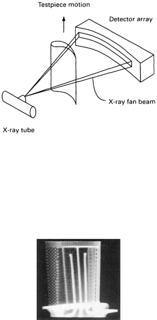
Fig. 25 Digital ra
diography mode. The component moves perpendicular to the fan beam, and the radiographic
data are acquired line-by-line.
The capabilities and use of these DR images are generally the same as discussed for radiographic inspection. The method
of data acquisition is different on the CT systems; the data are acquired as a sequence of lines or line segments. The use of
a thin fan beam with a slit-scan data acquisition is a very effective method of reducing the relative amount of measured
scatter radiation. This can significantly improve the overall image contrast (Fig. 26). In addition, the data acquisition
requirements for CT systems provide for a high sensitivity and very wide dynamic range detector system.
Fig. 26 Digital radiograph of an aircraft e
ngine turbine blade (nickel alloy precision casting) from an industrial
CT system
Image Processing and Analysis. Because the DR and CT images are stored as a matrix of numbers, the computer
can be used as a tool to obtain specific image information, to enhance images, or to assist in analyzing image data.
Display Features. A number of display features are used to obtain or present specific image information. A region-of-
interest program defines a portion of the image and provides information on the pixel values within the region, such as
minimum, maximum, and average CT number values; the standard deviation of the CT numbers; or the area or number of
pixels contained in the region. A histogram plot can display the relative frequency of occurrence of CT number values.
Cursor functions can be used to point to specific features, to annotate the images, and to determine coordinate locations,
distances between points, or angles between lines. A plot or profile can be generated from the CT numbers along a
defined line.
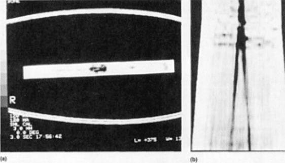
Image Processing. The image itself can be processed to enhance boundaries and details or to smooth the image to
reduce noise. Linear operators or filters used to sharpen the image, however, will increase the image noise, and smoothing
filters that reduce noise also blur or decrease the sharpness of the image. These processing steps, however, can assist in
improving the visibility of specific types of structures. Nonlinear processing techniques can be used to enhance or smooth
the image while minimizing the detrimental effects of the processing. Nonlinear techniques include median filtering and
smoothing limited to statistically similar pixel values; both techniques reduce the image noise while maintaining sharper
boundary edges. These nonlinear techniques, however, typically require more computation and can be difficult to
implement in fast array processors.
Image Analysis. Digital images can be analyzed for specific features or to measure definable parameters contained in
the image data. Where the image-processing capabilities are generic for any image, automated image analysis software is
written to analyze specific features in specific components. The software can verify the presence of specific necessary
components or can search for defined indications. Automated analysis is generally computational intensive, and
identifying a broad range of indications is a highly complex task. Consequently, the automated analysis of flaw
indications has generally been limited to a few, narrowly scoped analysis tasks.
The automated measurement of specific design parameters of components can be more readily defined and implemented.
The cross-sectional presentation of the component structure in the CT image allows for the measurement of critical
dimensions, wall thicknesses, cord lengths, and curvatures. The automated measurement of critical wall thicknesses on
complex precision castings is a standard feature on CT systems used in the manufacture of aircraft engine turbine blades
(Fig. 10).
Planar and Three-Dimensional Image Reformation. Having a stack of adjacent cross-sectional CT slices
characterizes the density distribution within the scanned volume in three dimensions. With this set of data in the
computer, alternate planes through the object can be defined, and the CT image data corresponding to these planes can be
assembled. These CT planar reformations allow the presentation of CT images in planes other than the planes in which
the data were originally acquired, including planes that CT data acquisition would not be feasible because of component
size and shape (Fig. 27). The CT image data can also be presented along other nonplanar surfaces. The CT data that
correspond to a conical surface on a rocket exit cone or along other structures can be presented. The data can also be
reformatted to alternate coordinates, such as presenting the data for a tubular section with the horizontal axis of the
display corresponding to an angular position and with the radial distance along the vertical axis of the display.
Fig. 27
CT image (a) across a sample helicopter tail rotor blade showing outer fiberglass airfoil and center
composite spar
. (b) Planar reformation through the composite spar from a series of CT slices. The dark vertical
lines are normal cloth layup boundaries, while the mottling at the top is due to interplanar waviness of the cloth
layup.
Structures within a scanned object can also be specifically characterized. Three-dimensional surface imaging identifies
the surface of a structure in a stack of CT images, defines surface tiles corresponding to this surface, and produces a
computer-assisted design perspective display of the structure, as shown in Fig. 11 (Ref 45). Because the CT images also
display interior surfaces, the computer can be used to slice open these surface models to display interior features. These

capabilities hold the potential for improving the component design cycle by documenting the configuration of physical
prototypes and operational components.
Computed tomography can also be used to define actual components, including their internal density distribution, as a
direct input for finite-element analysis (FEA). Automated meshing techniques are being analyzed that could significantly
reduce the time required to generate FEA models. In addition, this direct FEA modeling of actual components could assist
in the nondestructive analysis and characterization of components that are to be failure tested and may allow improved
calibration of the engineering models from the test data.
Dual Energy Imaging. In the range of x-ray energies used in industrial computed tomography, attenuation of the x-ray
photons predominantly occurs by either photoelectric absorption or Compton scattering. The probability of attenuation
due to photoelectric absorption decreases more rapidly than the probability of attenuation due to Compton scattering.
Consequently, photoelectric absorption predominates at low energies, and Compton scattering is the primary interaction
for high-energy photons. In addition, the probability for attenuation by photoelectic absorption is highly dependent on the
atomic number of the absorber, while Compton scattering is relatively independent of atomic number.
These differences between photoelectric and Compton attenuation cause difficulties in correcting x-ray transmission data
for beam hardening in structures having multiple materials. This difference in the attenuation process can be used,
however, to obtain additional information on the composition of the scanned object (Ref 46). If the object is scanned at
two separate energies, the data can be processed to determine a pair of images corresponding to the photoelectric and
Compton attenuation differences. The pair of images can be a photoelectric and Compton image or can correspond to
physical density and effective atomic number. This pair of basic images can be combined to form a CT image without
beam hardening variations.
The data for the basis images are a result of the differences in the high- and low-energy scans and are highly sensitive to
random variations. As a consequence, these basis images tend to have a very high noise level. The image noise level for
the combined pseudo-monoenergetic image, however, is equivalent to or lower than the noise in either of the original
single energy images. Accurate implementation of dual energy processing requires consistent data between the high- and
low-energy scans and characterization of the differences, such as that due to the detection of Compton scatter. Dual
energy imaging can also be implemented with the DR data to determine the basis data corresponding to the sum along the
measured ray paths.
Partial Angle Imaging. All of the CT imaging techniques discussed have considered the ability to obtain transmission
data from all angles in the plane of the cross-sectional slice. This is highly suitable for objects that can be readily
contained within a tight cylindrical workspace, but it can be impractical for large planar structures. Other components,
such as large ring and tubular structures, can be conventionally scanned given a large enough workspace, but it is
advantageous to minimize the source-to-detector distance and to image through a single wall in order to have reasonable
x-ray intensities and inspection throughput.
Methods have been investigated for medical imaging for reconstruction from partial data sets. Industrial imaging has the
advantage, however, in being able to apply a priori information from the component design or from measured external
contours to compensate for the missing data (Ref 47).
Another related approach is to use methods based on the focal-plane tomography or laminography techniques developed
in the early 1920s. Focal-plane tomography involves moving the x-ray source and film relative to the object such that
features in the object are blurred, except for the features in the focal plane. This method does not necessarily eliminate all
of the structures outside of the focal plane as in computed tomography, however, the data are obtained without circling
the object.
The capabilities of the conventional focal-plane tomography approach can be enhanced by using digital processing
techniques. With a series of digital radiographs, the images can be shifted and combined within the computer to yield a
series of focal planes at different levels in the object from one set of data. In addition, image filtering can be applied,
similar to the filtering in the filtered-backprojection CT reconstruction, to enhance the features in the focal plane and to
improve the elimination of out of plane structures.
References cited in this section

45.
H.E. Cline, W.E. Lorensen, S. Ludke, C.R. Crawford, and B.C. Teeter, Two Algorithms for the Three-
Dimensional Reconstruction of Tomograms, Med. Phys., Vol 15 (No. 3), 1988, p 320-327
46.
R. E. Alvarez and A. Macovski, Energy Selective Reconstructions in X-
Ray Computerized Tomography,
Phys. Med. Biol., Vol 21, 1976, p 733-744
47.
K.C. Tam, Limited-Angle Image Reconstruction in Non-Destructive Evaluation, in
Signal Processing and
Pattern Recognition in Nondestructive Evaluation of Materials,
C.H. Chen, Ed., NATO ASI Series Vol
F44, Springer-Verlag, 1988
Industrial Computed Tomography
Michael J. Dennis, General Electric Company, NDE Systems and Services
Appendix 1: CT Reconstruction Techniques
Computed tomography requires the reconstruction of a two-dimensional image from the set of one-dimensional radiation
measurements. It is useful to know some of the basic concepts of this process in order to understand the information
presented in the CT image and the factors that can affect image quality.
The reconstruction process consists of two basic steps:
•
The conversion of the measured radiation intensities into projection data that correspond to the sum or
projection of x-ray densities along a ray path
• The processing of the projection data with a reconstruction algorithm to determine the point-by-poin
t
distribution of the x-ray densities in the two-dimensional image of the cross-sectional slice
The development of projection data is common to all CT reconstruction techniques, while the reconstruction algorithm
can be approached by one of several mathematical methods (Ref 48, 49, 50).
Of the various reconstruction algorithms developed for computed tomography, there are two basic methods: transform
methods and iterative methods. Transform methods are based on analytical inversion formulas of the projection data
values given by Eq 9. Transform methods are fast compared to iterative methods and produce good-quality images. The
two main types of transform methods are the filtered-backprojection algorithm and the direct Fourier algorithm. The
filtered-backprojection technique is the most commonly used method in industrial CT systems.
Projection Data
The first step in the reconstruction of a CT image is the calculation of projection data. The measured transmitted intensity
data are normalized by the expected unattenuated intensity (intensity without the object). A logarithm is taken of these
relative intensity measurements to obtain the projection data. The projection data values correspond to the integral or sum
of the linear attenuation coefficient values along the line of the transmitted radiation. The reconstruction process seeks to
determine the distribution of linear attenuation coefficients (or x-ray densities) in the object that would produce the
measured set of transmission values.
The projection data values for a narrow monoenergetic beam of x-ray radiation can be theoretically modelled by first
considering Lambert's law of absorption:
I = I
0
exp(- s)
(Eq 6)
where I is the intensity of the beam transmitted through the absorber, I
0
is the initial intensity of the beam, s is the
thickness of the absorber, and is the linear attenuation coefficient of the absorber material. The linear attenuation
coefficient corresponds to the fraction of a radiation beam per unit thickness that a thin absorber will attenuate (absorb
and scatter). This coefficient is dependent on the atomic number of the materials and on the energy of the x-ray beam and
is proportional to the density of the absorber.
If instead of a single homogenous absorber there were a series of absorbers, each of thickness s, the overall transmitted
intensity would be:
I = I
0
exp[(
1
+
2
+
3
. . . +
i
)s]
(Eq 7)
where
i
is the linear attenuation coefficient of the ith absorber.
Considering a two-dimensional section through an object of interest, the linear attenuation coefficients of the material
distribution in this section can be represented by the function, (x, y), where x and y are the Cartesian coordinates
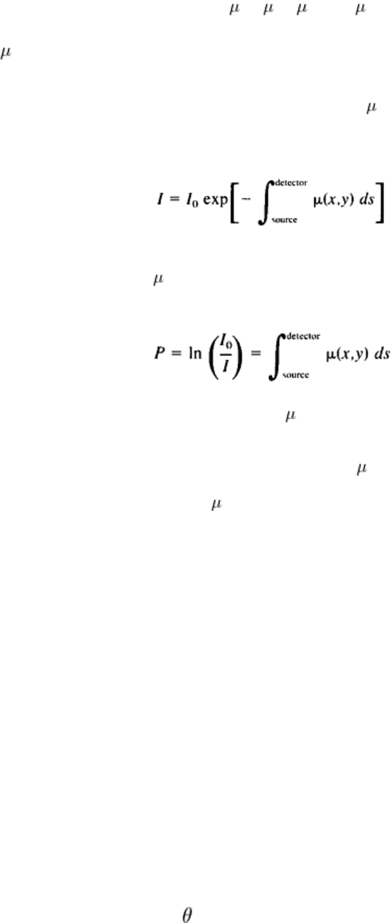
specifying the location of points in the section. Using the integral equivalent to Eq 7, the intensity of radiation transmitted
along a particular path is given by:
(Eq 8)
where ds is the differential of the path length along the ray. The objective of the reconstruction program in a CT system is
to determine the distribution of (x, y) from a series of intensity measurements through the section. Dividing both sides of
Eq 8 by I
0
and taking the natural logarithm of both sides of the equation yields the projection value, p, along the ray:
(Eq 9)
Equation 9, known as the Radon transformation of (x, y), is a fundamental equation of the CT process. It states that
taking the logarithm of the ratio of the unattenuated intensity to the transmitted intensity yields the line integral along the
path of the radiation through the two-dimensional distribution of linear attenuation coefficients. The inversion of Eq 9
was solved in 1917, when Radon demonstrated in principle that (x, y) could be determined analytically from an infinite
set of these line integrals. Similarly, given a sufficient number of projection values (or line integrals) in tomographic
imaging, the cross-sectional distribution, (x, y), can be estimated from a finite set of projection values.
The projection value as determined by Eq 9 is based on several assumptions that are not necessarily true in making
practical measurements. If an x-ray source is used, the x-ray photon energies or spectra range from very low energies up
to energies corresponding to the operating voltage of the x-ray tube. The energy of a beam can be characterized by an
effective or average energy. The effective energy, as well as the associated linear attenuation coefficients of the material,
will vary with the amount of material of filtration through which the beam passes. The increase in effective energy caused
by the preferential absorption of the less penetrating, lower-energy photons when passing through increasing thicknesses
of material is referred to as beam hardening. Beam hardening can cause nonlinearities in the measured projection values
relative to Eq 9 and can cause shading artifacts in the image. With knowledge of the type of material in the object being
imaged, corrections can be made to minimize this effect.
Other variations may also, occur in measuring the transmitted intensity values and determining the projection values.
These include the measurement of scatter radiation, the stability of the x-ray source, or any intensity or time-dependent
nonlinearities of the detector. To the extent that these systemic variations can be characterized or monitored, they can be
corrected in the data processing. Another variance that cannot be eliminated is the statistical noise of the measurement
due to the detection of a finite number of photons.
Direct Fourier Reconstruction Technique
The direct Fourier reconstruction technique utilizes the Fourier transformation of projection data. A Fourier transform is a
mathematical operation that converts the object distribution defined in spatial coordinates into an equivalent sinusoidal
amplitude and phase distribution in spatial frequency coordinates. The one-dimensional Fourier transformation of a set of
projection data at a particular angle, , is described mathematically by:
(Eq 10)
where r is the spatial position along the set of projection data and is the corresponding spatial frequency variable.
Fourier reconstruction techniques (and the filtered-backprojection method) are based on a mathematical relationship
known as the central projection theorem or central slice theorem. This theorem states that the Fourier transform of a one-
dimensional projection through a two-dimensional distribution is mathematically equivalent to the values along a radial
line through the two-dimensional Fourier transform of the original distribution. For example, given one set of projection
data measurements (Eq 9) through an object at a particular angle, taking the one-dimensional Fourier transform (Eq 10) of
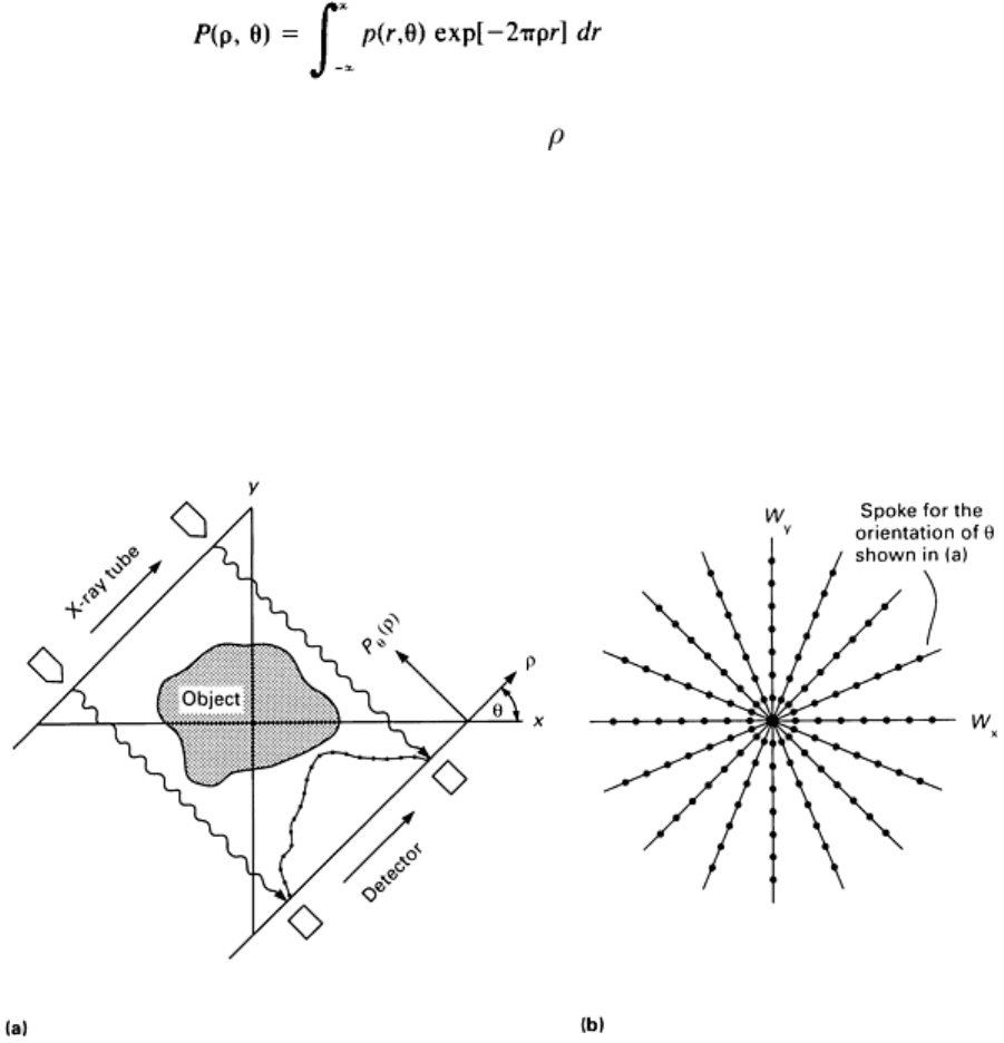
this data profile provides data values along one spoke in frequency space (Fig. 28). Repeating this process for a number of
angles defines the two-dimensional Fourier transform of the object distribution in polar coordinates (Fig. 28b). Taking the
inverse two-dimensional Fourier transform of this polar data array yields the object distribution in spatial coordinates.
Fig. 28
Data points in (a) direct space and (b) frequency space. The data points obtained in the orientation
shown in (a) correspond to the data points on one spoke in the two-dimensional Fourier transform space (b).
Although the direct Fourier technique is potentially the fastest method, the technique has not yet achieved the image
quality of the filtered-backprojection method, because of interpolation problems. Typical computer methods and display
systems are based on rectangular grids rather than polar distributions, and several variations of the direct Fourier
reconstruction technique exist for interpolating the data from polar coordinates to a Cartesian grid (usually interpolating
the data in the spatial frequency domain). The interpolation techniques can be complex, and the quality of the images
from measured data have generally been poorer than some of the other reconstruction methods. Interesting results have
been obtained in industrial multiplanar microtomography research, but this method is not typically used in commercial
systems (Ref 51).
Filtered-Backprojection Technique
The filtered-backprojection technique is the most commonly used CT reconstruction algorithm. Before discussing this
method, a brief discussion of filtering and the simple backprojection method is provided.
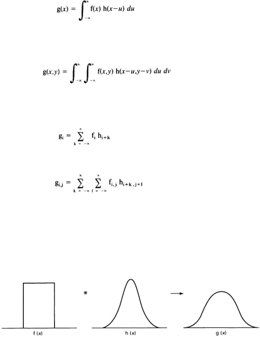
Convolutions and Filters. A convolution is a mathematical operation in which one function is smeared by another
function. Mathematically it can be defined as:
g(x) = f(x) * h(x)
(Eq 11)
(Eq 12)
for one dimension, or
g(x,y) = f(x,y) * h(x,y)
(Eq 13)
(Eq 14)
for two dimensions where * is the symbol for the convolution operation. If the system is a digital system with a discrete
number of samples, the corresponding equation to Eq 12 is:
(Eq 15)
and two-dimensional equivalent to Eq 13 is:
(Eq 16)
Convolutions are a common physical operation in data acquisition and imaging systems whether or not the term
convolution is used. All imaging systems have limitations in their ability to reproduce an object faithfully. One method of
quantifying the quality of an image is to define its point spread function or blurring function. A measured image is the
convolution of the object function with this blurring function (Fig. 29). This is represented in Eq 13, in which g(x, y) is
the resultant image formed by convoluting the object function f(x, y) with the blurring function h(x, y). If the object
function was an infinitely small target or a point, then f(x, y) is a delta function with a value of zero everywhere but at one
location, and the image g(x, y) will be equal to the PSF, h(x, y). This is how the point spread function is defined and
measured.
Fig. 29 Convolution operation (*) in which a distribution f(x) is blurred by a function h(x
) to form the blurred
distribution g(x). The function h(x
) is analogous to the PSF of an image system or a smoothing convolution
filter in image processing.
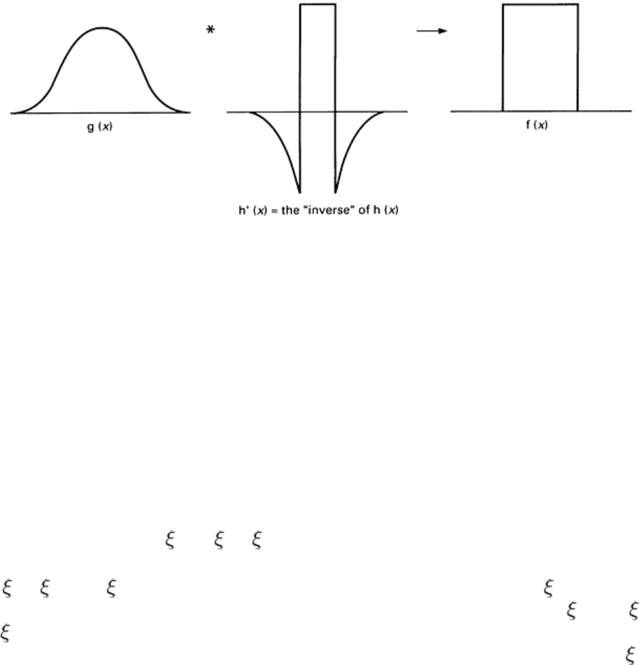
Convolutions can also be used in image processing to smooth (Fig. 29) or sharpen (Fig. 30) an image. Smoothing
convolution filters are typically square (averaging) or bell shaped, while sharpening convolution filters often have a
positive value in the center, with adjacent values being negative.
Fig. 30 Convolution operation with the sharpening filter h'(x). This sharpening of g(x) [or a restoration of f(x
)
with the "inverse" of h(x
)] is analogous to image process filtering or to the filtering of the projection data in the
filtered-backprojection reconstruction technique.
Reference has already been made to Fourier transforms, as in Eq 10, and to their ability to transform spatial data into
corresponding spatial frequency data. Filtering operations, such as image smoothing or sharpening, can be readily
performed on the data in the spatial frequency domain. According to the convolution theorem, convolution operations in
the spatial domain correspond to simple function multiplications in the spatial frequency domain (Fig. 31), that is:
g(x) = f(x) * h(x)
(Eq 17)
is equivalent to
G( ) = F( ) H( )
(Eq 18)
where G( ), F( ), and H( ) are the Fourier transformed functions of g(x), f(x), and h(x), where is the spatial frequency
conjugate of x. The functional multiplication in Eq 18 is simply the multiplication of the values of F( ) and H( ) at every
value of . The two-dimensional convolution of Eq 13 has its counterpart to Eq 18 where the functions G, F, and H are
the two-dimensional Fourier transforms of g(x, y), f(x, y), and h(x, y). Note that the spatial frequency variable, , has units
of 1/distance.
