ASM Metals HandBook Vol. 17 - Nondestructive Evaluation and Quality Control
Подождите немного. Документ загружается.

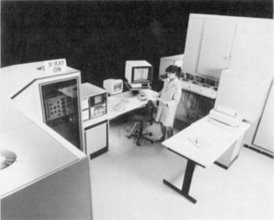
Fig. 13
Automated industrial computed tomography system for the production inspection of small components.
Parts-loading conveyor and cabinetized x-ray system are included in this setup.
In addition to these individual parts of a CT system, the overall capabilities of the total system are also important. The key
capabilities of a CT system are:
• The ability to handle the range of components to be inspected
• The throughput or rate at which the components can be tested
• The ability to provide sufficient image q
uality for the specified inspection task by considering the
selectable operational parameters of CT systems that affect image quality
CT Scanning Geometries
The CT scanning geometry is the approach used to acquire the necessary transmission data. In general, many closely
spaced transmission measurements from a number of angles are needed. Four types of CT scanning geometries are shown
in Fig. 14. Much of the historical progression of the data acquisition techniques has been driven by the need for
improvements in data acquisition speed for medical imaging. In medical CT systems, the radiation source and detector are
moved, not the patient. Industrial CT systems, however, often manipulate the component; this can simplify the
mechanical mechanism and help maintain precise source-detector alignment and positioning. In either case, the relative
positions of the object to the measured ray paths are equivalent. Some of the nomenclature derived from medical
computed tomography does not readily accommodate some of the manipulator configurations that are practical in
industrial imaging.
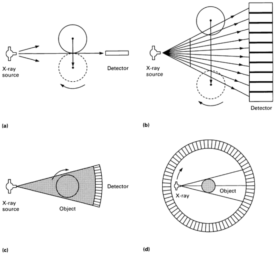
Fig. 14 Basic CT scanning geometries. (a) Single-detector translate-rotate, first-
generation system. (b)
Multidetector translate-rotate, second-generation system. (c) Rotate-only (rotate-
rotate), third generation
system. (d) Stationary-detector rotate-only, fourth-
generation system. In medical systems, the source and
detector are manipulated instead of the object.
Single-Detector Translate-Rotate Systems. The first commercial medical CT scanner developed by EMI, Ltd.,
uses a single detector to measure all of the data for a cross section. The x-ray source and detector are mounted on parallel
tracks on a rotating gantry (Fig. 14a). The source and detector linearly traverse past the specimen and make a series of x-
ray transmission measurements (240 measurements for a 160 × 160 image array). These measurements correspond to the
transmitted intensity through the object along a series of parallel rays. The source-detector mechanics are rotated by 1°,
and another linear traverse is made. This provides another set of parallel rays, but at a different angle through the object.
This process is repeated until data are obtained over a full 180° rotation of the source-detector system.
The original EMI scanner uses two detectors to collect data for two adjacent slices simultaneously; it is relatively slow,
with nearly a 5-min scan time to produce a low-resolution image. Despite its limitations, this system was a significant
break-through for diagnostic medicine.
Single-detector translate-rotate CT systems are sometimes referred to as first-generation systems. The use of the
generation nomenclature was originated by medical manufacturers to emphasize the newness of their designs and is
sometimes used as a matter of convenience to describe the operation of a system. Current commercial systems do not use
the single-detector approach, because of its limited throughput. The simplicity of this approach, however, makes it
suitable for applications in basic research.
Multidetector Translate-Rotate Systems. To improve the data acquisition speed, multiple detectors can be used to
make a number of simultaneous transmission measurements. The second-generation or multidetector translate-rotate
systems use a series of coarsely spaced detectors to acquire x-ray transmission profiles from a number of angles
simultaneously (Fig. 14b). The data measurements are still acquired during the translation motion, but the system makes a
much larger rotational increment. The distance translated needs to be somewhat longer than that for a single detector
system in order to have all source-detector rays pass over the entire object width, but fewer translations are needed to
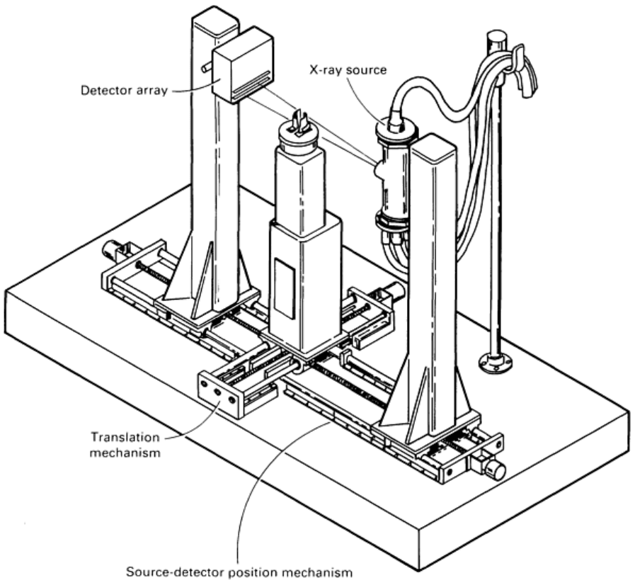
collect a full set of data. The mechanical system of a second-generation CT scanning geometry for industrial applications
may range from a design for small components (Fig. 15) to a design for large castings (Fig. 16).
Fig. 15
CT and DR scanner designed for the nondestructive testing and dimensional analysis of a variety of
parts up to 300 mm (12 in.) in diameter and 600 mm (24 in.) in length
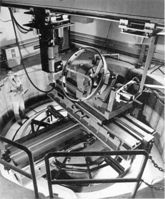
Fig. 16 Industrial computed tomography system x-
ray cell for the inspection of large fabricated components
and castings with multiaxis parts manipulator
Rotate-Only Systems. Further improvement in data acquisition speed can be achieved by using a broad fan beam of
radiation spanning the entire width of the object, eliminating the translation motion entirely. The fast scanning speed of
rotate-only systems is important in medical imaging to minimize patient motion during the scan and for high patient
throughput. Nearly all current medical scanners are rotate-only systems, with some of these systems making over 1
million transmission measurements for a complete scan in less than 2 s.
There are two basic configurations of rotate-only systems. Third-generation or rotate-rotate systems use many (sometimes
over 1000) closely spaced detector elements that are fixed relative to the x-ray beam (Fig. 14c). The source-detector
combination may rotate together around the specimen, as in medical scanners (hence the term rotate-rotate). In industrial
systems, the object can rotate in order to obtain data from all angles. The reconstruction process for these systems is
slightly different than for the other configurations because a transmission profile or view corresponds with a set of rays in
the shape of a fan. The use of the term rotate-only in this article will refer to third-generation systems.
Fourth-generation or stationary-detector rotate-only systems have a stationary ring of detectors encircling the specimen,
with the x-ray source rotating around the specimen (Fig. 14d). The x-ray fan beam exposes only a portion of the detectors
at any moment in time. The data are usually regrouped into fan beam sets, with the detector at the apex of the fan, or into
an equivalent parallel ray set. This configuration is generally less flexible, cannot utilize the ability to manipulate the
object, and is normally not found in systems designed for industrial use. Fourth-generation geometries require less
stringent detector element matching by a factor of 100 or so and are less susceptible to normalization and circular
artifacts.
Operational Differences and Variations. Each of the CT scanning geometries has its advantages and limitations.
The different approaches each have certain data acquisition parameters that tend to be fixed and others that are readily
adjustable.

One of the major factors in determining image resolution is the spacing between the measured transmitted rays. (Other
factors include source size, source-object and object-detector distances, detector aperture, and the reconstruction
algorithm.) The resolution at the center of the image field and in the radial direction at the periphery is highly dependent
on the ray spacing in a transmission profile. This ray spacing can be easily adjusted in translate-rotate systems by
adjusting the linear distance moved between measurements during translation.
On rotate-only (third-generation) systems, this ray spacing is fixed by the spacing of the detector elements. Consequently,
these systems use a densely packed detector array, with element spacings well below 1 mm (0.040 in.). The ray spacing
through the object can be improved by moving the object closer to the x-ray source or by acquiring interleaving data over
multiple rotations with the detector array shifting a fraction of the detector spacing.
The circumferential resolution at the periphery of the image is dependent on the angular separation between views and the
diameter of the image field. The ray spacing along the circumference or the number of angular views acquired is a readily
adjusted parameter on the rotate-only systems.
This angular separation is fixed by the detector array spacing on multidetector translate-rotate systems. Some variation
can be achieved by changing the source-detector spacing, or interleaved angular measurements can be obtained with small
angular increments.
Multidetector translate-rotate systems can adjust for object size by changing the translation distance to the object diameter
plus the width of the fan beam at the object. This tends to be inefficient for a wide fan beam system with small objects,
but gives considerable flexibility for large objects.
Rotate-only systems have a field-of-view defined principally by the detector array size and the source-object and object-
detector distances (object magnification). These systems can be used for larger fields-of-view by shifting the detector or
object between rotations to cover a wider field. However, this rotate-shift-rotate approach still acquires fan beam data
during the rotation motion, versus the parallel ray data collected during translation on a translate-rotate system.
Another difference is in acquiring data for DR mode imaging. The DR images are commonly used on CT systems to
locate the areas in a component where the CT slices should be obtained or can be used for radiographic inspection of the
component.
The high-resolution detector of the rotate-only systems normally requires a single z-axis translation to produce a high-
quality DR image. For even higher resolution, interleaved data can be obtained by repeating the rotational scan with a
shift of a fraction of the detector spacing. For large objects, a scan-shift-scan approach can be used.
Translate-rotate systems are typically less efficient at acquiring digital radiographic data if the wider detector spacing
requires multiple scans with shifts to provide adequate interleaving data. If the detector field-of-view does not cover the
full width of the object, the object can be translated and the sequence repeated.
Considerable flexibility can exist for various requirements. The mechanical packaging and implementation of the
particular data acquisition system are the limiting factors with regard to the range of capabilities. The different acquisition
approaches, however, can determine the efficiency of a system in acquiring appropriate data as well as the subsequent
imaging throughput.
Radiation Sources
Computed tomography reconstructions can be performed with any set of measured data along lines through an object.
This can include the use of ultrasound, neutrons, charged particle beams, nuclear tracer distributions (PET and SPECT
imaging), or emitted radio-frequency emissions from nuclei (NMR imaging). In most practical industrial applications,
however, x-ray or -ray radiation is used in computed tomography.
The three types of radiation sources typically used in industrial computed tomography are x-ray tubes, -ray sources, or a
high-energy x-ray source such as a linear accelerator. Each of these sources has advantages and disadvantages. The two
types of x-ray sources can provide a high-intensity beam, but they also produce a broad spectrum that complicates the
quantitative analysis of the projection data. Gamma-ray sources have a narrow energy spectrum but tend to be of low
intensity. Other factors include the size of the source and the energy of the beam.
The ideal radiation source would provide a high-intensity beam of x-ray photons at a single energy (or optimum spectral
spread) emanating from a very small area with an energy capable of transmitting a reasonable fraction of the x-rays
through the object.
Gamma-ray source are used in some industrial computed tomography systems. A major advantage of -ray sources is
that the high-energy photons produced by a radioactive source are all at specific energies, while x-ray sources produce
photons over a wide range of energies. Changes in the average energy transmitted through various thicknesses of material
can cause inconsistencies in the measured data, resulting in errors in the reconstructed image.
The most significant disadvantage of -ray sources is the limited intensity or number of gamma photons produced per
second. The intensity can be increased by using more radioactive material, but this requires a larger radioactive source,
which adversely affects the spatial resolution of the system. Also, because the energy of a -ray source is dependent on
the radioactive material, the effective energy cannot be readily changed for different imaging requirements.
X-Ray Sources. The number of high-energy photons included in the CT scan measurements is the primary factor
affecting the statistical noise in the reconstructed CT image. To collect the maximum number of photons in the least
amount of time, x-ray sources are generally used rather than radioactive -ray sources. For x-ray sources up to 500 kV in
operating voltage, x-ray tubes are used. When higher-energy x-rays are needed to penetrate thick or dense testpieces,
linear accelerators are often used.
The key characteristics of an x-ray source include the operating voltage range, the effective size of the focal spot, and the
operating power level. These characteristics are important in both radiography and computed tomography (see the article
"Radiographic Inspection" in this Volume). In computed tomography, however, the stability of the x-ray source is
especially important. Voltage variations are particularly disruptive in that they change the effective energy of the x-ray
beam and can cause image artifacts. Current variations are less of a problem because x-ray intensity can be monitored by
reference detectors.
X-ray collimators are radiation shields with open apertures that shape the x-ray beam striking the object and the
detector. For CT systems, the radiation field is typically a thin fan beam wide enough to cover the linear detector array. A
collimator (Fig. 1a) is located between the x-ray source and the object to shape the beam; normally, a second collimator is
also placed between the object and the detector array to further define the object volume being sampled. One or both of
the x-ray collimators may have an adjustable slot spacing to permit operator selection of the slice thickness.
One of the benefits of using a thin fan beam of radiation for CT and DR imaging is that most of the radiation scattered by
the object will miss the detector array and not be measured. This improves the quality of the measured data over that
obtained by large-field radiography.
Systems that use coarsely spaced detectors, such as multidetector translate-rotate systems, may also have detector aperture
width collimators. These collimators reduce the effective size of the detector element, thus improving the transmission
data resolution.
X-Ray Detectors
Detector Characteristics. The ability to measure the transmitted x-ray intensity efficiently and precisely is critical to
x-ray CT imaging. The features of the detector that are important to imaging performance include efficiency, size,
linearity, stability, response time, dynamic range, and energy range of effectiveness.
Detector efficiency is a quantitative measure of the effectiveness of the detector in intercepting, capturing, and
converting the energy of the x-ray photons into a measurable signal. This efficiency is a factor in the image quality for a
given x-ray source output, or the exposure time required to collect a sufficient amount of radiation. The three components
of overall detector efficiency are described below.
The geometrical efficiency is the fraction of the transmitted beam passing through the measured slice volume that is
incident on the active detectors. It is equal to the active detector element width in the plane of the slice divided by the
center-to-center spacing between detector elements. The collection efficiency is the fraction of the energy incident on an
active detector area that is absorbed in the detector. It is dependent on the atomic number and density of the detector
material and on the size and depth of the detector. Lack of absorption occurs because of x-ray photons passing through the
detector without interacting or because of interactions in which some of the energy is lost due to scatter or characteristic
x-ray emissions from the detector material. Conversion efficiency is the fraction of absorbed energy that is converted into
a measurable signal.
Detector size consists of both the detector element width and height. The detector height determines the maximum
slice thickness that can be measured. Increasing the slice thickness increases the collected x-ray intensity, thus reducing
the image noise, but decreases z-axis (interplane) resolution and may increase partial volume blurring and partial volume
artifacts. The measured slice thickness is adjusted by the slice thickness collimators to be less than the detector height.
The detector element width is a factor in determining the resolution of the CT image. On some CT systems, particularly
the multidetector translate-rotate systems, the detector aperture width can also be adjustable with a collimator. This offers
higher potential resolution with a lower geometrical efficiency.
Detector Consistency. Detector linearity is the ability to produce a signal that is proportional to the incident x-ray
intensity over a wide range of intensities. Detector stability concerns the ability to produce a consistent response to a
signal without drifting over time. Channel-to-channel uniformity relates to the consistency between the detector elements
in their signal response, noise, aperture size, and other characteristics. These parameters are important for CT detector
selection to produce the consistent data set required for image reconstruction. For reproducible characteristics, some
degree of inconsistency may be correctable by computer processing of the measured data.
The detector response time is the effective time for the signal to settle and not be significantly influenced by prior
incident intensities. The response time is a critical factor in determining how rapidly independent samples can be
collected and the quality of the data. Some CT systems interrogate each detector at rates up to 10,000 times per second.
Dynamic range is the range of intensities over which accurate measurement can be made. It is usually specified as a
ratio of the maximum signal output to the minimum output. The minimum signal output is limited by the electronic noise
of the detector and its electronics. The level of electronic noise determines how finely the signal can be effectively
sampled for processing by the computer.
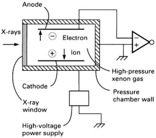
Gas ionization detectors consist of an ionization chamber with a positive and negative electrode (Fig. 17). When
radiation interacts with the gas inside the detector, the effect is to knock orbital electrons out of atoms, thus ionizing these
atoms. The freed electrons in this gas are pulled to the positive electrode, and the positively charged atoms (anions) are
pulled to the negative electrode. This principle is used in basic radiation survey meters, which use an ionization chamber
to measure the amount of ionization occurring within a volume of air or gas.
Gas ionization detectors have been shown to be
particularly effective in rotate-only CT scanners.
Rotate-only systems require a closely spaced
detector array with good detector uniformity to
provide the finely sampled projection data for
image reconstruction. These systems may contain
well over 1000 individual detector elements in a
contiguous array.
Ion chamber detectors define the individual array
elements by the placement of the collecting
electrodes of each detector element. Medical CT
systems arrange the collecting electrodes between
the detector elements, with a typical detector
spacing of 1.2 mm (0.05 in.). Higher-resolution
industrial detectors place the discrete electrodes
above and below the x-ray fan beam, with typical
detector spacings of 0.25 mm (0.01 in.). The
spread of the resolution response function for
tightly spaced detector systems, such as high-
resolution ionization detectors, may be greater
than the physical detector spacing because a small
percentage of the intensity incident on a detector
Fig. 17 Typical configuration of a xenon gas ionization detector
element may scatter into adjacent detector elements, contributing to crosstalk between detectors.
Because of the high packing density, with little or no gap between active detector elements, the geometrical efficiency of
gas ionization detector arrays is very good. Because a gas is used to stop the x-ray beam, the collection efficiency is
typically lower than a high-density solid detector material of the same dimensions. The conversion efficiency of
ionization detectors is very good in that few of the ions produced by the radiation are lost due to recombination before
reaching the collecting electrode.
To improve the efficiency of the detector at stopping and measuring the ionizing radiation, a high atomic number gas at
high pressure is used. The entire detector array is contained in a single pressure vessel that provides a common gas
mixture and pressure for all detector elements and aids in producing a uniform detector response. Xenon, an inert gas, is
used because of its high atomic number (Z = 54). High pressures are used that can increase the physical density of the gas
up to 1.5 g/cm
3
(0.9 oz/in.
3
).
Xenon ionization detectors provide high linearity over a wide range of x-ray intensities and have a reasonably fast
response time associated with the travel time of the ions in the electric field of the chamber. Of particular benefit for
third-generation rotate-only scanners is that xenon detectors are highly stable and do not exhibit radiation damage effects
or other long-time-constant exposure effects that may distort the measured data.
Scintillation detectors, which have been used for many years to measure radionuclide emissions, are used in both
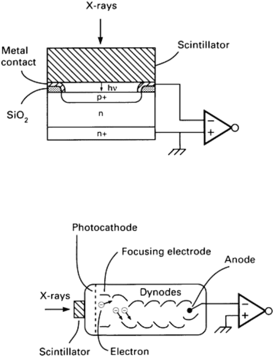
translate-rotate and rotate-only CT systems. The two basic designs are shown in Fig. 18 and 19. The most preferred and
widely used design in CT systems is the scintillation crystal-photodiode detector system (Fig. 18).
Fig. 18 Configuration for a scintillation crystal-photodiode detector
Fig. 19 Configuration for a scintillation crystal-photomultiplier tube detector

Scintillators. As with the gas ionization detectors, the x-ray radiation causes electrons in the atoms in the absorbing
material to be excited or ejected from the atom. These electrons rapidly recombine with the atoms and return to their
ground-state configuration, emitting a flash of visible light photons in the process. The term scintilla means spark or flash.
Scintillation detectors typically use relatively large transparent crystals. The scintillator is encased in a light-reflective
coating and is optically coupled to a sensitive light detector, such as a photomultiplier tube (PMT) (Fig. 19) or photodiode
(Fig. 18).
Sodium iodide doped with thallium [NaI(Tl)] coupled to a photomultiplier tube is a standard scintillation detector used for
nuclear radiation measurements. This type of detector was used on the original EMI medical scanner. However, new
scintillating materials have resulted in improved performance for certain detector characteristics. The scintillation
materials of choice are cadmium tungstate (CdWO
4
) and bismuth germanate (BGO). The properties of the material are
important to the efficiency and speed of the detector and its ability to provide accurate data. Table 3 lists some of these
materials along with some of their key properties.
Table 3 Scintillator materials
Material
Decay
constant, s
Afterglow,
% at 3 ms
Index of
refraction
Density, g/cm
3
Conversion
efficiency, %
(versus NaI)
NaI(Tl) 0.23 0.5-5 1.85 3.67
100
CsI(Tl) 1.0 0.5-5 1.8 4.51
45
BGO 0.3 0.005 2.15 7.13
8
CaWO
4
0.5-20 1-5 1.92 6.12
50
CdWO
4
0.5-20 0.0005 2.2 7.90 65
The relatively high density and effective atomic number are key attributes of many scintillator materials, providing a
highly effective ability to stop and absorb the x-ray radiation. The scintillation conversion efficiency, or fraction of x-ray
energy converted to light, is about 15% for NaI(Tl). Table 3 lists the conversion efficiencies relative to sodium iodide
with a standard light detector. The efficiency of the scintillator in producing light is important in that some of this light is
lost by absorption in the scintillator, reflective walls, and optical coupling material. A high index of refraction increases
the difficulty of optically coupling the scintillator.
The principal decay constant (time for the signal to decay to 37% of the maximum) is also listed in Table 3. Other lower-
intensity but longer-duration decay constants may also exist. Long afterglow decay constants (from 1 to 100 s) in sodium
iodide are one of its significant deficiencies for high-speed data acquisition. In addition to afterglow, the scintillator
materials may also exhibit temporary radiation damage, such as reduced optical transparency, which also has an
associated time constant for repair. The effect of prior radiation exposure on the measured signal is especially important
when a wide range of intensities are to be measured by the detector. Careful selection and characterization of the
scintillation detector material are necessary for high-speed CT data acquisition.
Photomultiplier Tube. The light detector is another important element in scintillation detector systems. Early systems
utilized PMT optical sensors (Fig. 19). A photomultiplier tube is a vacuum tube with a photocathode that produces free
electrons when struck by light. These electrons are accelerated and strike a positive electrode (dynode) that releases
additional free electrons. A series of dynodes is used to obtain very high levels of amplification with low back-ground
noise. For use on CT systems with high x-ray intensities, the photomultiplier tube is used to produce a signal current
proportional to the rate at which x-ray energy is absorbed in the scintillator.
Scintillation crystal-photodiode array detectors have important advantages over photomultiplier tubes in CT
applications. Although photomultiplier tubes are relatively efficient, they are subject to drifts in gain and are relatively
bulky. Crystals with photodiodes are more stable and permit the use of small, tightly packed detector arrays. Therefore,
photodiodes are commonly used as the optical sensor for CT scintillation detectors. Photodiodes produce a small current
when light strikes the semiconductor junction. This current is then amplified by a low-noise current-to-voltage converter
to produce the measured signal.
Very high resolution detector systems have been fabricated by coupling very thin slabs of scintillation material to high-
resolution photodiode arrays. These photodiode arrays may have 40 or more individual photodiodes per millimeter on a
single chip. Detectors with very small, tightly packed scintillation crystals may be particularly appropriate for the
materials evaluation of small samples at low x-ray energies.
Quantum Noise (Mottle) and the Detective Quantum Efficiency. The beam intensity being measured consists
of a finite number of photons. Even with an ideal detector, repeated measurement will contain a certain degree of random
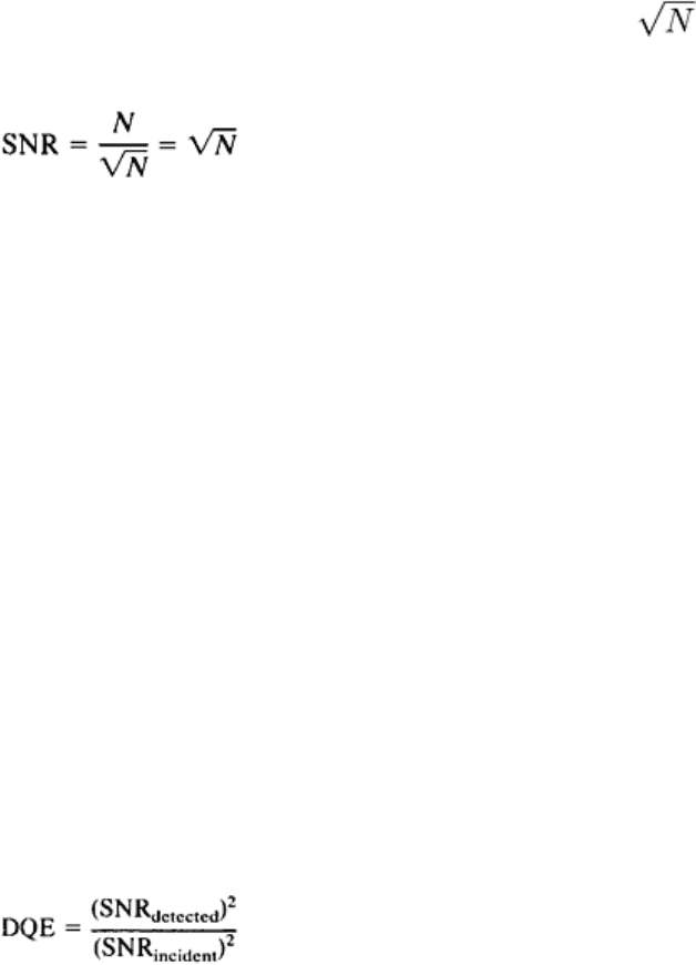
variation, or noise. The standard deviation of the number of photons detected per measurement is , where N is the
average number of photons detected per measurement. Therefore, the signal-to-noise ratio (SNR) can be given by:
(Eq 1)
Just as fast films that require less exposure tend to produce grainy images, the fewer the number of photons detected, the
lower the signal-to-noise ratio. Because this type of noise is due to the measurement of a finite number of discrete
particles, or quanta, it is referred to as quantum noise.
Because practical radiation detectors are imperfect devices, not all x-ray photons are absorbed in the detector. In gas
ionization detectors, each photon absorbed produces a finite number of measured electrons with their own quantum
statistics. In scintillation detectors, a finite number of light photons reach the light sensor, and a finite number of electrons
flow in the sensor. Each of these steps has its own quantum statistics (that is, quantum noise). The detector electronics,
particularly the initial amplifiers, introduce additional noise into the measured signal, which adds to the quantum noise.
The overall efficiency of the detector, including the noise added to the signal in the detection process, can be
characterized by comparing its performance to that of an ideal detector. The detective quantum efficiency (DQE) of a
detector is the ratio of the lower x-ray beam intensity needed with an ideal detector to the intensity incident on the actual
detector for the same signal-to-noise ratio. If the only increase in noise versus an ideal detector is due to the absorption
efficiency, that is, the fraction of photons absorbed in the detector, the DQE would equal the absorption efficiency.
Other losses of the signal occur in the detection process, and other sources of noise can be introduced into the signal to
further reduce the signal-to-noise ratio for actual measurements. The result in the quality of the measured signal is
effectively the same as detecting fewer photons. The DQE can be determined by comparing the measured signal-to-noise
ratio to that calculated for an ideal detector as follows:
(Eq 2)
The designers of CT detector systems strive to maximize overall detector efficiency while meeting the size, speed,
stability, and other operational requirements of a practical CT system.
Data Acquisition System
The data acquisition system is the electronic interface between the detector system and the computer. It provides further
amplification of the detector signals, multiplexes the signals from a series of detector channels, and converts these analog
voltage or current signals to a binary number that is transmitted to the computer for processing.
