ASM Metals HandBook Vol. 17 - Nondestructive Evaluation and Quality Control
Подождите немного. Документ загружается.


Some of the factors key to this electronics package include low noise, high stability, ability to calibrate offset and gain
variations, linearity, sensitivity, dynamic range, and sampling rate. The electronics of the data acquisition system should
be well matched to the detector system to minimize degradations to the data accuracy and scanning performance.
The dynamic range of the detector system is characterized by the ratio of the maximum signal to the noise. The dynamic
range of the data acquisition system is given by the range of numbers that can be transmitted to the computer, often
specified by the number of bits per measurement. It is not uncommon for CT systems to have a dynamic range of 20 bits
(2
20
), or 1 × 10
6
to 1.
Although it is difficult to build practical analog-to-digital converters with a 20-bit dynamic range, this electronics problem
has been solved in several different ways. One way is to use an autoranging or floating-point converter. In this case, a
linear converter is preceded by an adjustable-gain amplifier. One of several ranges can be selected by changing the gain
of the amplifier, usually by a factor of two. If an autoranging amplifier having four range settings corresponding to gains
of 1, 2, 4, and 8 precedes a 16-bit linear converter, the overall dynamic range is 20 bits, while the sensitivity varies from 1
to 2
16
for high intensities to 1 in 2
20
for low intensities relative to the maximum measurable intensity.
Computer System
Computer systems vary considerably in architecture, with computational performance steadily increasing. The availability
of practical minicomputer systems was the enabling technology for the development of computed tomography in the early
1970s. Computed tomography imaging has benefited from the computational improvements by permitting increasing
amounts of transmission data to be measured to produce larger, more detailed image matrices with less processing time.
High-speed computer systems are also enabling increasingly sophisticated data processing for image enhancement,
alternate data presentations, and automated analysis.
Evaluating computer system performance parameters is difficult, especially with the use of distributed processing. It is
common to have several processing systems controlled by a central processor or operating relatively independently in a
network configuration. This distributed processing may include programmable controllers and numerical controllers for
automated parts handling, manipulator control, and safety monitoring; specialized array processors for image
reconstruction; image processor for image enhancement and analysis; and display processors for presentation.
Because computed tomography uses large amounts of data (millions of transmission measurements to produce an image
containing up to several million image values) and because several billion mathematical operations are required for image
reconstruction, high-speed array processors are used for image reconstruction. Array processors are very efficient devices
for performing standard vector or array operations. The CT reconstruction technique commonly used also requires a
backprojection operation in which the projection values are mapped back into the two-dimensional image matrix.
Consequently, most CT systems contain specialized processors designed to perform this operation efficiently in addition
to standard array operations.
Data handling and archiving can also become significant considerations because of the size of the data files. Optical disks
can be beneficial for compact archiving as an alternative to magnetic tape. High-speed data networking can also provide
improved utility in transferring data to alternate workstations for further review and processing.
Industrial Computed Tomography
Michael J. Dennis, General Electric Company, NDE Systems and Services
Image Quality
Flaw sensitivity and image quality are the two basic criteria for evaluating the ability of any imaging system to define
sufficiently certain structures for a range of test object situations. Although image quality is a requirement in achieving
adequate flaw sensitivity, the evaluation of flaw sensitivity can be ambiguous in relation to inspection task requirements.
The analysis for the sensitivity of defined features in specific components can be quite complex and may involve
probability-of-detection or relative-operating-curve analysis. These analyses are also dependent on the perception and
skill of the interpreter and on the decision criteria, which may emphasize maximizing true positive or minimizing false
negative indication detection.
To evaluate and monitor the image quality and performance of an imaging system, a number of simpler methods can be
used to characterize the resolution, sensitivity, and accuracy of a system. The three basic factors affecting image quality
and system performance are:
• Spatial resolution
• Image contrast
• Image artifacts
These three factors are discussed in the following sections.
Spatial Resolution
Spatial resolution is a measure of the ability of an imaging system to identify and distinguish small details. As in
measuring the resolution of a radiographic system, an image of a regularly repeating high-contrast pattern is typically
evaluated. The equivalent to a radiography line pair gage for computed tomography is a bar pattern resolution test
phantom.
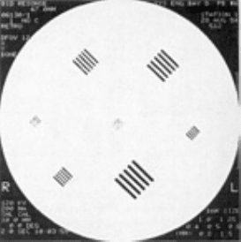
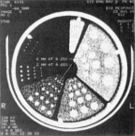
Resolution Test Patterns. A CT resolution bar phantom consists of a stack of alternating high- and low-density
layers of decreasing thickness. This phantom is scanned such that the layers are perpendicular to the slice plane. The
resultant CT image displays a bar pattern, and the minimum resolvable bar size (layer thickness) is determined (Fig. 20).
This measure can be reported as the bar size or as line pairs per millimeter.
Fig. 20 CT image
on a medical CT system of a resolution bar phantom. The center pattern has 0.5 mm (0.020
in.) bars on 1.0 mm (0.040 in.) spacings.
Alternatively, resolution hole patterns can be used. This type of phantom is a uniform block of material (usually
cylindrical) containing a series of drill holes. The holes are often arranged in rows, with the hole spacings twice the hole
diameter. Hole patterns can also be placed in test specimens to demonstrate resolution in the materials to be scanned. As
with the bar pattern, the minimum resolvable hole size is determined as a measure of the resolution.
Some differences may exist in the measured results for a system with the different test patterns described. Bar patterns
will generally indicate a higher-resolution performance than holes, probably because of the improved perceptibility of the
larger structures.
If the system resolution is approaching the display resolution for an image, the relative position of the test structures to the
voxels may affect the results. This can be avoided by orienting the test pattern at a 45° angle to the pixel row pattern of
the displayed image.
Point Spread Function (PSF). The image produced by a system is a blurred reproduction of the actual object
distribution. A more complex way of characterizing resolution and ability to reproduce the object structure is by
measuring the degree of image blurring or the PSF of the system. The PSF is the image response to a very small or
pointlike feature. This measurement can be made on a CT system by imaging a fine wire oriented perpendicular to the
slice plane (additional information on the point spread function is available in the section "Convolutions and Filters" in
Appendix 1 in this article).
If the pixel spacing is close to the resolution of the system, the extent of the PSF may extend for only a few pixels and be
poorly characterized. The PSF can also be determined from measurements of the line spread function (LSF) or edge
response function (Ref 31). The LSF is a normalized plot of the image across a line, such as a very thin sheet of metal
oriented perpendicular to the slice plane. The edge response function is the plot of the image across a low-density to high-
density boundary.
It is helpful in making LSF and edge response function measurements to orient the line or edge slightly out of alignment
with the columns of displayed pixels. By determining the relative position of data from several rows, one can obtain a
finer sampling of these functions. These functions can be partially characterized as a single parameter by reporting the full
width at half the maximum value of the spread function.
Modulation Transfer Function (MTF). Fourier theory states that a signal or an object can be described by a series of
sinusoidal functions. The MTF is a plot of the ability of the imaging system to transfer a sinusoidal signal versus the
frequency of that signal. The MTF for an imaging system is equivalent to evaluating the frequency response of an audio
system, except that the MTF is in terms of spatial frequency (cycles per unit distance).
The bar resolution test measurements provide an approximation to the MTF values. Thick bars show maximum contrast
between the high- and low-density materials, corresponding to the difference in linear attenuation coefficient of the
materials. As the bars become thinner, the overlap of the blurring at the edges tends to reduce the apparent CT number of
the high-density material and raise the values for the low-density material. The contrast or difference in CT number
diminishes and can be reported as a fraction of the large bar contrast.
The MTF is similar to a plot of contrast versus cycles per millimeter, except that it is for an idealized test object with a
sinusoidal density variation. Because fabricating such a sinusoidal distribution is impractical, the MTF is normally
determined by taking the normalized Fourier transform of a measured PSF. In reporting specific values from the MTF
function, the spatial frequency (line pairs per millimeter) at which the contrast transfer falls below a specified value (such
as 50, 20, or 10%) is sometimes given.
Factors Affecting Resolution. One of the major factors in determining resolution is the spacing between the
measured transmitted rays. The resolution at the center of the image field and in the radial direction at the periphery is
highly dependent on the ray spacing in a transmission profile. This ray spacing can be easily adjusted in translate-rotate
systems by adjusting the linear distance moved between measurements during translation.
Other factors that affect resolution include source size, degree of geometric magnification, detector aperture, and the
reconstruction algorithm. The resulting resolution of the reconstructed image may also be limited by the display system.
The measured spatial information is dependent on the sample spacing of the data and on the degree of blurring that occurs
in these data. Practical measurements are made with radiation sources of a finite size and a detector with a definable
aperture width. A single transmission measurement is therefore a type of average over a ray of some width. This ray
profile is dependent on the size and shape of the focal spot, the width of the detector aperture, and the relative position
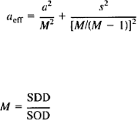
between the source and the detector. An approximation to the effective beam width, known as the effective aperture size,
is given by (Ref 32):
(Eq 3)
where a is the detector aperture and s is the width of the x-ray focal spot. The variable, M, is the magnification factor
given by:
(Eq 4)
where SDD is the source-detector distance and SOD is the source-object distance. If the magnification factor is close to 1,
as in contact film radiography, the detector resolution is the critical factor. As the magnification increases, a greater
burden is placed on having a small x-ray source in order to produce a high-resolution image.
If the sampled data are at a finer spacing than the effective aperture, deconvolution processing of the measured data can
reduce the effective aperture somewhat, but the result will be an increase in noise in the data (see the section
"Convolutions and Filters" in Appendix 1 of this article). Sample spacings through the object below one-half the effective
aperture, however, have little benefit because of the lack of appropriately detailed (high-frequency) information in the
measurements (Ref 31).
The sample spacing can also be a limiting factor to the resolution of the system. The Nyquist sampling theorem states that
sampling frequency must be at least twice the maximum spatial frequency of the structure being measured. This becomes
clear if one thinks of making sampled measurements across the bar resolution pattern. To define the one line-pair per
millimeter bars, two samples per millimeter are required--one for the high-density bar and the other for the low-density
space between bars.
Given an adequately small effective aperture, the sample spacing within a view is the limiting resolution factor in the
radial dimension from the center of the image. However, the circumferential resolution, perpendicular to the radius, is
primarily associated with the angular separation between views. This can present particular difficulties near the periphery
of the object, where the angular separation of the data corresponds to an increasingly large linear distance along the
circumference. The larger the diameter of the object, the larger the number of angular views required to maintain a given
circumferential resolution at the edges.
The measured transmission values are processed by a reconstruction algorithm to obtain the image, with the filtered
backprojection technique being the common method used. The filter function can be modified or the image postprocessed
to yield a certain degree of smoothing in the image in order to minimize the image noise. This smoothing is obviously an
additional factor affecting the system resolution.
Also part of the reconstruction process is the backprojection of the projection data into the image matrix. The rays
corresponding to the measured data generally do not pass through the center of each affected pixel; therefore,
interpolation between measured values is required. The method of interpolation can also affect the image resolution and
noise.
The reconstructed image is a two-dimensional array of data with a defined pixel spacing in terms of object dimensions.
The Nyquist sampling theorem also applies in this case, and details smaller than the pixel spacing cannot be accurately
defined. The pixel spacing can be a significant limitation where a large object is viewed in a single image. The quality of
the display system or recording media can also further degrade the resolution contained in the resultant image.
Many factors can affect the final resolution of the images. Appropriate system design and scan parameter selection are
required for optimizing the image quality for a given task.
Image Contrast
As with other imaging systems, the amount of contrast on a CT image is governed by object contrast, the contrast
resolution of the detector-readout system, and the size of a discontinuity with respect to the spatial resolution of the
system. The following sections describe these factors affecting image contrast, along with the use of contrast-detail-dose
diagrams for gaging these interrelationships in a particular scanning situation.
Object contrast refers to the contrast generated by variations in the attenuation of the radiation propagating through the
testpiece. Object contrast is a function of the relative differences in the linear attenuation coefficients and is affected by
the x-ray energy and the composition and density of the materials.
In order for a feature to be detected or identified, it must have a displayed density noticeably different from that of the
surrounding material. Computed tomography images are a scaled display of the linear attenuation coefficients of the
materials within the specimen. The relative contrast between a feature and the surrounding background material is the
normalized difference between the two linear attenuation coefficients, that is:
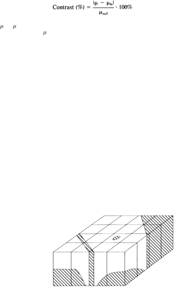
(Eq 5)
where and
b
are the linear attenuation coefficient of the feature of interest and the background material, respectively.
The reference coefficient,
ref
, is normally that of the background material, but the maximum coefficient of the two
materials can be used, especially if the background material is air or has a very low relative value.
The linear attenuation coefficient values are a function of the physical density of the material, the composition or
effective atomic number of the material, and the effective energy of the x-ray beam. The effect of the atomic number on
the linear attenuation coefficient is especially important at lower energies (sub-MeV) and for high atomic number
materials; in these cases, photo-electric absorption interactions are a significant factor in the overall attenuation. This
effect of atomic number can yield a much larger relative contrast at the low energies relative to high-energy scanning;
however, sufficient beam energy is required for penetrating the full thickness of the object and collecting an adequate
photon signal.
At very high energies, where Compton scattering interactions overwhelmingly dominate, atomic number has relatively
little effect. Under these conditions, the linear attenuation coefficient tends to be proportional to the electron density of
the material, which corresponds quite closely to the physical density of the material. For situations in which the
composition of the material is uniform, the linear attenuation coefficient is directly proportional to the physical density of
the material.
Size and Partial Volume Effect. In addition to the true object contrast, other factors also influence the measured and
displayed contrast between structures. When the voxel or sampled volume corresponding to a pixel contains two or more
separate structures, the resulting pixel value is a volume average of the structures. This is one of the causes for the
rounded profile or intermediate CT numbers found along boundaries. The edge pixels correspond to a combination of the
high- and low-density materials.
With small features, such as subresolution inclusions or cracks, only part of the voxel is occupied by this structure (Fig.
21). In these cases, the feature may be detectable, but the measured or apparent contrast is less than the difference
between the two materials. The smaller the feature, the less the imaged contrast. For situations in which the CT number
values are known for the two materials, this partial volume effect can be used to estimate the size of the feature or to
measure structural dimensions with subresolution accuracy.
Fig. 21 Sche
matic of structures partially filling a sampled voxel. The structures yield a measured CT value that
correspond to a volume average of the two materials.
The resolution characteristics of the imaging system and the degree of blurring in the image also reduce the apparent
contrast difference for small structures. Where the ideal image of a small structure would be a single pixel with a CT
number corresponding to the average linear attenuation coefficient within the voxel, the image blurring spreads this
measured signal over a large area. This can be thought of as an extension of the partial volume effect, in which the
volume corresponding to a pixel is the three-dimensional sensitivity distribution of object locations affecting the pixel
value.
Noise and Contrast Sensitivity. The contrast sensitivity of the imaging system is defined by the signal-to-noise ratio
and is affected by unwanted random variations in the measured data and the reconstructed image. Image noise is a
primary limiting factor in distinguishing low-contrast structures and in detecting high-contrast subresolution features that
appear in the image as low-contrast structures. The random uncertainty of the CT numbers causes subtle features to be
lost in the texture of the noise.
A primary contributor to image noise is the quantum noise (or mottle) in the measured data due to measurement of a finite
number of x-ray photons (Eq 1). Additional sources of noise are the data acquisition system electronics and roundoff
errors in the computer processing.
Measurement of Noise. The typical measurement of noise is the standard deviation of the CT number values over an
area of many pixels. The CT number standard deviation is commonly measured near the center of the cylindrical test
object and may vary with radial position. Because the results are dependent on the quantity of x-rays transmitted through
the object, the size and material of the test object should be specified. The noise is often reported as a percentage of the
CT number difference between the test object and air.
A more complete method of characterizing the noise is to determine the noise power spectrum of the system over a series
of images. The noise power spectrum defines the noise content in the image versus spatial frequency. As a consequence
of the filtering operation of the reconstruction process, CT images predominantly contain high-frequency noise.
Factors Affecting Noise. Because x-ray photon statistics (that is, quantum noise) is a major contributor to the noise,
factors that increase the number of photons detected tend to reduce the noise (Eq 1). The factors affecting the number of
detected photons are:
• The intensity of the radiation source
• The source-to-detector distance
• The attenuation of radiation passing through the te
stpiece (which in turn is a function of the thickness
and material of the testpiece and the energy of the incident radiation)
• The overall detection efficiency of the detector
The overall efficiency with which the useful x-ray beam is captured and measured depends on the geometrical,
absorption, and conversion efficiencies of the detector. The geometrical efficiency increases with the number of detectors
and the detector aperture width. The absorption and conversion efficiencies depend on the detector design, including the
size and material composition of the detector. The number of photons detected is also directly proportional to the scanned
slice thickness.
In addition to the transmitted primary photons, which constitute the x-ray signal, a certain amount of the detected
radiation is scattered radiation. This scattered radiation reduces contrast by creating a field of background noise.
Reduction of scatter, by tight collimation or increasing the object-to-detector distance, reduces this contribution to noise.
The reconstruction process also affects the resultant image noise. For ideal noise-less data, the shape of the reconstruction
filter is a high-pass ramp function, which amplifies the high-frequency noise. Because the signal content at these high
frequencies may be low due to other factors (such as the effective detector aperture), image quality can be improved for
certain imaging tasks by windowing this ramp function and by reducing the amplification of the high-frequency signal
and noise. This is effectively the same as smoothing the final image. A CT system may have selectable filters or
algorithms that provide variable degrees of smoothing.
The high-frequency content of the noise caused by the reconstruction filter is negatively correlated. This means that if one
pixel has a large positive noise fluctuation, surrounding pixels are more likely to have a negative fluctuation. Because the
image noise is negatively correlated, filter windowing or smoothing can be particularly advantageous in improving the
detectability of low-contrast structures (Ref 33). For situations in which the resolution is limited by the reconstruction
process and the effect of the effective aperture is small, the number of detected photons required to maintain a given
signal-to-noise ratio is inversely proportional to the resolution cubed (Ref 34). Under these conditions, improving the
resolution by a factor of two has the same effect on the signal-to-noise ratio as increasing the number of photons detected
by a factor of eight.
Other limitations in system implementation can also contribute to the noise. The high-sensitivity detector and data
acquisition system electronics adds some electronic noise to the signal. The reconstruction process consists of a large
number of computational operations, and the roundoff errors in these calculations can also contribute to image noise. In
addition, noise included in reference measurements used to normalize the scan projection data adds to the noise of the
reconstructed image.
Contrast-Detail-Dose (CDD) Diagram. The perception of a feature is dependent on the contrast between the feature
and the background, the size of the feature, the noise in the image, and the display window settings used to view the
image data. One method of gaging the range of structures that may be resolved is by determining the CDD characteristics
for a particular scanning situation (Ref 35, 36).
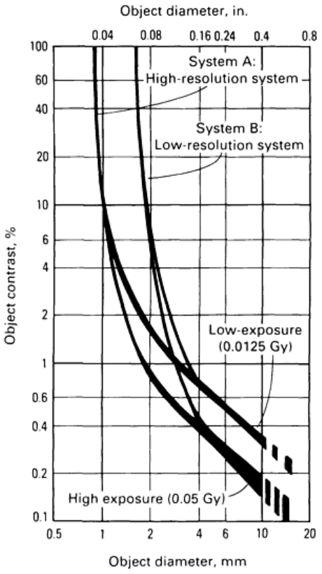
The CDD diagram is a plot of the imaged percent contrast versus the diameter of resolvable cylindrical test structures.
Figure 22 shows a CDD diagram for two similar systems with different detector spacings. Figure 22 also shows the effect
of changing the object dose, or number of photons collected, on the ability to resolve features.
Fig. 22 CDD diagram for two similar CT systems. The resolvability is limited by system resolution for high-
contrast objects and by noise for low-contrast structures.
For high-contrast structures, the ability to resolve the features has little dependence on the dose or image noise, but is
dependent on the resolution characteristics of the system. System A, which resolves smaller-diameter features, has a
smaller detector aperture and sample spacing than system B (Fig. 22).
As the contrast of the features decreases and the feature contrast approaches the noise variation in the image, larger
structures are required in order to distinguish them from the surrounding noise (Ref 37, 38). With the low-contrast
structures, the importance of the overall resolution capability of the system is reduced, and the level of noise in the image
becomes more critical. The two systems in Fig. 22 have similar performance for low-contrast features, with the object
dose and the level of noise in the image becoming the dominant factor.
System resolution can be measured for different contrast structures with a low-contrast detectability test phantom such as
the one shown in Fig. 23. This image demonstrates the ability of computed tomography to display very low contrast,
large-area structures. The measured results of these tests depend on factors that affect the structure contrast versus the
noise, including the size and the material of the test phantom.
Fig. 23 CT image of low-contrast detectability test phantom
Image Artifacts
Artifacts are image features that do not correspond to physical structures in the object. All imaging techniques are subject
to certain types of artifacts. Computed tomography imaging can be especially susceptible to artifacts because of its
sensitivity to small object differences and because each image point is calculated from a large number of measurements
(Ref 39).
The types of artifacts that may be produced and the factors causing them must be understood in order to prevent their
interpretation as physical structures and to know how scan operating parameters can be adjusted to reduce or eliminate
certain image artifacts. In general, the two major causes of CT image artifacts are the finite amount of data for generating
the reconstructed image and the systemic errors in the CT process and hardware. Some of the factors that can cause the
generation of artifacts include inaccuracies in the geometry, beam hardening, aliasing, and partial penetration (Ref 40, 41,
42).
Aliasing and Gibbs Phenomena. Because of the finite number of measurements, certain restrictions are placed on
the resolvable feature size. A series of adjacent x-ray transmission measurements allows the characterization of object
structures that are larger than this sample spacing. Coarse sampling of detailed features can create falsely characterized
data. The effect of this undersampling of the data is termed aliasing, in which high-frequency structures mimic and are
recorded as low-frequency data. This effect can cause image artifacts that appear as a series of coarsely spaced waves or
herringbone-type patterns over sections of the image (Fig. 24).
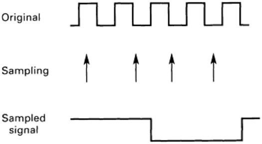
Fig. 24 Illustration of undersampling of a distribution containing high-
frequency information causing the false
appearance of low-frequency information or aliasing
Computed tomography systems do not measure the transmission data for infinitely thin rays. The detector has an aperture
of some finite thickness, and very fine structures within this broad ray tend to be smoothed out or eliminated from the
signal before being measured. This effective smoothing of the data prior to sampling acts to limit the spatial frequency
content prior to sampling and minimizes the potential for aliasing artifacts.
Aliasing can also occur as a result of the reconstruction process. If one attempts to reconstruct an image in which the pixel
spacing is much greater than the finely sampled projection data, the high-frequency information in the projection data can
cause aliasing artifacts in the image. This can be eliminated through appropriate selection of the reconstruction filter
function to filter out or eliminate spatial frequencies higher than that contained in the reconstructed image matrix.
The Gibbs phenomenon is also a type of artifact associated with the limited spatial frequency response of the system. It is
a consequence of a sharp frequency cutoff in the reconstruction filter function. This causes overshooting or oscillations
along high-contrast boundaries. The range and spacing of these oscillations decrease as the cutoff frequency increases.
This type of artifact can be minimized by windowing the reconstruction filter function such that it has a smooth roll-off,
rather than a sharp cutoff, at the high frequencies.
View aliasing is a form of aliasing resulting from an insufficient number of angular views. This artifact is typically a
radial streaking pattern that is apparent toward the outer edges of the image or emanates from a high-contrast structure
within the cross-sectional slice. The number of angular views required is dependent on the diameter of the object, the
circumferential resolution of the image, and the effective aperture width.
Partial Volume Artifacts. The partial volume effect is the volume averaging of the linear attenuation coefficients of
multiple structures within a voxel. Because of the exponential nature of the x-ray attenuation, the actual averaging process
can cause nonlinearities or inconsistencies in the measured projection data that produce streaking artifacts in the image.
In Eq 8, 9, and 10 in Appendix 1 , the attenuation through a series of materials is shown as the exponential of the sum of
individual material attenuation properties. This relationship is used to obtain the line integral of the linear attenuation
coefficients of the object along a ray, called a projection value. With the finite size of the measured x-ray beam, if two
materials are positioned such that part of the measured ray passes through one material and the other part of the ray passes
through the other material, the attenuation equation is the sum of exponential terms rather than the exponential of a sum.
Situations in which only part of a measured ray passes through a structure include rays tangential to high-contrast
boundaries and object structures that may penetrate only partially into the measured slice thickness (Fig. 21). If the two
materials have considerably different attenuation coefficient values, this inconsistency in the measured data can become
significant and cause streaks emanating along edges, from high-contrast features, or between high-contrast features.
Artifacts can be particularly prevalent between multiple high-contrast features that only partially intersect the slice
thickness.
Partial volume artifacts can be reduced by decreasing the effective ray size with thinner slice thickness and narrower
effective aperture. Smoothing or windowing the reconstruction filter also reduces the sensitivity to these artifacts. Another
approach is to reduce the contrast of the boundaries by immersing or padding the object with a material having
intermediate or similar attenuation values.
Beam Hardening. X-ray sources produce radiation with a range of photon energies up to the maximum energy of the
electron beam producing the radiation. The lower-energy, or soft, photons tend to be less penetrating and are attenuated to
a greater degree by an object than the higher-energy photons. Consequently, the effective energy of a beam passing
through a thick object section is higher than that of a beam traversing a thin section. This preferential transmission of the
higher-energy photons and the resulting increase in effective energy is referred to as beam hardening.
Changes in the effective energy of the x-ray beam due to the degree of attenuation in the object cause inconsistencies in
the measured data (Ref 43). X-rays that pass through the center of a cylindrical object will have a higher effective energy
than those traversing the periphery. This leads to a lower measured linear attenuation coefficient and lower CT number
values in the center of the reconstructed image. This CT number shading artifact is referred to as cupping, corresponding
to the shape of a plot of CT numbers across the object.
The effect of beam hardening can be reduced with several techniques. The original EMI scanner used a constant-length
water bath that yielded a relatively uniform degree of attenuation from the center to the periphery. The addition of x-ray
beam filtration reduces the soft x-rays and reduces the degree of beam hardening. Compensating or bow tie shaped x-ray
filters are sometimes used in medical CT systems to increase the beam filtration toward the periphery and to have a
smaller attenuation variation across a cylindrical-shaped object. Normalizing the data with a cylindrical object of similar
size and material as the test object also reduces the effect of the beam hardening.
Beam hardening is often compensated for in the processing software. If the material being scanned is known, the
measured transmission value can be empirically corrected. Difficulty occurs, however, when the object consists of several
materials with widely varying effective atomic numbers. The extent of the beam hardening depends on the relative degree
the attenuation is from the highly energy dependent and atomic number dependent photoelectric absorption versus the less
energy dependent and atomic number independent Compton scattering process. Different materials have differing degrees
of attenuation of the low-energy versus high-energy photons for a given overall level of attenuation.
Other processing techniques for minimizing beam hardening artifacts are sometimes used. If the object is composed of
two specific materials that are readily identifiable in the image, an iterative beam hardening correction can be
implemented (Ref 44). This approach identifies the distribution of the second material in the image and implements a
correction of the projection data for an improved second reconstruction of the data. Dual energy techniques can also be
used. These techniques use data obtained at multiple energies to determine the relative photoelectric and Compton
attenuations and can produce images that are fully corrected for beam hardening.
Scatter Radiation. Detected scatter radiation produces a false detected signal that does not correspond to the
transmitted intensity along the measured ray. The amount of scatter radiation detected is much lower than that
encountered with large-area radiographs because of the thin fan beam normally used in computed tomography. The
sensitivity of computed tomography, however, makes even the low levels of scatter a potential problem.
The scatter contribution across the detector array tends to be a slowly varying additive signal. The effect on the measured
data is most significant for highly attenuated rays, in which the scatter signal is relatively large compared to the primary
signal. The additional scattered photons measured make the materials along the measured ray appear less attenuating,
which is the same effect caused by beam hardening.
The types of artifacts caused by scatter radiation are similar to and often associated with beam hardening. In addition to
the cupping-type artifact, beam hardening and scatter can also cause broad, low CT number bands between high-density
structures. The rays that pass through both of the high-density structures are highly attenuated, and the increase in
effective energy and the scatter signal makes the materials along these rays appear to be less attenuating than is
determined from other view angles.
Because of the similarities of the effect, the basic beam hardening correction often provides some degree of compensation
for scatter. The amount of scatter radiation detected can be modeled, and specific scatter correction processing can be
implemented. Fundamental dual energy processing, however, does not correct for the detected scatter radiation.
Control of Scattered Radiation. System design can also minimize the detected scatter radiation. A tightly
collimated, thin x-ray beam minimizes the amount of scatter radiation produced. Increasing the distance between the
