Журнал - Проблемы криобиологии 2009 №1
Подождите немного. Документ загружается.

70
PROBLEMS
OF CRYOBIOLOGY
Vol. 19, 2009, ¹1
ÏÐÎÁËÅÌÛ
ÊÐÈÎÁÈÎËÎÃÈÈ
Ò. 19, 2009, ¹1
5.
6.
7.
8.
9.
10.
Салех Дж. М. Абу Жаяб. Коррекция андрогенной функции
у экспериментальных животных путем трансплантации
нативных и криоконсервированных органных культур
семенников: Дис. …канд. мед. наук. – Киев, 2004. – 129 с.
Benton L., Shan L.X., Hardy M.P. Differentiation of adult Leydig
cells // J. Steroid Biochem. Mol. Biol. – 1995. – Vol. 53, N1–6.–
P. 61–68.
Chen G.R., Ge R.S., Lin H. et al. Development of a cryo-
preservation protocol for Leydig cells // Hum. Reprod. – 2007.–
Vol. 22, N8. – P. 2160–2168.
Keros V., Rosenlund B., Hultenby K. et al. Optimizing cryopre-
servation of human testicular tissue: comparison of protocols
with glycerol, propanediol and dimethylsulphoxide as cryo-
protectants // Hum. Reprod.– 2005.– Vol. 20, N6.– P. 1676–
1687.
Tai J., Tze W.J., Johnson H.W. Cryopreservation of rat Leydig
cells for in vitro and in vivo studies // Horm. Metab. Res.–
1994.– Vol. 26, N3.– P. 145–147.
Tchoukalova Y.D., Harteneck D.A., Karwoski R.A. et al. A
quick, reliable, and automated method for fat cell sizing // J.
Lipid Res.– 2003.– Vol. 44, N9.– P. 1795–1801.
Поступила 02.09.2008
Рецензент В.Е. Чадаев
Pushkar N.S. Cryoprotectants.– Kiev: Naukova Dumka.–
1978.– 201 p.
Salekh J. M. Abu Jajab. Correction of androgen function in
experimental animals by transplantation of native and
cryopreserved organ cultures of testes: Author’s abstract
of the thesis of Candidate of Medical Sciences.– Kiev, 2004.–
129 p.
Benton L., Shan L.X., Hardy M.P. Differentiation of adult Leydig
cells // J. Steroid Biochem. Mol. Biol. – 1995. – Vol. 53, N1–6.–
P. 61–68.
Chen G.R., Ge R.S., Lin H. et al. Development of a cryo-
preservation protocol for Leydig cells // Hum. Reprod. – 2007.–
Vol. 22, N8. – P. 2160–2168.
Keros V., Rosenlund B., Hultenby K. et al. Optimizing
cryopreservation of human testicular tissue: comparison of
protocols with glycerol, propanediol and dimethylsulphoxide
as cryoprotectants // Hum. Reprod.– 2005.– Vol. 20, N6.–
P. 1676–1687.
Tai J., Tze W.J., Johnson H.W. Cryopreservation of rat Leydig
cells for in vitro and in vivo studies // Horm. Metab. Res.–
1994.– Vol. 26, N3.– P. 145–147.
Tchoukalova Y.D., Harteneck D.A., Karwoski R.A. et al. A
quick, reliable, and automated method for fat cell sizing // J.
Lipid Res.– 2003.– Vol. 44, N9.– P. 1795–1801.
Accepted in 02.09.2008
4.
5.
6.
7.
8.
9.
10.

71
PROBLEMS
OF CRYOBIOLOGY
Vol. 19, 2009, ¹1
ÏÐÎÁËÅÌÛ
ÊÐÈÎÁÈÎËÎÃÈÈ
Ò. 19, 2009, ¹1
УДК 616.419-089.843-001.28-092.9:616.4-003.93
À.À. ÖÓÖÀÅÂÀ, Ñ.Ñ. ×ÅÐÍÎÓÑÎÂÀ*, Ò.À. ÃËÓØÊÎ, Ë.Å. ØÀÒÈËÎÂÀ,
Â.Â. ÂÎËÈÍÀ, Ë.Â. ÑÎÊÎË, Ë.Þ. ÈÂÀÕÍÅÍÊÎ, Ë.Ã. ×ÅÐÍÛØÅÍÊÎ, Å.Â. ÁÐÎÂÊÎ
Âëèÿíèå òðàíñïëàíòàöèè êðèîêîíñåðâèðîâàííîãî êîñòíîãî ìîçãà
íà äèíàìèêó âîññòàíîâëåíèÿ ìîðôîôóíêöèîíàëüíûõ ñâîéñòâ
ëèìôîèäíûõ, ìèåëîèäíûõ è ýíäîêðèííûõ îðãàíîâ
ó ëåòàëüíî îáëó÷åííûõ ðåöèïèåíòîâ
UDC 616.419-089.843-001.28-092.9:616.4-003.93
A.A. TSUTSAYEVA, S.S. CHERNOUSOVA*, T.A. GLUSHKO, L.E. SHATILOVA,
V.V. VOLINA, L.V. SOKOL, L.YU. IVAKHNENKO, L.G. CHERNYSHENKO, E.V. BROVKO
Effect of Cryopreserved Bone Marrow Transplantation on Dynamics
of Recovery of Morphofunctional Properties of Lymphoid, Myeloid
and Endocrine Organs in Lethally Irradiated Recipients
Комплексно изучали морфофункциональные свойства лимфоидных, миелоидных и эндокринных органов у летально
облученных мышей на разных этапах после трансплантации им криоконсервированных клеток сингенного костного мозга
(КСКМ). Показано, что трансплантация КСКМ летально облученным животным стимулирует процессы восстановления
органов нейрогуморальной системы. При этом изменения уровня гормонов трийодтиронина (Т
3
), тироксина (Т
4
),
кортикостерона и инсулина в периферической крови, протекающие волнообразно, хоть и не достигают уровня физиоло-
гической нормы, однако обеспечивают выживаемость в течение 3-х месяцев летально облученных животных, погибающих
без трансплантации к 10 суткам.
Ключевые слова: летальное облучение, трансплантация, криоконсервированный сингенный костный мозг, лимфоидные,
миелоидные, эндокринные органы, гормоны.
Комплексно вивчали морфофункціональні властивості лімфоїдних, мієлоїдних та ендокринних органів у летально
опромінених мишей на різних етапах після трансплантації їм кріоконсервованих клітин сингенного кісткового мозку (КСКМ).
Показано, що трансплантація КСКМ летально опроміненим тваринам стимулює процеси відновлення органів нейро-
гуморальної системи. При цьому зміни рівня гормонів трийодтироніну (Т
3
), тироксину (Т
4
), кортикостерону та інсуліну в
периферичній крові, які протікають хвилеподібно, хоч і не досягають рівня фізіологічної норми, проте забезпечують виживання
на протязі 3-х місяців летально опромінених тварин, що гинуть без трансплантації на 10 добу.
Ключові слова: летальне опромінення, трансплантація, кріоконсервований сингенний кістковий мозок, лімфоїдні,
мієлоїдні, ендокринні органи, гормони.
There was realised a combined study of morphofunctional properties of lymphoid, myeloid and endocrine organs in lethally
irradiated mice at different stages after cryopreserved syngeneic bone marrow (CSBM) cell transplantation. The CSBM cell
transplantation to lethally irradiated animals demonstrated the stimulation of recovery processes in neurohumoral organs. At the same
time the wave-like changes in triiodothyronine (T
3
), thyroxin (T
4
), corticosterone and insulin hormone levels in peripheral blood,
even if not reaching the level of physiological norm, provide the survival within 3 months for lethally irradiated animals, dying
without transplantation to the 10
th
day.
Keywords: lethal irradiation, transplantation, cryopreserved syngeneic bone marrow, lymphoid, myeloid, endocrine organs, hormones.
* Àâòîð, êîòîðîìó íåîáõîäèìî íàïðàâëÿòü êîððåñïîíäåíöèþ:
óë. Ïåðåÿñëàâñêàÿ, 23, ã. Õàðüêîâ, Óêðàèíà 61015; òåë.:+38
(057) 373-31-26, ôàêñ: +38 (057) 373-30-84, ýëåêòðîííàÿ ïî÷òà:
cryo@online.kharkov.ua
* To whom correspondence should be addressed: 23, Pereyaslavskaya
str., Kharkov, Ukraine 61015; tel.:+380 57 373 3126, fax: +380 57
373 3084, e-mail: cryo@online.kharkov.ua
Institute for Problems of Cryobiology and Cryomedicine of the Na-
tional Academy of Sciences of Ukraine, Kharkov, Ukraine
Èíñòèòóò ïðîáëåì êðèîáèîëîãèè è êðèîìåäèöèíû
ÍÀÍ Óêðàèíû, ã. Õàðüêîâ
Известно, что гомеостаз зависит от состояния
ведущих систем организма: кроветворной, иммун-
ной, эндокринной и нервной [4]. Поскольку функ-
ционирование этих систем взаимосвязано, то нару-
шение функций одной из них может повлечь за
собой изменение функций других систем [7].
ÊÐÈÎÌÅÄÈÖÈÍÀ,
ÊËÈÍÈ×ÅÑÊÀß È ÝÊÑÏÅÐÈÌÅÍÒÀËÜÍÀß
ÒÐÀÍÑÏËÀÍÒÎËÎÃÈß
CRYOMEDICINE,
CLINICAL AND EXPERIMENTAL
TRANSPLANTOLOGY
Homeostasis is known to be dependent on the state
of leading systems of an organism such as: hemo-
poietic, immune, endocrine and nervous ones [4]. Sin-
ce the functioning of these systems is interrelated, the
disorder in one of them may involve the changes in
functions of other systems [7].
72
PROBLEMS
OF CRYOBIOLOGY
Vol. 19, 2009, ¹1
ÏÐÎÁËÅÌÛ
ÊÐÈÎÁÈÎËÎÃÈÈ
Ò. 19, 2009, ¹1
Ionising radiation depending on the doze and effect
duration causes differently pronounced morphofunc-
tional disorders in organs of lymphoid and myeloid
complexes and organs of neuroendocrine system,
being the cause of hemo- and immune depressions in
lethally irradiated animals and humans, that may result
either in their death or development of different
pathological processes [14, 20, 25]. The transplanta-
tion of syngeneic or HLA-DR-DQ-compatible
allogenic bone marrow was established to be the only
way to recover hemopoietic processes after lethal
irradiation [10, 15, 24]. The researches, devoted to
studying the recovery dynamics of morphofunctional
properties of neuroendocrine organs after lethal irra-
diation at different stages after bone marrow transplan-
tation, are single and their findings are not deprived
of contradictions [1, 3, 16, 19, 23, 26, 27].
The research was aimed to study the recovery
dynamics of morphofunctional properties of lympho-
myeloid organs (bone marrow, blood, lymph nodes,
thymus, spleen, peritoneal cavity) and endocrine ones
(hypophysis, thyroid and adrenal glands, pancreas) in
lethally irradiated recipients at different stages after
transplantation of cryopreserved cells of syngeneic
bone marrow.
Materials and methods
Experiments were performed in 2 months’ male
mice (CBA×C57B1)F
1
with 18–20 g (n=600) in
autumn-winter period. Animals were divided into
3 groups: the 1
st
group comprised lethally irradiated
animals; the 2
nd
one did lethally irradiated animals with
introduced cryopreserved syngeneic bone marrow
(CSBM) cells; and the intact animals made the 3
rd
one
(control). Bone marrow cells were isolated from
femoral bones and cryopreserved by the method, repor-
ted in the paper [14]. After cryopreservation more than
90% integral cells and 87% CFUs and CFUc were
determined in suspension. Donor cryopreserved bone
marrow cells were intravenously introduced in 0.2 ml
volume under 1×10
7
cell/ml dose. Animals were irra-
diated in morning hours with RUМ-17 device in 7.55
Gy dose, under 180 kV voltage, 10 mA current inten-
sity, 0.5±0.1 mm with LD
100/10
filters. Animals were
decapitated with ether narcosis in 1, 2, 3, 4, 5, 7, 10,
15, 20, 25, 30, 40, 60, 70, 80 and 90 days after expe-
riment beginning. Bone marrow cells were transplan-
ted 1 hr later irradiation.
The experiments were carried-out according to the
“General ethical principles of experiments in animals”,
approved by the 2
nd
National Congress on Bioethics
(Kiev, 2004).
Thymus and lymph node cellularities were
determined with Goryaev’s chamber by calculating the
number of nucleated cells, obtained after soft homo-
Ионизирующая радиация в зависимости от
дозы и продолжительности воздействия вызывает
разной степени выраженности морфофункцио-
нальные нарушения органов лимфоидного и мие-
лоидного комплекса и органов нейроэндокринной
системы, являющиеся причиной возникновения у
летально облученных животных и человека гемо-
и иммунодепрессий, которые могут приводить к
их гибели либо к развитию разных патологи-
ческих процессов [14, 20, 25]. Установлено, что
единственным способом восстановления процес-
сов кроветворения после летального облучения
является трансплантация сингенного, либо сов-
местимого по HLA-DR-DQ аллогенного костного
мозга [10, 15, 24]. Работы, посвященные исследова-
нию динамики восстановления морфофункцио-
нальных свойств нейроэндокринных органов пос-
ле летального облучения на разных этапах после
трансплантации костного мозга, единичны, а их
результаты не лишены противоречий [1, 3, 16, 19,
23, 26, 27].
Цель данной работы – изучение динамики
восстановления морфофункциональных свойств
органов лимфомиелоидного комплекса (костного
мозга, крови, лимфатических узлов, тимуса,
селезенки, перитонеальной полости) и эндокрин-
ных органов (гипофиза, щитовидной железы,
надпочечников, поджелудочной железы) у леталь-
но облученных реципиентов на разных этапах
после трансплантации криоконсервированных
клеток сингенного костного мозга (КСКМ) .
Ìàòåðèàëû è ìåòîäû
Эксперименты проводили на 600 линейных
мышах-самцах (CBA×C57Bl)F
1
массой 18–20 г в
возрасте 2 мес. в осенне-зимний период. Животные
были разделены на 3 группы: 1 – летально облучен-
ные; 2 – летально облученные животные, которым
вводили криоконсервированные клетки синген-
ного костного мозга; 3 – интактные животные
(контроль). Клетки костного мозга выделяли из
бедренных костей и подвергали криоконсерви-
рованию по методу [14]. После криоконсервиро-
вания в суспензии определялось больше 90%
сохранных клеток и 87% КОЕс и КОЕк. Клетки
криоконсервированного донорского костного моз-
га вводили внутривенно в объеме 0,2 мл в дозе
1×10
7
кл/мл. Облучали животных в утренние часы
на установке РУМ-17 в дозе 7,55 Гр, при напряже-
нии 180 кВ, силе тока 10 мА, фильтры 0,5±0,1 мм
Al c ЛД
100/10
.
Декапитировали животных под эфир-
ным наркозом через 1, 2, 3, 4, 5, 7, 10, 15, 20, 25,
30, 40, 60, 70, 80 и 90 суток после начала экспери-
мента. Клетки костного мозга трансплантировали
через 1 ч после облучения.
73
PROBLEMS
OF CRYOBIOLOGY
Vol. 19, 2009, ¹1
ÏÐÎÁËÅÌÛ
ÊÐÈÎÁÈÎËÎÃÈÈ
Ò. 19, 2009, ¹1
Эксперименты проведены в соответствии с
“Общими принципами экспериментов на живот-
ных”, одобренными ІІ Национальным конгрессом
по биоэтике (Киев, 2004).
Клеточность тимуса и лимфоузлов определяли
в камере Горяева путем подсчета количества ядер-
ных клеток, полученных после мягкой гомогени-
зации предварительно взвешенного соответствую-
щего органа. Количество антителообразующих
клеток (АОК) в селезенке облученных животных
определяли по стандартному методу [18]. Исследо-
вали активность реакции бласттрансформации
Т-лимфоцитов мышей на митогены фитогемагглю-
тинин (ФГА) и конканавалин А (Кон А) (“Difco”,
США; “Wellcome”, Великобритания) и В-лимфо-
цитов – на липополисахарид (ЛПС) (“Sigma”,
США). Трансформирующую активность клеток
определяли по уровню включения
3
Н-тимидина на
счетчике SL-40 (Intertechniquе, Франция). Индекс
бласттрансформации (индекс стимуляции) клеток
вычисляли по формуле: уровень включения
3
Н-ти-
мидина в клетки, культивируемые в присутствии
митогенов, в отношении к уровню включения
3
Н-тимидина в клетки, культивируемые без митоге-
нов [21]. Моноцитарно-макрофагальные клетки
получали из экссудата перитонеальной полости
животных [12]. Мазки окрашивали азур ІІ – эози-
ном и подсчитывали процентное содержание раз-
личных видов клеток. Фагоцитарную активность
перитонеальных макрофагов определяли через 1 ч
после инкубации клеток со стафилоккоком-209,
убитым нагреванием, подсчетом количества фаго-
цитирующих клеток. Кислую фосфатазу и неспе-
цифическую эстеразу в клетках экссудата перито-
неальной полости определяли гистохимическим
методом [17, 22], средний гистохимический коэф-
фициент (СГК) вычисляли по формуле [13].
Гистологические препараты эндокринных органов
(гипофиза, щитовидной железы, надпочечников,
поджелудочной железы) окрашивали гематоксили-
ном и эозином. В гипофизе подсчитывали хромо-
фильные и хромофобные ряды клеток передней
доли гипофиза (аденогипофиз); вычисляли про-
центное содержание этих клеток, измеряли пло-
щадь клеток и их ядер с помощью окуляр-микро-
метра и определяли ядерно-цитоплазменное
отношение в этих группах клеток [2]. Для гистоло-
гического исследования использовали правый
надпочечник, а левый – для определения липидов
в криостатных срезах, которые окрашивали суда-
ном III и суданом черным В по Лизону [8]. Степень
суданофилии оценивали с помощью гистохими-
ческого коэффициента [13]. В плазме крови живот-
ных радиоиммунологическими методами опреде-
ляли концентрацию тиреоидных гормонов –
тироксина (Т
4
) и трийодтиронина (Т
3
), кортикосте-
рона и инсулина (наборы ХОП ИБОХ НАН Бела-
genisation of a preliminary suspended corresponding
organ. The number of antibody-forming cells (AFCs)
in spleen of irradiated animals was defined according
to the standard method [18]. The activity of murine
T-lymphocyte blast-transformation response to phyto-
hemagglutinin (PHA) and concanavalin A (Con A)
mitogens (Difco, USA; Wellcome, UK) and B-lym-
phocyte to lypopolysaccharide (LPS) (Sigma, USA),
was under study. Transforming activity of cells was
determined by the
3
H-thymidine inclusion level using
the SL-40 counter (Intertechnique, France). The cell
blast-transformation index (stimulation index) was
calculated by the formulae: the level of
3
H-timidine
inclusion into the cells, cultured in mitogen presence,
towards to the level of
3
H-timidine inclusion into the
mitogen-free cultured ones [21]. Monocyte-macro-
phage cells were derived from the animal peritoneal
cavity exudate [12]. Smears were azure II-eosin stained
and the percentage of different cell types was calcula-
ted. Phagocyte activity of peritoneal macrophages was
defined 1 hr following cell incubation with heat-killed
staphylococcus-209 by calculating the number of
phagocytic cells. Acid phosphatase and non-specific
esterase in exudate cells of peritoneal cavity was deter-
mined using the histochemical methods [17, 22], the
average histochemical coefficient (AHC) was calcula-
ted by the formula [13]. Histological preparations of
endocrine organs (hypophysis, thyroid, adrenal glands,
pancreas) were stained with hematoxylin and eosin.
In hypophysis there were calculated the chromophilic
and chromophobic cell rows of anterior lobe of hypo-
physis (adenohypophysis) and the percentage of these
cells; the area of cells and their nuclei was measured
by means of ocular-micrometer and the nucleus-cyto-
plasm ratio in these cell groups was determined [2].
Right adrenal gland was used for histological study
and the left one for examining lipids in cryostat sec-
tions, stained with sudan III and sudan black B by
Lison [8]. The sudanophily extent was estimated with
histochemical coefficient [13]. In animal blood plasm
there was radioimmunologically determined the con-
centration of thyroid hormones: thyroxin (T
4
) and
triiodothyronine (T
3
), corticosterone and insulin (kits
of Institute of Bioorganic Chemistry of the National
Academy of Sciences of Belarus, Belarus). Sugar con-
tent in blood plasma was examined by the method [9].
The data were statistically processed by the Student-
Fisher method [6] using Excel and Statistica software.
Results and discussion
Cryopreserved bone marrow cell transplantation
to the lethally irradiated animals in syngeneic system
completely protected them against death. Histological
structure of thymus and lymph nodes in the irradiated
animals and CSBM cell protected ones recovered to
60 and 30 days, correspondingly. An increase in lymph
node and thymus mass started from the 15
th
day, more-
а a
74
PROBLEMS
OF CRYOBIOLOGY
Vol. 19, 2009, ¹1
ÏÐÎÁËÅÌÛ
ÊÐÈÎÁÈÎËÎÃÈÈ
Ò. 19, 2009, ¹1
руси, Беларусь). Содержание сахара в плазме крови
определяли по методу [9].
Статистическую обработку данных проводили
по методу Стьюдента-Фишера [6] с использова-
нием пакетов программ MS Excel и Statistica.
Ðåçóëüòàòû è îáñóæäåíèå
Трансплантация криоконсервированного кост-
ного мозга летально облученным животным в син-
генной системе полностью защищала их от гибели.
Гистологическое строение тимуса у облученных
и защищенных клетками КСКМ животных восста-
навливалось к 60-м, лимфоузлов – к 30-м суткам.
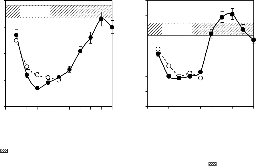
Увеличение массы лимфоузлов и тимуса начина-
лось с 15-х суток, причем более интенсивно – лим-
фоузлов, а в интервале между 30–40-ми сутками
масса лимфоузлов превышала значения ее в
контроле. Масса тимуса возрастала менее интен-
сивно и только к 60-м суткам достигала значений
нормы (рис. 1).
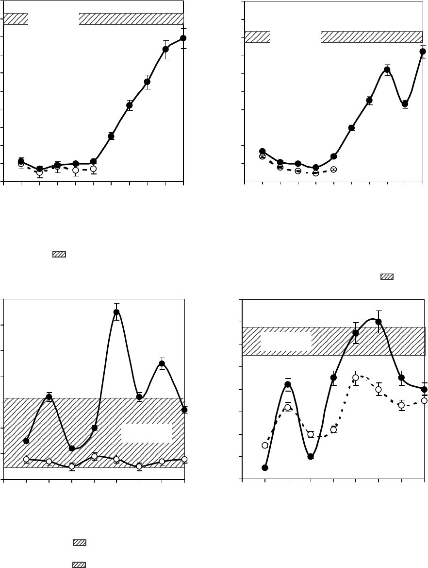
Клеточность и в тимусе, и в лимфатических
узлах у животных 2 группы также начинало возрас-
тать с 15-х суток (более интенсивно – в лимфати-
ческих узлах) и почти достигало значения этого
показателя у интактных животных к 90-м суткам
наблюдения (рис. 2).
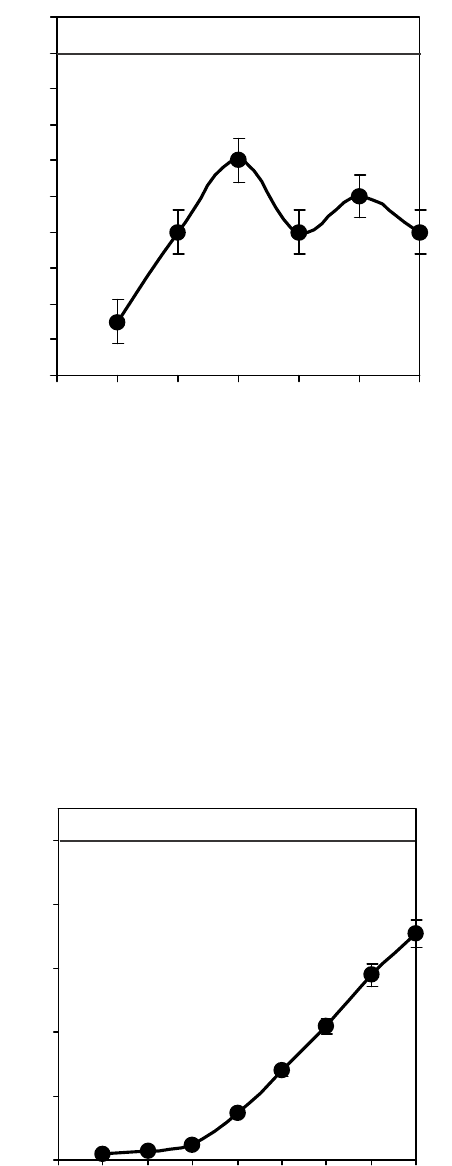
Количество клеток, отвечающих на ФГА, в ти-
мусе достигало уровня их содержания у интактных
животных на 20-е сутки и с незначительными коле-
баниями сохранялось на этом уровне до конца наб-
людения (90 суток). В лимфоузлах количество
over it was more intensive in lymph nodes, but within
the interval of 30–40 days the lymph node mass
exceeded its control values. Thymus mass increase
was less active and only to the 60
th
day achieved the
norm values (Fig. 1).
The cellullarity both in thymus and lymph nodes
in animals of the 2
nd
group started to rise from the 15
th
day (more intensively in lymph nodes) and almost
achieved the values of this index in the intact animals
to the 90
th
observation day (Fig. 2).
The number of cells, responding to PHA in thymus
reached the level of their content in the intact animals
to the 20
th
day and was kept at this level with slight
variation up to the observation end (90 days). In lymph
nodes the number of lymphocytes with positive
response to PHA changed wave-like without reaching
to the 90
th
day their number in the intact animals (Fig.
3, a).
The amount of T-lymphocytes, responding in blast-
formation reaction (BFR) to Con A started its statis-
tically significant increase in lymph nodes and thymus
from the 20
th
day, then it was a wave-like change, but
in lymph nodes the number of functionally active
T-lymphocytes to the 60
th
day did not differ from their
content in the intact animals, but reduced again to the
70
th
-90
th
days. The number of functionally active
T-lymphocytes in thymus was lower, than that in these
cells at all observation stages (Fig. 3, b).
The number of B-lymphocytes in lymph nodes,
responding to BTR to LPS in the groups of experimen-
tal animals began increasing to the 20
th
day and to the
Рис. 1. Изменение массы тимуса (а) и лимфоузлов (б) у летально облученных животных на разных этапах после
трансплантации КСКМ: – летально облученные животные; – летально облученные животные, защищенные
КСКМ; – контроль.
Fig. 1. Change in thymus (a) and lymph nodes (b) mass in lethally irradiated animals at different stages after CSBM cell
transplantation: – lethally irradiated animals; – CSBM protected lethally irradiated animals; – control.
0
1
2
3
4
5
6
7
Сроки наблюдения, сутки
Observation term, days
Масса органа, мг
Organ mass, mg
0 1 2 3 5 7 15 30 40 60 90
0
10
20
30
40
Контроль
Control
Сроки наблюдения, сутки
Observation term, days
Масса органа, мг
Organ mass, mg
0 1 2 3 5 7 15 30 40 60 90
Контроль
Control
б b
Контроль
Control
а aб b
75
лимфоцитов, положительно реагирующих на ФГА,
изменялось волнообразно, не достигая на 90-е
сутки количества у интактных животных (рис. 3, а).
Количество Т-лимфоцитов, отвечающих в реак-
ции бласттрансформации (РБТ) на Кон А, начина-
ло достоверно возрастать в лимфоузлах и тимусе
с 20-х суток, далее – изменялось волнообразно, но
в лимфоузлах количество функционально актив-
PROBLEMS
OF CRYOBIOLOGY
Vol. 19, 2009, ¹1
ÏÐÎÁËÅÌÛ
ÊÐÈÎÁÈÎËÎÃÈÈ
Ò. 19, 2009, ¹1
60
th
one was statistically and significantly lower than
the one of functionally active B-lymphocytes in the
intact animals (Fig. 4).
During post-transplantation period the AFCs
number in spleen of lethally irradiated and CSBM-
protected animals began to augment uniformly from
the 20
th
day, but reduced to the 60
th
one compared to
their content in the intact animals (Fig. 5).
0
10
20
30
40
50
60
70
80
90
100
Сроки наблюдения, сутки
Observation term, days
Количество ядерных клеток, ×10
6
Number of nucleated cells, ×10
6
0 1 2 3 5 7 15 30 40 60 90
Контроль
Control
0
10
20
30
40
50
60
70
80
90
100
Сроки наблюдения, сутки
Observation term, days
Количество ядерных клеток, ×10
6
Number of nucleated cells, ×10
6
0 1 2 3 5 7 15 30 40 60 90
Контроль
Control
Рис. 2. Количество ядерных клеток в тимусе (а) и лимфоузлах (б) ) у летально облученных животных на разных
этапах после трансплантации КСКМ: – летально облученные животные; – летально облученные животные,
защищенные КСКМ; – контроль.
Fig. 2. Number of nucleated cells in thymus (a) and lymph nodes (b) in lethally irradiated animals at different stages after
CSBM cell transplantation: – lethally irradiated animals; – CSBM protected lethally irradiated animals; – control.
0
10
20
30
40
50
60
70
0 1020304050607080
Контроль
Control
Рис. 3. Ответ на ФГА (а) и Кон А (б) клеток тимуса () и лимфоузлов () в зависимости от сроков после облучения
и трансплантации КСКМ. – контроль.
Fig. 3. Response to PHA (a) and Con A (b) of thymus () and lymph node () cells depending on terms after irradiation
and CSBM transplantation; – control.
а aб b
Сроки наблюдения, сутки
Observation term, days
Сроки наблюдения, сутки
Observation term, days
0
10
20
30
40
50
60
70
80
0 1020304050607080
Количество ядерных клеток, ×10
6
Number of nucleated cells, ×10
6
Количество ядерных клеток, ×10
6
Number of nucleated cells, ×10
6
76
PROBLEMS
OF CRYOBIOLOGY
Vol. 19, 2009, ¹1
ÏÐÎÁËÅÌÛ
ÊÐÈÎÁÈÎËÎÃÈÈ
Ò. 19, 2009, ¹1
ных Т-лимфоцитов к 60-м суткам не отличалось
от их содержания у интактных животных, однако
снова снижалось к 70–90-м суткам. В тимусе
количество функционально активных Т-лимфоци-
тов было ниже количества этих клеток на всех
этапах наблюдения (рис. 3, б).
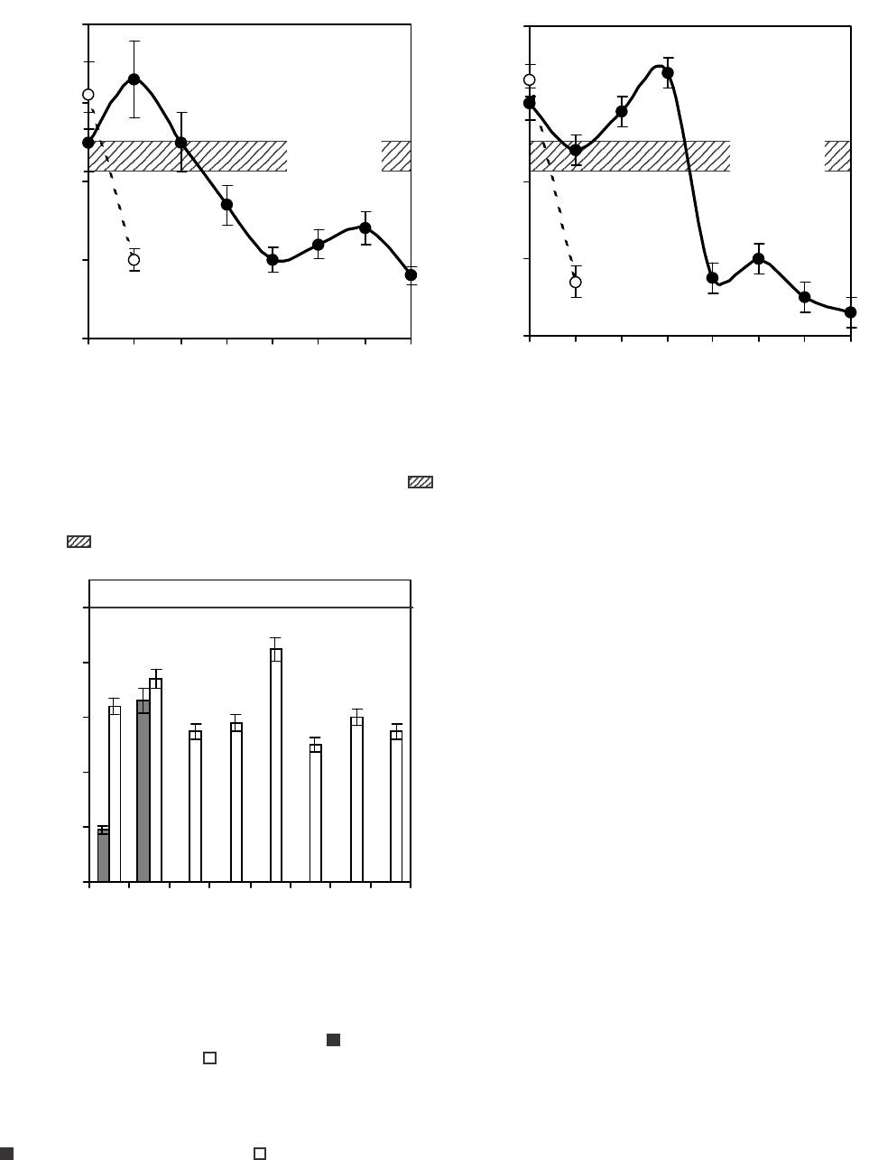
Количество В-лимфоцитов в лимфоузлах, отве-
чающих в РБТ на ЛПС, в группах опытных живот-
ных начинало возрастать на 20-е сутки, а к 60-м
суткам было достоверно ниже количества функ-
ционально активных В-лимфоцитов у интактных
животных (рис. 4).
В посттрансплантационный период количество
АОК в селезенках животных, летально облучен-
ных и защищенных КСКМ, начинало равномерно
повышаться с 20-х суток, но к 60-м суткам было
снижено по сравнению с их содержанием у интакт-
ных животных (рис. 5).
Относительное количество макрофагов в интер-
вале между 7-ми и 20-ми сутками было либо
достоверно выше, либо не отличалось от их содер-
жания у интактных животных, а начиная с 15-х
суток, снижалось по сравнению с их содержанием
у интактных животных (рис. 6, а).
Относительное количество моноцитов в пери-
тонеальной полости животных опытной группы в
первые 20 суток после облучения и транспланта-
ции костного мозга снижалось, затем возрастало
(рис. 6, б).
Количество фагоцитирующих клеток у живот-
ных опытной группы было достоверно ниже на
всех этапах наблюдения по сравнению с их содер-
жанием у интактных животных (рис. 7).
Активность ферментов, определяющих перева-
ривающую активность фагоцитов (неспецифичес-
кая эстераза и кислая фосфатаза) либо не изме-
нялась, либо изменялась волнообразно, достигая
исходного уровня на 60-е сутки (рис. 8).
Далее будут рассмотрены морфофункциональ-
ные свойства нейроэндокринных органов живот-
ных на разных этапах после летального облучения
и после трансплантации им КСКМ.
Гипофиз. У летально облученных животных
морфологические изменения в ткани передней
доли гипофиза наблюдали, начиная с первых суток
после облучения. Возрастало количество секретор-
ных клеток с пикнотичными ядрами и вакуолизи-
рованной цитоплазмой, преобладали клетки не-
больших размеров, сосуды были расширены и
переполнены кровью. Начиная с 3-х суток, отме-
чались увеличение количества ацидофильных кле-
ток и снижение количества базофильных и хромо-
фобных клеток по сравнению с их содержанием у
интактных животных (таблица). К моменту гибели
животных (7–10-е сутки) нарастали дегенератив-
ные изменения в секреторных клетках, большин-
A relative number of macrophages within the
interval between 7 and 20 days was either statistically
and significantly higher or with no difference from
their content in the intact animals, but starting from
the 15
th
day decreased compared to their content in
the intact animals (Fig. 6, a).
0
1
2
3
4
5
6
7
8
9
10
0 102030405060
Сроки наблюдения, сутки
Observation term, days
Индекс стимуляции
Stimulation index
Контроль
Control
Рис. 4. Ответ клеток лимфоузлов на ЛПС в зависимости
от сроков после облучения и трансплантации КСКМ.
Fig. 4. Response of lymph node cells to LPS depending on
terms after irradiation and CSBM cell transplantation.
Рис. 5. Содержание антителообразующих клеток (АОК)
в селезенке летально облученных и защищенных живот-
ных после трансплантации КСКМ.
Fig. 5. Number of antibody-forming cells (AFCs) in spleen
of lethally irradiated and protected animals after CSBM cell
transplantation.
0
20
40
60
80
100
Контроль
Control
Количество АОК, % к контролю
AFCs number, % of the control
0 1 7 15 20 25 30 40 60
Сроки наблюдения, сутки
Observation term, days
77
PROBLEMS
OF CRYOBIOLOGY
Vol. 19, 2009, ¹1
ÏÐÎÁËÅÌÛ
ÊÐÈÎÁÈÎËÎÃÈÈ
Ò. 19, 2009, ¹1
A relative number of monocytes in peritoneal cavity
of experimental group’s animals reduced, then
enhanced during first 20 days after irradiation and bone
marrow transplantation (Fig. 6, b).
The number of phagocytic cells in animals of
experimental group was statistically and significantly
lower at all observation stages compared to their
content in the intact animals (Fig.7).
The enzyme activity, determining a digesting activi-
ty of phagocytes (non-specific esterase and acid
phosphatase) were either unchanged or had the wave-
like changes by achieving the initial level to the 60
th
day (Fig. 8).
Hereinafter we will focus to the morphofunctional
properties of animals neuroendocrine organs at diffe-
rent stages after lethal irradiation and CSBM trans-
plantation.
Hypophysis. In the lethally irradiated animals the
morphological changes in tissue of hypophysis anterior
lobe were observed starting from the first days after
irradiation. The number of secretory cells with pycno-
tic nuclei and vacuolated cytoplasm increased, there
were mostly the cells of small size, the vessels were
extended and congested. Starting from the 3
rd
day we
noted the augmentation of acidophilic cell number and
a decrease in basophilic and chromophobic cells com-
pared to their content in the intact animals (Table). To
the moment of animal death (7–10 days) the degene-
rative changes intensified in secretory cells, the majo-
rity of which destroyed with revealing cell detritus at
their place. The value of nucleus-cytoplasm ratio to
Контроль
Control
0
10
20
30
40
0
10
20
30
40
а a
Сроки наблюдения, сутки
Observation term, days
Количество клеток, %
Cell number, %
2 7 15 20 30 40 50 60
Контроль
Control
Сроки наблюдения, сутки
Observation term, days
Количество клеток, %
Cell number, %
2 7 15 20 30 40 50 60
б b
Рис. 6. Количество макрофагов (а) и моноцитов (б) в перитонеальной полости летально облученных мышей в
зависимости от сроков после облучения и трансплантации КСКМ: – летально облученные животные; – ле-
тально облученные животные, защищенные КСКМ; – контроль.
Fig. 6. Macrophage (a) and monocyte (b) number in peritoneal cavity of lethally irradiated mice depending on terms after
irradiation and CSBM cell transplantation: – lethally irradiated animals; – CSBM protected lethally irradiated
animals; – control.
Рис. 7. Количество фагоцитирующих моноцитарно-
макрофагальных клеток в перитонеальной полости
летально облученных мышей в зависимости от сроков
после облучения и трансплантации КСКМ: – летально
облученные животные; – летально облученные
животные, защищенные КСКМ.
Fig. 7. Number of phagocyte monocyte-macrophage cells
in peritoneal cavity of lethally irradiated mice depending
on terms after irradiation and CSBM cell transplantation:
– lethally irradiated animals; – CSBM protected
lethally irradiated animals.
0
20
40
60
80
100
2 7 15 20 30 40 50 60
Сроки наблюдения, сутки
Observation term, days
СГК, усл. ед
AHS, units
Контроль
Control
ство из них разрушались и на их месте определялся
клеточный детрит. Величина ядерно-цитоплазмен-
ного отношения на первые сутки в базофилах гипо-
а aб b
78
PROBLEMS
OF CRYOBIOLOGY
Vol. 19, 2009, ¹1
ÏÐÎÁËÅÌÛ
ÊÐÈÎÁÈÎËÎÃÈÈ
Ò. 19, 2009, ¹1
физа достоверно снижалась по сравнению с ана-
логичными показателями у интактных животных.
На 3-и сутки у ацидофильных клеток эта величина
достоверно увеличивалась. К 7–10-м суткам вели-
чина ядерно-цитоплазменного отношения у ацидо-
филов и базофилов не достигала ее уровня у ин-
тактных животных.
У летально облученных животных через сутки
после трансплантации КСКМ гистологическое
строение передней доли гипофиза было сравнимо
с таковым у контрольных животных, однако при
этом отмечали увеличение количества ацидофилов
и тенденцию к снижению количества базофильных
и хромофобных клеток. Начиная с 3-х суток, в аде-
ногипофизе увеличивались размеры и кровенапол-
нение синусов, появлялись секреторные клетки с
гиперхромными и пикнотичными ядрами. Увели-
чение количества ацидофильных клеток и умень-
шение количества хромофобных клеток сохраня-
лись на протяжении всего срока наблюдения, а
количество базофильных клеток колебалось от
минимального значения на 10- и 40-е сутки до
максимального − на 20-е сутки, но не превышало
значения этого показателя у интактных животных.
При анализе ядерно-цитоплазменного отношения
во всех секреторных клетках передней доли гипо-
физа выявлялась тенденция к снижению этого по-
казателя на протяжении всего срока наблюдения у
животных 2-й группы с достоверным снижением
по сравнению со значением его у интактных жи-
the first day in hypophysis basophils statistically and
significantly reduced compared to similar indices in
the intact animals. To the 3
rd
day this value statistically
and significantly augmented in acidophilic cells. To
the 7
th
–10
th
days the value of nuclear-cytoplasm ratio
in acidophils and basophils did not achieve its level
in the intact animals.
In the lethally irradiated animals one day after
CSBM transplantation the histological structure of
hypophysis anterior lobe was comparable to that in
the control animals, but at the same time there was
noted an increase in acidophil number and the tenden-
cy to reduction in basophilic and chromophobic cells.
Starting from the 3
rd
day in adenohypophysis there
was an increase in size and blood-filling of sinuses,
the secretory cells with hyperchromic and pycnotic
nuclei appeared. The enhancement of acidophilic cell
number and the reduction of chromophobic cell one
were preserved within all observation term and a
number of basophilic cells varied from the minimum
value by the 10
th
and 40
th
days up to the maximum one
to the 20
th
day, but not exceeded this index for the
intact animals. When analysing the nucleus-cytoplasm
ratio in all secretory cells of hypophysis anterior lobe
there was found out the tendency to this index
reduction within the all observation term in animals
of the 2
nd
group with a statistically significant decrease
compared to its value in the intact animals: to the 3
rd
,
20
th
, 60
th
and 90
th
days in acidophils; to the 7
th
, 10
th
,
20
th
and 60
th
ones in basophils; to the 10
th
and 30
th
0,0
0,5
1,0
1,5
2,0
2,5
2 7 15 20 30 40 50 60
0,0
0,5
1,0
1,5
2,0
2,5
3,0
2 7 15 20 30 40 50 60
Сроки наблюдения, сутки
Observation term, days
СГК, усл. ед
AHS, rel. units
Контроль
Control
Контроль
Control
Сроки наблюдения, сутки
Observation term, days
СГК, усл. ед
AHS, rel. units
Рис. 8. Активность неспецифической эстеразы (а) и кислой фосфатазы (б) в моноцитарно-макрофагальных клетках
перитонеальной полости летально облученных мышей в зависимости от сроков после облучения и трансплантации
КСКМ: – летально облученные животные; – летально облученные животные, защищенные КСКМ; –
контроль; * – статистически достоверные различия с контрольной группой (Р < 0,05).
Fig. 8. Activity of non-specific esterase (a) and acid phosphatase (b) in monocyte-macrophage cells of peritoneal cavity
of lethally irradiated mice depending on terms after irradiation and SCBM cell transplantation: – lethally irradiated ani-
mals; – SCBM protected lethally irradiated animals; – control; * – data with statistically significant differences
with the control group (P < 0.05).
*
*
*
*
*
*
*
*
*
*
*

79
PROBLEMS
OF CRYOBIOLOGY
Vol. 19, 2009, ¹1
ÏÐÎÁËÅÌÛ
ÊÐÈÎÁÈÎËÎÃÈÈ
Ò. 19, 2009, ¹1
вотных: у ацидофилов – на 3-, 20-, 60- и 90-е сутки;
у базофилов – на 7-, 10-, 20- и 60-е сутки; у
хромофобных клеток – на 10- и 30-е сутки. Начиная
с 10-х суток, развивались процессы восстановле-
ния гистологического строения аденогипофиза,
увеличивались размеры секреторных клеток, осо-
бенно на 40-е сутки. К 90-м суткам гистологи-
ческое строение ткани передней доли гипофиза у
летально облученных и защищенных КСКМ жи-
вотных восстанавливалось.
ones in chromophobic cells. From the 10
th
day the
recovery processes of adenohypophysis histological
structure developed and the size of secretory cells
augmented, especially to the 40
th
day. To the 90
th
day
a histological structure of tissue of anterior hypophysis
lobe in lethally irradiated and CSBM-protected ani-
mals recovered.
Thyroid gland. In the lethally irradiated animals
one day after irradiation the size of thyroid gland follic-
les differed by heterogeneity (from average to large
Соотношение количества секреторных клеток в аденогипофизе и их ядерно-цитоплазменное отношение у
летально облученных мышей линии (CBA×C57Вl)F
1
после трансплантации КСКМ
Secretory cell content in adenohypophysis and their nucleus to cytoplasm ratio
in lethally irradiated mice of (CBA×C57B1)F
1
line after CSBM cell transplantation
Примечание: * – достоверные различия данных по сравнению с контролем (P < 0,05).
Notes: * – statistically significant differences comparing to the control data (P < 0,05).
èêîðÑ
,ÿèíåäþëáàí
èêòóñ
,smretnoitavresbO
syad
éåøûìåçèôîïèãîíåäàâêîòåëêûïèÒ
sisyhpopyhonedaenirumnisepytlleC
îâòñå÷èëîK
%,êîòåëê
%,rebmunlleC
åûíüëèôîäèöÀ
cilihpodecA
åûíüëèôîçàÁ
cilihposaB
åûíáîôîìîðÕ
cibohpomorhC
îíðåäß -
åîííåìçàëïîòèö
,åèíåøîíòî
.äå.íòî
otsuelcuN
,oitarmsalpotyc
stinu.ler
îâòñå÷èëîK
%,êîòåëê
%,rebmunlleC
îíðåäß -
åîííåìçàëïîòèö
,åèíåøîíòî
.äå.íòî
otsuelcuN
,oitarmsalpotyc
stinu.ler
îâòñå÷èëîK
%,êîòåëê
%,rebmunlleC
îíðåäß -
åîííåìçàëïîòèö
,åèíåøîíòî
.äå.íòî
otsuelcuN
,oitarmsalpotyc
stinu.ler
)åûíòîâèæåûíòêàòíè(üëîðòíîK
)slaminatcatni(lortnoC
32,1±0,4502,192,0±5,751,121,3±5,8359,0
åèíå÷óëáîåîíüëàòåË
noitaidarrilahteL
113,2±3,9502,113,0±7,415,0 * 71,2±0,7389,0
339,1±1,36 * 07,1 * 71,0±2,536,0 * 83,2±7,1309,0
710,2±2,67 * 65,0 * 15,0±1,646,0 * 72,1±7,71 * 28,0
0190,2±9,67 * 15,0 * 27,0±1,516,0 * 51,1±0,81 * 18,0
MKÑKÿèöàòíàëïñíàðÒ
noitatnalpsnartllecMBSC
171,1±0,36 * 20,171,0±0,560,110,2±0,2329,0
359,1±0,7558,0 * 11,0±9,3 * 90,115,1±1,92 * 08,0
713,1±5,56 * 71,113,0±5,667,0 * 37,1±0,82 * 39,0
0172,1±0,46 * 90,152,0±0,2 * 08,0 * 71,2±0,9205,0 *
0268,1±0,9506,0 * 81,0±0,797,0 * 10,1±7,32 * 09,0
0350,2±8,06 * 99,090,0±9,678,099,2±0,72*56,0
0459,1±7,8510,111,0±2,2 * 59,050,1±6,52 * 19,0
0610,3±8,9508,0 * 60,0±7,3 * 76,0 * 11,1±5,72 * 07,0
0908,1±4,1608,0 * 21,0±9,499,054,1±7,72 * 06,0
