Журнал - Проблемы криобиологии 2009 №1
Подождите немного. Документ загружается.

40
PROBLEMS
OF CRYOBIOLOGY
Vol. 19, 2009, ¹1
ÏÐÎÁËÅÌÛ
ÊÐÈÎÁÈÎËÎÃÈÈ
Ò. 19, 2009, ¹1
Quintanilha A.T., Racker L. Surface localization of sites of
reduction of nitroxide spin-labeled molecules in mitochondria //
Proc. Nat. Acad. Sci. USA.– 1977.– Vol. 74, N2.– P. 570–574.
Sheleg S., Hixon H., Cohen B. et al. Cardiac mitochondrial
membrane stability after deep hypothermia using a xenon
clathrate cryostasis protocol – an electron microscopy
study // Int. J. Clin. Exp. Pathol.– 2008.– Vol. 1, N5.– P. 440–
447.
Steichman L., Avi-Dor I. The effect of osmotic shock on
swelling pattern and respiratory control of rat liver mito-
chondria // Biochem. J.– 1967.– Vol. 104, N1.– P. 71–77.
Поступила 13.01.2009
Рецензент Е.А. Гордиенко
Minetti M., Di Stasi A.M. Involvement of erythrocyte skeletal
proteins in the modulation of membrane fluidity by phenothia-
zines // Biochemistry.– 1987.– Vol. 26, N25.– P. 8133–8137.
Mitchell P. Protonmotive chemiosmotic mechanism in oxidative
and photosynthetic phosphorylation // Trends Biochem. Sci.–
1978.– Vol. 31, N3.– P. 60–61.
Quintanilha A.T., Racker L. Surface localization of sites of
reduction of nitroxide spin-labeled molecules in mitochondria //
Proc. Nat. Acad. Sci. USA.– 1977.– Vol. 74, N2.– P. 570–574.
Sheleg S., Hixon H., Cohen B. et al. Cardiac mitochondrial
membrane stability after deep hypothermia using a xenon
clathrate cryostasis protocol – an electron microscopy
study // Int. J. Clin. Exp. Pathol.– 2008.– Vol. 1, N5.– P. 440–
447.
Steichman L., Avi-Dor I. The effect of osmotic shock on
swelling pattern and respiratory control of rat liver mito-
chondria // Biochem. J.– 1967.– Vol. 104, N1.– P. 71–77.
Accepted in 13.01.2009
23.
24.
25.
21.
22.
23.
24.
25.

41
PROBLEMS
OF CRYOBIOLOGY
Vol. 19, 2009, ¹1
ÏÐÎÁËÅÌÛ
ÊÐÈÎÁÈÎËÎÃÈÈ
Ò. 19, 2009, ¹1
УДК 57.043:577.352.4
Î.Â. ÑÀÊÓÍ, Â.Â. ÌÀÐÓÙÅÍÊÎ, ².Ô. ÊÎÂÀËÅÍÊÎ,
À.Þ. ѲÐÅÍÊÎ, ².Ï. ÂÈÑÅÊÀÍÖÅÂ, Î.Â. ÄÀÂÈÄÎÂÀ, Î.². ÃÎÐIJªÍÊÎ*
Âïëèâ òåìïåðàòóðè íà êîåô³ö³ºíòè ïðîíèêíîñò³ ìåìáðàí äð³æäæ³â
Saccharomyces cerevisiae äëÿ âîäè ³ êð³îïðîòåêòîð³â
UDC 57.043:577.352.4
O.V. SAKUN, V.V. MARUSCHENKO, I.F. KOVALENKO,
A.YU. SIRENKO, I.P. VYSEKANTSEV, O.V. DAVYDOVA, O.I. GORDIYENKO*
Temperature Effect on Membrane Permeability Coefficients
of Yeast-Like Fungi Saccharomyces cerevisiae
for Water and Cryoprotectant s
Визначені біофізичні параметри клітин дріжджів Saccharomyces cerevisiae: осмотично неактивний об’єм, коефіцієнт
фільтрації в середовищах з кріопротекторами (гліцерин, 1,2-пропандіол та диметилсульфоксид) та коефіцієнти проникності
для цих кріопротекторів при температурах 25 та 10°С. Для визначення вказаних параметрів використовували удосконалені
рівняння масопереносу крізь мембрани клітин, отримані у наближенні лінійної термодинаміки необоротних процесів.
Розраховані величини енергії активації проникання молекул води та кріопротекторів крізь клітинні мембрани.
Ключові слова: Saccharomyces cerevisiae, проникність, кріопротектори, енергія активації.
Определены биофизические параметры клеток дрожжей Saccharomyces cerevisiae: осмотически неактивный объем,
коэффициент фильтрации в средах с криопротекторами (глицерин, 1,2-пропандиол, диметилсульфоксид) и коэффициенты
проницаемости для этих криопротекторов при температурах 25 и 10°С. Для определения указанных параметров использовали
усовершенствованные уравнения массопереноса через мембраны клеток, полученные в приближении линейной термодинамики
необратимых процессов. Рассчитаны величины энергии активации проникновения молекул воды и криопротекторов через
клеточные мембраны.
Ключевые слова: Saccharomyces cerevisiae, проницаемость, криопротекторы, энергия активации.
The biophysical parameters of yeast cell Saccharomyces cerevisiae, osmotically inactive volume, filtration coefficients in the
cryoprotectant containing media and permeability coefficients for the cryoprotectants glycerol, 1,2-propane diol, dimethyl sulfoxide,
were determined at the temperatures of 25 and 10°C. For determination of the parameters the improved equations of mass transfer via
cell membranes were used. The equations are based on the approach of linear thermodynamics of irreversible processes. The values
of activation energy of water and cryoprotectants molecules penetration via cell membranes were calculated.
Keywords: Saccharomyces cerevisiae, permeability, cryoprotectants, activation energy.
* Àâòîð, ÿêîìó íåîáõ³äíî íàïðàâëÿòè êîðåñïîíäåíö³þ:
âóë. Ïåðåÿñëàâñüêà, 23, ì. Õàðê³â, Óêðà¿íà 61015; òåë.:+38 (057)
373-38-71, ôàêñ: +38 (057) 373-30-84, åëåêòðîííà ïîøòà:
gordienko@gala.net
* To whom correspondence should be addressed: 23, Pereyaslavskaya
str., Kharkov, Ukraine 61015; tel.:+380 57 373 3871, fax: +380 57
373 3084, e-mail: gordienko@gala.net
Institute for Problems of Cryobiology and Cryomedicine of the Na-
tional Academy of Sciences of Ukraine, Kharkov, Ukraine
²íñòèòóò ïðîáëåì êð³îá³îëî㳿 ³ êð³îìåäèöèíè
ÍÀÍ Óêðà¿íè, ì. Õàðê³â
Найбільш поширеним способом підтримки
мікроорганізмів є періодичні їх пересіви на свіже
живильне середовище [1, 2]. Проте проблема
ефективної за кінцевими результатами, а також
обґрунтованої за трудомісткістю і вартістю кон-
сервації культур мікроорганізмів обумовлена
збільшенням кількості та різноманітністю культур,
що зберігаються [5]. Кріоконсервування клітинних
суспензій, зокрема мікроорганізмів, залишається
основним методом їх тривалого збереження без
можливої зміни генетичного складу. Правильний
вибір режимів низькотемпературного консерву-
вання дозволяє тривалий час підтримувати мікро-
організми у стані, що забезпечує їх повернення до
життєдіяльності без зміни початкових власти-
востей і зниження кількості життєздатних клітин.
Оптимальними режимами заморожування для
The most widely spread way to support microorga-
nism culture is their periodic replating on a fresh nut-
rient medium [1, 2]. However the problem of micro-
organism culture preservation to be efficient by the
final results, as well substantiated by labour intensity
and cost, is stipulated by the augmentation of quantity
and variety of cultures under preservation [5]. The
cryopreservation of cell suspensions, namely micro-
organisms, remains the principal method for their long-
term preservation without any possible changes in ge-
netic composition. Correct selection of low temperatu-
re preservation regimens enables a long-term support
of microorganism in the state, providing their return
to vital activity without any changes in the initial
properties and a decrease in viable cell number. Either
slow freezing rates (1–10°C/min) or two-step freezing
programs with a slow freezing at the first stage and
42
PROBLEMS
OF CRYOBIOLOGY
Vol. 19, 2009, ¹1
ÏÐÎÁËÅÌÛ
ÊÐÈÎÁÈÎËÎÃÈÈ
Ò. 19, 2009, ¹1
дріжджових клітин вважають повільні швидкості
заморожування (1–10°С/хв) або двохетапні про-
грами заморожування з повільним заморожуван-
ням на першому етапі і наступним зануренням у
скраплений азот [11, 12, 14]. В роботі [14] показано,
що виживання клітин, зокрема дріжджів, залежить
також від швидкості відігрівання. При швидкому
відігріванні (500°С/хв) знайдено два максимуми
виживання: при повільній швидкості охолодження
(від 1до 10°С/хв) та при надшвидкому охолодженні
(∼10
4
°С/хв) [9].
Існування оптимальної швидкості охолодження
було пояснено в межах двохфакторної теорії
Мейзура [15]. При різній швидкості охолодження
змінюється внесок двох конкуруючих типів пошко-
джень, пов’язаних із збезводненням клітин, з одно-
го боку, та внутрішньоклітинною кристалізацією,
з іншого. Зі збільшенням збезводнення клітини
зменшується величина ефективного переохоло-
дження протоплазми та імовірність утворення
кристалів усередині клітини. Як надмірне збезвод-
нення, так і внутрішньоклітинна кристалізація
приводять до пошкодження клітинних структур
[3]. Отже, для кожного типу клітин існує оптималь-
на (забезпечує максимальну збереженість) швид-
кість охолодження, що прямо пов’язана зі швидкіс-
тю, з якою внутрішньоклітинна вода може вихо-
дити з клітин, тобто з коефіцієнтом проникності
клітинної мембрани для води. Оскільки проник-
ність для молекул води мембран різних видів
клітин може відрізнятись на декілька порядків, то
і значення оптимальної швидкості охолодження
для цих клітин суттєво відрізняються. Так, наприк-
лад, для еритроцитів людини оптимальна швид-
кість охолодження становить біля 3000°С/хв, а
ооцитів миші – 0,1°С/хв. Надзвичайно високе зна-
чення оптимальної швидкості охолодження для
еритроцитів людини з погляду двохфакторної тео-
рії пояснюється дуже високою проникністю їх
мембран для води і великим поверхнево-об’ємним
відношенням [3].
Додавання кріопротектора до клітинної суспен-
зії, як правило, приводить до зсуву оптимальної
швидкості охолодження у бік менших значень. Це
пов’язано зі здатністю кріопротекторів утрудню-
вати процеси утворення та росту кристалів, досяг-
нення клітиною мінімального об’єму. Важливого
значення при цьому набувають здатність кріо-
протектора проникати в клітини та швидкість
проникання. Отже, іншим суттєвим кріобіологіч-
ним параметром клітинної мембрани є її проник-
ність для кріопротекторів.
Однією з важливих характеристик транспорт-
них процесів є їх залежність від температури.
Вплив температури на константи швидкостей, що
характеризують хімічні або біологічні процеси,
часто аналізують в термінах емпіричної енергії ак-
following immersion into a liquid nitrogen are consi-
dered as the optimal freezing regimens for yeast cells
[11, 12, 14]. As reported [14], the survival of cells,
especially yeast, is also dependent on thawing rate.
Under rapid thawing (500°C/min) there were found
out the two survival maxima: under slow cooling rate
(from 1 to 10°C/min) and ultrarapid cooling one
(~10
4
°C/min) [9].
The existence of optimal cooling rate was explai-
ned within the frames of Mazur’s two-factor theory
[15]. The contribution of two competitive types of da-
mages, associated to cell dehydration from one side,
and intracellular crystallisation from another one,
changes under different cooling rates. With increasing
cell dehydration, there are reduced the value of proto-
plasm efficient overcooling and statistically signifi-
cance of crystal formation inside cell. Both excessive
dehydration and intracellular crystallisation result in
cell structure damaging [3]. Thus, for each cell type
there is the optimal (providing maximum preservation)
cooling rate, directly associated to the one, with which
intracellular water may release from cells, i. e. with
cell membrane permeability coefficient for water.
Since the water molecules’ permeability through mem-
branes of different cell types may be several order
different, the values of optimal cooling rate for cells
are significantly distinguished. Thus, for human
erythrocytes and murine oocytes the optimal cooling
rate is about 3,000 and 0.1°C/min, correspondingly.
Ultrahigh value of optimal cooling rate for human
erythrocytes from the point of view of two-factor
theory is explained by a very high permeability of their
membranes for water and a high surface-volume ratio
[3].
Addition of cryoprotectant to cell suspensions
generally results in a shift of optimal cooling rate
towards lower values. This is associated to the
capability of cryoprotectants to complicate the proces-
ses of crystal formation and growth, as well the
reaching the minimum volume by a cell. At the same
time of importance are the capability of cryoprotectant
to penetrate into cells and the penetration rate. Thus,
another essential cryobiological parameter of cell
membrane is its penetration for cryoprotectants.
One of the important characteristics of transport
processes is their dependence on temperature. Tempe-
rature effect on rate constants, characterising either
chemical or biological processes, are often analysed
in terms of empirical activation energy (E
A
). Penetra-
tion of water and dissolved substances through the
artificial and natural membranes by the different,
structurally stipulated ways, is characterised by the
distinctive values of visible activation energy. The va-
lues of activation energy within 12–25 kJ/mol range,
corresponding to those of diffusive activation energy
in a volume solution, are generally consistent with the
substance penetration by a channel mechanism. During
43
PROBLEMS
OF CRYOBIOLOGY
Vol. 19, 2009, ¹1
ÏÐÎÁËÅÌÛ
ÊÐÈÎÁÈÎËÎÃÈÈ
Ò. 19, 2009, ¹1
тивації (Е
A
). Проникання води і розчинених речо-
вин крізь штучні та природні мембрани різними
структурно обумовленими шляхами характери-
зуються відмінними значеннями видимої енергії
активації. Прониканню речовин за канальним ме-
ханізмом відповідають, як правило, значення енер-
гії активації в діапазоні 12–25 кДж/моль, які узго-
джуються з величинами енергії активації дифузії
в об’ємному розчині. При прониканні молекул
шляхом розчинення та дифузії в ліпідному матрик-
сі енергія активації набуває значень 30–85 кДж/моль
і навіть більших [16, 18]. Тобто визначення вели-
чин енергії активації проникності для молекул різ-
них речовин є можливістю з’ясувати механізми їх
проникання крізь плазматичні мембрани конкрет-
них клітин. В той же час, для описання процесів,
що відбуваються з клітиною впродовж охолоджен-
ня, важливо враховувати залежність різних пара-
метрів, зокрема проникності плазматичних мем-
бран для молекул води та кріопротекторів, від тем-
ператури.
Мета роботи – визначення коефіцієнтів проник-
ності клітин дріжджів Saccharomyces cerevisiae для
молекул води і кріопротекторів та величин енергії
активації процесу їх проникання.
Ìàòåð³àëè ³ ìåòîäè
Клітини дріжджів S. cerevisiae вирощували на
скошеному сусло-агаровому середовищі при 22°С
впродовж 48 годин. Культура дріжджів на цьому
етапі знаходиться у стаціонарній фазі росту. Клі-
тини змивали з агарової підложи фізіологічним
розчином та використовували в експерименті.
Коефіцієнт фільтрації та коефіцієнти проник-
ності мембран дріжджів для кріопротекторів
визначали вольюмометричним методом. Клітини
вміщували у бінарний розчин (0,15 М NaCl, 1М
кріопротектор), об’єм якого на порядок перевищу-
вав початковий об’єм клітинної суспензії. Дослі-
джували кінетику зміни розмірів клітин у розчинах
трьох кріопротекторів (гліцерин, 1,2-пропандіол
(1,2-ПД), диметилсульфоксид (ДМСО)) при темпе-
ратурах 25 та 10°С за допомогою мікроскопа
Axio Observer Z1 (Carl Zeiss, Німеччина) з викорис-
танням масляно-імерсійного об’єктива ×63. Об’єм
клітин апроксимували об’ємом розтягнутого еліп-
соїда обертання. Лінійні розміри клітин (довжину
великої та малої осі еліпсоїда) у різних часових
точках визначали за допомогою програми Axio
Vision Rel. 4.6 (Carl Zeiss, Німеччина). Експеримен-
тально визначені часові залежності об’єму клітин
при їх контакті з гіпертонічними розчинами кріо-
протекторів апроксимували чисельними рішен-
нями системи нелінійних рівнянь, що описують цю
залежність у наближенні лінійної термодинаміки
необоротних процесів [3].
molecule penetration via dissolution and diffusion in
a lipid matrix, the activation energy gets values of 30–
85 kJ/mol and even higher [16, 18]. So, the determi-
ning of activation energy values of permeability for
molecules of different substances is the possibility to
reveal the mechanisms of their penetration through
plasma membranes of specific cells. At the same time,
in order to describe the processes, occurring with a
cell during cooling, of importance is to take into ac-
count the dependency of different parameters on a tem-
perature, especially permeability of plasma membra-
nes for molecules of water and cryoprotectants.
The research was aimed to determine the permea-
bility coefficients for Saccharomyces cerevisiae yeast
cells for water and cryoprotectant molecules and the
activation energy values of their penetration process.
Materials and methods
The S. cerevisiae yeast cells were cultured on wort-
agar slope at 22°C for 48 hrs. Yeast culture at this
stage is in a stationary growth phase. Cells were
washed from an agar substrate with physiological
solution and used in the experiment.
Filtration coefficient and those of yeast membrane
permeability for cryoprotectants were volumomet-
rically determined. Cells were placed in a binary solu-
tion (0.15 M NaCl, 1M cryoprotectant), which volume
was higher order than initial one of cell suspension.
The kinetics of cell size change in three cryoprotective
solutions (glycerol, 1,2-propane diol (1,2-PD), dime-
thyl sulfoxide (DMSO)) at 25 and 10°C using Axio
Observer Z1 (Carl Zeiss, Germany) oil-immersion
lens, ×63, was under study. Cell volume was approxi-
mated by the prolate spheroidal volume. Linear dimen-
sion of cells (transversal line and minor axis lengths)
in different time points were determined using the Axio
Vision Rel. 4.6 (Carl Zeiss) software. The experimen-
tally defined time dependencies of cell volume during
contact with hypertonic cryoprotective solutions were
approximated by computational solutions of the
system of nonlinear equations, describing this depen-
dency in approximating linear thermodynamics of
irreversible processes [3].
Values of activation energy E
A
of water and cryo-
protective molecule transfer were determined by the
slope of plot In k vs. 1/T (Arrhenius plot), where k is
the constant of process rate (in our case K is
permeability coefficient or L
p
is filtration coefficient).
Data were statistically processed using MS Excell
software and Student’s t-criterion.
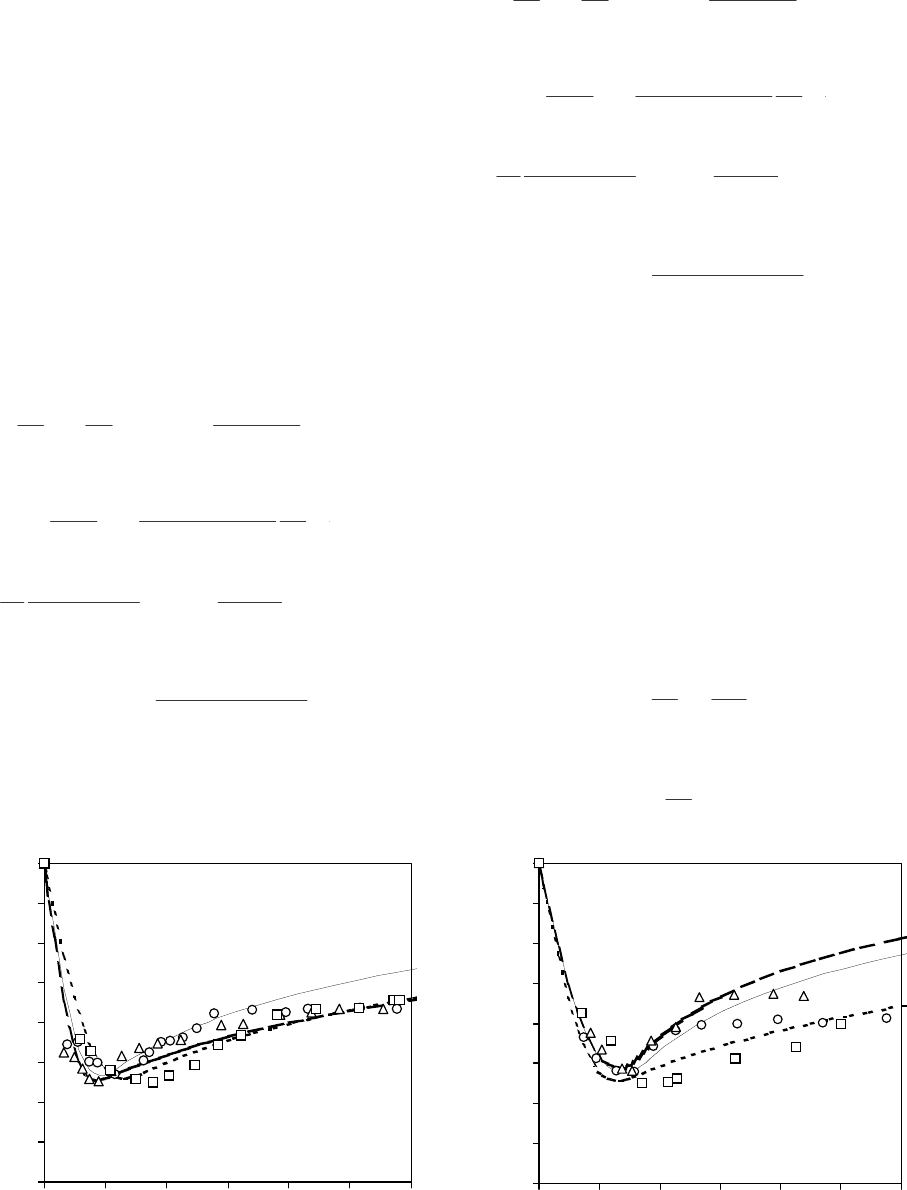
Results and discussion
The coefficients L
p
and K of yeast membranes for
cryoprotectants were obtained by optimal approxi-
mation of experimental points by theoretic curves, i.e.
equation system solutions [3] (Figure):
0,2
0,3
0,4
0,5
0,6
0,7
0,8
0,9
1
0 100 200 300 400 500 600
a a
Час, хв Time, min
Відносний об’єм клітини V/V
0
Normalized cell volume V/V
0
б bЧас, хв Time, min
0,2
0,3
0,4
0,5
0,6
0,7
0,8
0,9
1
0 50 100 150 200 250 300
Відносний об’єм клітини V/V
0
Normalized cell volume V/V
0
44
PROBLEMS
OF CRYOBIOLOGY
Vol. 19, 2009, ¹1
ÏÐÎÁËÅÌÛ
ÊÐÈÎÁÈÎËÎÃÈÈ
Ò. 19, 2009, ¹1
Величини енергії активації (E
A
) процесів пере-
носу молекул води та кріопротекторів визначали з
нахилу графіків ln k від 1/T (графік Ареніуса), де k
є константа швидкості процесу (в нашому випадку
K – коефіцієнт проникності або L
p
– коефіцієнт
фільтрації).
Статистичну обробку результатів здійснювали
за допомогою програмного забезпечення MS Excel
з використанням t-критерію Стьюдента.
Ðåçóëüòàòè òà îáãîâîðåííÿ
Коефіцієнти L
p
і K мембран дріжджів для кріо-
протекторів отримували шляхом оптимальної
апроксимації експериментальних точок теоретич-
ними кривими, тобто рішеннями системи рівнянь
[3] (рисунок):
∆
+
+
+∆−=
∑
≠=
n
swkk
k
in
s
in
ss
ss
w
dt
dy
),(1
ˆ
ˆ
1
ˆ
1
ˆ
1
π
π
πσ
πσ
τ
;
+
−
+
−=
in
s
in
ss
in
s
dt
dy
ydt
d
)(
ˆ
)
ˆ
1(
ˆ
τ
α
ππσπ
∆
+
+∆
−
+
+
∑
≠=
n
swkk
k
in
s
in
s
s
in
s
in
s
s
y
),(1
ˆ
ˆ
1
ˆ
ˆ
)(
ˆ
)
ˆ
1(
1
π
π
π
π
α
ππ
τ
;
))(
ˆ
1(
)1)(
ˆ
1(
ˆˆ
0
0
απ
απ
ππ
−+
−+
=
y
in
s
in
s
in
k
in
k
,
де
in
s
π
і
in
k
π
– осмотичний тиск проникаючої і не
проникаючої крізь клітинну мембрану внутрі-
∆
+
+
+∆−=
∑
≠=
n
swkk
k
in
s
in
ss
ss
w
dt
dy
),(1
ˆ
ˆ
1
ˆ
1
ˆ
1
π
π
πσ
πσ
τ
;
+
−
+
−=
in
s
in
ss
in
s
dt
dy
ydt
d
)(
ˆ
)
ˆ
1(
ˆ
τ
α
ππσπ
∆
+
+∆
−
+
+
∑
≠=
n
swkk
k
in
s
in
s
s
in
s
in
s
s
y
),(1
ˆ
ˆ
1
ˆ
ˆ
)(
ˆ
)
ˆ
1(
1
π
π
π
π
α
ππ
τ
;
))(
ˆ
1(
)1)(
ˆ
1(
ˆˆ
0
0
απ
απ
ππ
−+
−+
=
y
in
s
in
s
in
k
in
k
,
where
in
s
π
and
in
k
π
are osmotic pressures of penetra-
tive and non-penetrative through cell membrane
intracellular substances, correspondingly;
in
s0
π
and
in
k 0
π
are the initial values; t is time;
out
s
π
and
out
k
π
are
the osmotic pressures of penetrative and non-penetra-
tive through cell membrane extracellular substances,
correspondingly; V is cell volume; V
0
is initial value
of cell volume; y = V/V
0
is a relative cell volume;
σ
s
is
reflection coefficient of cell membrane to a dissolved
substance, peneteating through it;
α
is a volume part
of non-penetrative through cell membrane substances
inside a cell;
1
0
−
=
s
pw
RT
L
V
S
υ
τ
,
1
0
−
=
ss
k
V
S
τ
–
Експериментальні (точки) та теоретичні (лінії) залежності відносного об’єму клітин дріжджів Saccharomyces
cerevisiae від часу в гіпертонічних розчинах кріопротекторів при температурах 25 та 10°С: – гліцерин; –
1,2-ПД; – ДМСО.
Experimental (points) and theoretical (lines) dependencies of a relative volume of S. cerevisiae yeast cells on time in
hypertonic solutions of cryoprotectants at 25 (a) and 10°C (b): – glycerol; – 1,2-PD; – DMSO.

45
шньоклітинних речовин відповідно;
in
s0
π
і
in
k 0
π
–
початкові значення цих величин;
τ
– час;
out
s
π
і
out
k
π
– осмотичний тиск проникаючої і не прони-
каючої крізь клітинну мембрану позаклітинних ре-
човин відповідно; V – об’єм клітини; V
0
– початкове
значення об’єму клітини, y = V/V
0
– відносний
об’єм клітини;
σ
s
– коефіцієнт відбиття клітинної
мембрани до проникаючої крізь неї розчиненої
речовини;
α
– об’ємна частка не проникаючих
крізь клітинну мембрану речовин усередині клі-
тини;
1
0
−
=
s
pw
RT
L
V
S
υ
τ
,
1
0
−
=
ss
k
V
S
τ
–
величини, що мають розмірність часу; S – площа
поверхні клітинної мембрани; L
p
– коефіцієнт
фільтрації клітинної мембрани; R – універсальна
газова константа; T – абсолютна температура;
υ
s
–
парціальний молярний об’єм проникаючої крізь
мембрани клітин розчиненої речовини; k
s
– кое-
фіцієнт проникності клітинної мембрани до прони-
каючої крізь неї розчиненої речовини;
RT
sk
k
υπ
π
=
ˆ
–
приведений осмотичний тиск k-ї розчиненої
речовини;
s
π
ˆ
∆ i
i
π
ˆ
∆ – трансмембранний перепад
приведеного осмотичного тиску проникаючої та
не проникаючої крізь клітинну мембрану речовин
відповідно; індекси s і w позначають величини, які
відносяться до розчиненої речовини і розчинника.
У попередній роботі [6] ми визначили осмотич-
но неактивний об’єм
α
: експериментальні дані
залежності асимптотичного (за t >>
τ
w
) відносного
об’єму клітин дріжджів S. cerevisiae y
∞
від обер-
неного осмотичного тиску розчину хлориду натрію
апроксимували методом найменших квадратів
рівнянням
xy
out
k
in
k
)1(
ˆ
)1(
ˆ
0
αα
π
απ
α
−+=
−
+=
∞
,
де
out
k
in
k
x
π
π
ˆ
ˆ
0
= .
Отримане значення величини осмотично неак-
тивного об’єму становило
α
= 0,27. Значення кое-
фіцієнта фільтрації у середовищах з різними кріо-
are the values with time dimension; S is surface area
of cell membrane; L
p
is the cell membrane filtration
coefficient; R is universal gas constant; T is absolute
temperature;
υ
s
is a partial molar volume of dissolved
substance, penetrating through cell membranes; k
s
is
the permeability coefficient of cell membrane to a
dissolved substance, penetrating through it;
RT
sk
k
υπ
π
=
ˆ
is the normalised osmotic pressure of the k-th dissolved
substance;
s
π
ˆ
∆ and
i
π
ˆ
∆ are the transmembrane fall
of the normalised osmotic pressure of penetrative and
non-penetrative through cell membrane substance,
correspondingly; s and w indices designate the values,
related to a dissolved substance and a solvent.
In the previous research [6] we have determined
an osmotically inactive volume
α
: experimental data
of dependency of asymptotic (t >>
τ
w
) relative volume,
y
∞
,
of S. cerevisiae yeast cells on a reverse osmotic
pressure of sodium chloride solution were approxima-
ted using the least square method by the equation:
xy
out
k
in
k
)1(
ˆ
)1(
ˆ
0
αα
π
απ
α
−+=
−
+=
∞
,
where
out
k
in
k
x
π
π
ˆ
ˆ
0
= .
The obtained value of osmotically inactive volume
was
α
= 0.27. The filtration coefficient value in the
media with different cryoprotectants and the calculated
values of activation energy of water molecule pene-
tration through yeast cell membranes in these media
are presented in the Table 1, the permeability coef-
ficients for cryoprotectants and the activation energy
values are done in the Table 2.
The determined values for filtration coefficients
are in a reasonable agreement with those reported [13].
As the result showed, the values of filtration coef-
ficient in the media with glycerol and DMSO were
practically consistent and statistically and significantly
lower, than in 1,2-PD containing medium. This result
was obtained by the both studied temperatures. The
energy of activation process of water molecule trans-
membrane transfer is also the same in two first cases
and twice lower in the medium with 1,2-PD. This
testifies to its possible effect on cell membranes,
resulting in increasing such important characteristic,
as filtration coefficient. The 1,2-PD effect on plasma
membrane state was noted for murine oocytes as well
[10].
The permeability coefficients for all the studied
cryoprotectants are not statistically and significantly
PROBLEMS
OF CRYOBIOLOGY
Vol. 19, 2009, ¹1
ÏÐÎÁËÅÌÛ
ÊÐÈÎÁÈÎËÎÃÈÈ
Ò. 19, 2009, ¹1

46
PROBLEMS
OF CRYOBIOLOGY
Vol. 19, 2009, ¹1
ÏÐÎÁËÅÌÛ
ÊÐÈÎÁÈÎËÎÃÈÈ
Ò. 19, 2009, ¹1
протекторами і розраховані величини енергії акти-
вації проникання молекул води крізь мембрани клі-
тин дріжджів в цих середовищах подані в табл. 1,
коефіцієнти проникності для кріопротекторів та
величини енергії активації їх проникання – у
табл. 2.
Визначені величини коефіцієнтів фільтрації
задовільно узгоджуються з [13]. Результати пока-
зали, що значення коефіцієнта фільтрації, визна-
чені в середовищах з гліцерином та ДМСО, прак-
тично збігаються і є вірогідно меншими, ніж в
середовищі з 1,2-ПД. Такий результат отримано за
обох досліджених температур. Енергія активації
процесу трансмембранного переносу молекул води
також збігається у двох перших випадках і у 2 рази
менша в середовищі з 1,2-ПД. Це свідчить про
можливу його негативну дію на мембрани клітин,
що призводить до збільшення такої важливої
характеристики, як коефіцієнт фільтрації. Вплив
1,2-ПД на стан плазматичних мембран був відмі-
чений також і для ооцитів миші [10].
Коефіцієнти проникності для всіх досліджених
кріопротекторів вірогідно не відрізняються. Якщо
порівнювати проникність мембран дріжджів з
проникністю мембран еритроцитів людини, то у
останніх проникність для гліцерину значно менша,
ніж проникність для 1,2-ПД та ДМСО. Така
різниця для еритроцитів людини обумовлена
неоднаковими розмірами молекул та ступенем їх
гідрофобності або гідрофільності. Показано, що
гліцерин не проникає крізь мембрани еритроцитів
людини гідрофільними каналами, які є шляхом
проникання для молекул води [4]. Проте відомо,
що гліцерин – суб’єкт метаболізму дріжджових
клітин, мембрани яких мають спеціалізовані кана-
different. If comparing the permeability of yeast mem-
branes with that for human erythrocyte membranes,
the latter has much lower permeability for glycerol,
than for 1,2-PD and DMSO. This difference for human
erythrocytes is stipulated by unequal sizes of
molecules and the extent of their hydrophobicity or
hydrophilicity. Glycerol was shown as non-penetrative
through human erythrocyte membranes via hydrofoil
channels, being the penetration ways for water
molecules [4]. However, glycerol is known as the
subject of yeast cell metabolism, which membranes
have special channel for glycerol and water transport
[8, 17, 19]. The molecule of 1,2-PD differs from
glycerol one by the presence of methyl group instead
of third hydroxyl group in glycerol molecule. There-
fore 1,2-PD molecule has a higher hydrophobicity,
affecting the distribution coefficient of this substance
between hydrophobic phase and water, which is
several time higher than the one for glycerol (0.076
and 0.005, correspondingly). At the same time the sizes
of 1,2-PD molecule are much lower compared to
glycerol ones [4]. Thus, 1,2-PD may penetrate through
the yeast cell membranes, via the same channels as
glycerol. However, the physical and chemical
properties of 1,2-PD molecules, being different from
those for glycerol, may affect the state of glycerol
channels and their penetration for other molecules,
especially water. The existence of this effect confirms
a significant reduction of the value of activation energy
of water molecule penetration in the medium with
1,2-PD. At the same time, the values of activation
energy for glycerol and 1,2-PD molecule penetration
are not statistically and significantly different, mean-
while for DMSO molecules this parameter is statis-
tically and significantly higher.
The hydrophobicity of DMSO molecules is much
higher compared to both glycerol and 1,2-PD ones.
àùèâîäåðåñäàëêÑ
noitisopmocmuideM
L
p
× 01
41
ì,
3
ñ·í/
L
p
× 01
41
m,
3
s·N/
E
A
,
üëîì/æÄê
E
A
,
lom/Jk
C°01C°52
lCaN-àäîâ-íèðåöiëÃ
lCaN-retaw-lorecylG
21,0±35,081,0±9,06,42
2,1 - lCaN-àäîâ-ÄÏ
2,1 - lCaN-retaw-DP
1,0±97,041,0±30,14,21
lCaN-àäîâ-ÎÑÌÄ
lCaN-retaw-OSMD
80.0±45,02,0±29,05,42
Таблиця 1. Коефіцієнти фільтрації клітинних
мембран дріжджів S. cerevisiae і енергія активації
процесу переносу води крізь них, визначені
в середовищах різного складу (M±m)
Table 1. Filtration coefficients of S. cerevisiae yeast cell
membranes and activation energy of water transfer
through them are determined in the media with different
compositions (M±m)
Таблиця 2. Коефіцієнти проникності для
кріопротекторів і енергія активації процесу
їх переносу крізь клітинні мембрани
дріжджів S. cerevisiae (M±m)
Table 2. Permeability coefficients for cryoprotectants
and activation energy of their transfer through
cell membranes of S. cerevisiae yeast (M±m)
àíèâî÷åÐ
ecnatsbuS
K× 01
8
ñ/ì,
K× 01
8
s/m,
E
A
,
üëîì/æÄê
E
A
,
lom/Jk
C°01C°52
íèðåöiëÃ
lorecylG
1,0±24,021,0±47,032,62
2,1 - ÄÏ
2,1 - DP
31,0±4,02,0±7,057,52
ÎÑÌÄ
OSMD
70,0±93,04,0±88,07,73
47
PROBLEMS
OF CRYOBIOLOGY
Vol. 19, 2009, ¹1
ÏÐÎÁËÅÌÛ
ÊÐÈÎÁÈÎËÎÃÈÈ
Ò. 19, 2009, ¹1
ли транспорту гліцерину і води [8, 17, 19]. Моле-
кула 1,2-ПД відрізняється від молекули гліцерину
наявністю метильної групи замість третьої гідро-
ксильної групи в молекулі гліцерину. Тому молеку-
ла 1,2-ПД більш гідрофобна, що впливає на
коефіцієнт розподілу цієї речовини між гідрофоб-
ною фазою і водою, який на порядок перевищує
такий для гліцерину (0,076 и 0,005 відповідно). В
той же час розміри молекули 1,2-ПД є значно мен-
шими порівняно з молекулами гліцерину [4]. Отже,
1,2-ПД може проникати крізь мембрани дріж-
джових клітин тими ж каналами, що і гліцерин.
Проте фізико-хімічні властивості молекул 1,2-ПД,
які відрізняються від таких для гліцерину, можуть
впливати на стан гліцеринових каналів та їх
проникність для інших молекул, зокрема води.
Існування такого впливу підтверджує значне змен-
шення значення енергії активації проникання
молекул води у середовищі з 1,2-ПД. В той же час
значення енергії активації проникання молекул
гліцерину та 1,2-ПД вірогідно не відрізняються,
тоді як для молекул ДМСО цей параметр вірогідно
більший.
Відомо, що гідрофобність молекул ДМСО є
суттєво вищою порівняно як з молекулами гліце-
рину, так і 1,2-ПД. Коефіцієнт розподілу для даної
речовини становить 0,25 [7], що надає можливість
молекулам ДМСО з більшою вірогідністю прони-
кати крізь ліпідний бішар. Це неминуче приводить
до збільшення енергії активації проникання моле-
кул цієї речовини крізь мембрану, оскільки саме
проникання крізь ліпідний бішар має суттєво біль-
шу енергію активації. Не виключено, що молекули
ДМСО можуть проникати крізь клітинні мембрани
дріжджів обома шляхами, як і у випадку еритро-
цитів людини. Щоб визначити внесок того чи ін-
шого механізму проникання, необхідно провести
додаткове дослідження з використанням блокато-
рів білкових каналів.
Âèñíîâêè
1. Визначені коефіцієнти фільтрації для клітин-
них мембран дріжджів Saccharomyces cerevisiae у
середовищах, що містять кріопротекторні речови-
ни. Показано, що у середовищі з 1,2-ПД значення
коефіцієнта фільтрації є вірогідно більшим, а зна-
чення енергії активації проникання молекул води
у два рази меншим, ніж у середовищах з гліцери-
ном та ДМСО. Це свідчить про негативний вплив
1,2-ПД на мембрани дріжджів.
2. Визначені коефіцієнти проникності мембран
дріжджів для молекул кріопротекторів (гліцерину,
1,2-ПД, ДМСО) вірогідно не відрізняються. Енер-
гія активації проникання молекул ДМСО є вірогід-
но більшою, ніж для молекул гліцерину та 1,2-ПД,
що свідчить про принаймні часткове проникання
молекул ДМСО крізь ліпідний бішар.
The distribution coefficient for this substance is 0.25
[7], that helps DMSO molecule to penetrate through
a lipid layer with higher probability. This inevitably
results in the augmentation of penetration activation
energy of this substance molecules through a mem-
brane, since namely the penetration through a lipid
bilayer has much higher activation energy. It is not
improbable, that DMSO molecules may penetrate
through yeast cell membranes by two ways as in case
with human erythrocytes. In order to determine the
contribution of any penetration mechanism it is neces-
sary to carry out an additional study using protein
channel blockers.
Conclusions
1. There were determined the filtration coefficients
for Saccharomyces cerevisiae yeast cell membranes
in the media with cryoprotective substances. In me-
dium with 1,2-PD the value of filtration coefficient
was shown to be statistically and significantly higher,
and that for energy activation of water molecule
penetration twice lower, than in the media with
glycerol and DMSO. This testifies to the 1,2-PD nega-
tive effect on yeast membranes.
2. The determined coefficients of yeast membrane
permeability for cryoprotectant molecules (glycerol,
1,2-PD, DMSO) are not statistically and significantly
different. The activation energy of penetration of
DMSO molecules is statistically and significantly
higher, than for glycerol and 1,2-PD ones, testifying
to at least a partial penetration of DMSO molecules
through a lipid bilayer.
References
Arkadieva E.A. Factors, affecting the microorganisms’ viability
and properties under different methods of storage // Nauchn.
Dokl. Vyssh. Shk. Biol. Nauki.– 1983.– N4.– P. 93–105.
Gerna R. Storage of microorganisms // In: Methods of general
bacteriology / Ed. by F. Gerhardt.– Moscow: Mir, 1983.–
Vol. 1.– P. 512–534.
Gordienko E.A., Pushkar N.S. Physical grounds for low
temperature preservation of cell suspensions.– Kiev: Naukova
dumka, 1994.– 143 p.
Gordienko O.I. Mechanisms of passive penetration to non-
electrolytes and sphericity index of human erythrocytes: Thesis
of doctor of sciences (physics and mathematics).– Kharkov,
2005.– 277 p.
Kudokotseva O.V. The effect of physical and chemical factors
of low temperature preservation on Saccharomyces cerevisiae
yeast strains: Thesis of candidate of biological sciences.–
Kharkov, 1986.– 171p.
Sakun O.V., Kovalenko I.F., Sirenko A.Yu. et al. Permeability
coefficients of Saccharomyces cerevisiae yeast membranes
for water and cryoprotectants // Visnyk of V.N. Karazin Kharkov
National University. Series: Biology.– Vol. 814, N8.– 2008.–
P. 140–147.
Shevchenko N.A., Stribul T.F., Rozanov L.F. Effect of polyatom
alcohols, amides and DMSO on the integrity of grape and
potato meristems // Problems of Cryobiology.– 2004.– N3.–
P. 79–85.
1.
2.
3.
4.
5.
6.
7.
48
PROBLEMS
OF CRYOBIOLOGY
Vol. 19, 2009, ¹1
ÏÐÎÁËÅÌÛ
ÊÐÈÎÁÈÎËÎÃÈÈ
Ò. 19, 2009, ¹1
Література
Аркадьева Э.А. Факторы, влияющие на жизнеспособность
и свойства микроорганизмов при различных методах
хранения // Научн.докл.высш. школы. Биол.науки.– 1983.–
№4.– С. 93–105.
Герна Р. Хранение микроорганизмов // Методы общей
бактериологии / Под. ред. Ф. Герхардта.– М.: Мир, 1983.–
Т. 1.– С. 512–534.
Гордиенко Е.А., Пушкарь Н.С. Физические основы
низкотемпературного консервирования клеточных суспен-
зий. – К.: Наук. думка, 1994.– 143 с.
Гордієнко О.І. Механізми пасивної проникності до
неелектролітів та індекс сферичності еритроцитів людини:
Дис. ... докт. фіз.-мат. наук.– Харків, 2005.– 277 с.
Кудокоцева О.В. Влияние физико-химических факторов
низкотемпературного консервирования на расы дрожжей
Saccharomyces cerevisiae: Дис. ... канд. биол. наук.–
Харьков, 1986.– 171 с.
Сакун О.В., Коваленко І.Ф., Сіренко А.Ю. та інш. Коефі-
цієнти проникності мембран дріжджів Saccharomyces cere-
visiae для води і кріопротекторів // Вісник ХНУ ім.
В.Н.Каразіна. Серія: біологія.– №814.– 2008.– C. 140–147.
Шевченко Н.А., Стрибуль Т.Ф., Розанов Л.Ф. Действие
многоатомных спиртов, амидов и ДМСО на сохранность
меристем винограда и картофеля // Пробл. криобиологии.–
2004.– №3.– С. 79–85.
Andre L., Hemming A., Adler L. Osmoregulation in Saccha-
romyces cerevisiae. Studies on the osmotic induction of
glycerol production and glycerol 3-phosphate dehydrogenase
(NAD
+
) // FEBS Letters.– 1991.– Vol. 286, N1–2.– P. 13–17.
Aurich F., Peterson B. Cooling rate measurement and conti-
nuous rapid freezing of cell suspensions by a cryospinning
wheel method // Cryo-Letters.– 1985.– Vol. 6, N1.– P. 35–42.
Huang J.Y.J., Chen H.-Y., Tan S.L., Chian R.-C. Effects of
osmotic stress and cryoprotectant toxicity on mouse oocyte
fertilization and subsequent embryonic development // Cell
Preservation Technoljgies.– 2006.– Vol. 4, N3.– P. 149–160.
Kirsop B., Henry J. Development of a miniaturised cryopreser-
vation method for the maintenance of a wide range of
yeast //Cryo-Letters.– 1984.– Vol. 5, N3.– P. 194–200.
Lepock J.R., Keith A.D., Kruuv J. Permeability changes in
years after freeze-thaw damage: comparison to reproductive
survival // Cryo-Letters.– 1984.– Vol. 5, N4.– P. 277–280.
Levin R. L., Ushiyama M., Cravalho E.G. Water permeability
of yeast cells at subzero temperatures. Volumetric changes
in yeast cells during freezing constant cooling rates // J. Membr.
Biol.– 1979.– Vol. 46, N2.– P. 91–124.
Mazur P., Schmidt J.J. Interactions of cooling velocity,
temperature and warming velocity on the survival of frozen
and thawed yeast // Cryobiology.– 1968.– Vol. 5, N1.– P. 1–17.
Mazur P. Theoretical and experimental effects of cooling and
warming velocity on the survival of frozen and thawed cells//
Cryobiology.– 1966.– Vol. 2, N4.– P. 181–192.
McElhaney R.N. The effect of membrane lipids on permeability
and transport in procariotes // Structure and Properties of cell
membranes.Vol. II. Molecular basis of selected transport sys-
tems / Ed. by G. Benga.– Florida:CRC Press, 1985.– P. 20–51.
Meyrial V., Laize V., Gobin R. et al. Existence of a tightly
regulated water channel in Saccharomyces cerevisiae // Eur.
J. Biochem.– 2001.– Vol. 268, N2.– P. 334–343.
Solomon A.K., Chasan B., Dix J.A. et al. The aqueous pore in
the red blood cell membrane: Band 3 as a channel for anions,
cations,nonelectrolytes and water // Ann. N.-Y. Acad. Sci.–
1983.– Vol. 414.– P. 97–124.
Tanghe A., Van Dijck P., Colavizza D., Thevelein J.M.
Aquaporin-mediated improvement of freeze tolerance of
Saccharomyces cerevisiae is restricted to rapid freezing
conditions // Appl. Environ. Microbiol.– 2004.– Vol. 70, N6.–
P. 3377–3382.
Надійшла 04.11.2008
Рецензент В.В. Рамазанов
1.
2.
3.
4.
5.
6.
7.
8.
9.
10.
11.
12.
13.
14.
15.
16.
17.
18.
19.
Andre L., Hemming A., Adler L. Osmoregulation in Saccha-
romyces cerevisiae. Studies on the osmotic induction of
glycerol production and glycerol 3-phosphate dehydrogenase
(NAD
+
) // FEBS Letters.– 1991.– Vol. 286, N1–2.– P. 13–17.
Aurich F. Peterson B. Cooling rate measurement and
continuous rapid freezing of cell suspensions by a cryospinning
wheel method // Cryo-Letters.– 1985.– Vol. 6, N1.– P. 35–42.
Huang J.Y.J., Chen H.-Y., Tan S.L., Chian R.-C. Effects of
osmotic stress and cryoprotectant toxicity on mouse oocyte
fertilization and subsequent embryonic development // Cell
Preservation Technoljgies.– 2006.– Vol. 4, N3.– P. 149–160.
Kirsop B., Henry J. Development of a miniaturised cryopreser-
vation method for the maintenance of a wide range of
yeast //Cryo-Letters.– 1984.– Vol. 5, N3.– P. 194–200.
Lepock J.R., Keith A.D., Kruuv J. Permeability changes in
years after freeze-thaw damage: comparison to reproductive
survival // Cryo-Letters.– 1984.– Vol. 5, N4.– P. 277–280.
Levin R. L., Ushiyama M., Cravalho E.G. Water permeability
of yeast cells at subzero temperatures. Volumetric changes
in yeast cells during freezing constant cooling rates // J. Membr.
Biol.– 1979.– Vol. 46, N2.– P. 91–124.
Mazur P., Schmidt J.J. Interactions of cooling velocity,
temperature and warming velocity on the survival of frozen
and thawed yeast // Cryobiology.– 1968.– Vol. 5, N1.– P. 1–17.
Mazur P. Theoretical and experimental effects of cooling and
warming velocity on the survival of frozen and thawed cells//
Cryobiology.– 1966.– Vol. 2, N4.– P. 181–192.
McElhaney R.N. The effect of membrane lipids on permeability
and transport in procariotes // Structure and Properties of cell
membranes.Vol. II. Molecular basis of selected transport sys-
tems / Ed. by G. Benga.– Florida:CRC Press, 1985.– P. 20–51.
Meyrial V., Laize V., Gobin R. et al. Existence of a tightly
regulated water channel in Saccharomyces cerevisiae // Eur.
J. Biochem.– 2001.– Vol. 268, N2.– P. 334–343.
Solomon A.K., Chasan B., Dix J.A. et al. The aqueous pore in
the red blood cell membrane: Band 3 as a channel for anions,
cations,nonelectrolytes and water // Ann. N.-Y. Acad. Sci.–
1983.– Vol. 414.– P. 97–124.
Tanghe A., Van Dijck P., Colavizza D., Thevelein J.M.
Aquaporin-mediated improvement of freeze tolerance of
Saccharomyces cerevisiae is restricted to rapid freezing
conditions // Appl. Environ. Microbiol.– 2004.– Vol. 70, N6.–
P. 3377–3382.
Accepted in 04.11.2008
8.
9.
10.
11.
12.
13.
14.
15.
16.
17.
18.
19.

49
PROBLEMS
OF CRYOBIOLOGY
Vol. 19, 2009, ¹1
ÏÐÎÁËÅÌÛ
ÊÐÈÎÁÈÎËÎÃÈÈ
Ò. 19, 2009, ¹1
УДК 57.043:591.111.1
Í.Ì. ØÏÀÊÎÂÀ*, Í.À. ÏÈÑÀÐÅÍÊÎ, Í.Â. ÎÐËÎÂÀ
Âëèÿíèå èíêóáàöèè ýðèòðîöèòîâ ëîøàäè ïðè 49°Ñ
íà ýôôåêòèâíîñòü àìôèôèëüíûõ âåùåñòâ â óñëîâèÿõ
ãèïåðòîíè÷åñêîãî ñòðåññà
UDC 57.043:591.111.1
N.M. SHPAKOVA*, N.A. PISARENKO, N.V. ORLOVA
Effect of Equine Erythrocyte Incubation at 49°C on Amphiphilic
Substance Efficiency Under Hypertonic Stress Conditions
Исследовано влияние амфифильных соединений (С10, ДМ, ТФП) и тепловой предобработки эритроцитов лошади (49°C)
на чувствительность клеток к гипертоническому шоку (4,0 М NaCl). Показано, что все амфифильные соединения снижают
уровень гипертонического гемолиза эритроцитов лошади. Инкубация эритроцитов при 49°С, приводящая к денатурации
спектрина, уменьшает уровень гипертонического повреждения эритроцитов при 37 и повышает при 0°С. В указанных условиях
наблюдается снижение эффективности всех исследуемых амфифильных соединений.
Ключевые слова: эритроциты лошади, гипертонический гемолиз, спектрин, амфифильные соединения.
Досліджено вплив амфіфільних сполук (С10, ДМ, ТФП) та теплової обробки еритроцитів коня (49°C) на чутливість клітин
до гіпертонічного шоку (4,0 М NaCl). Показано, що всі амфіфільні сполуки зменшують рівень гіпертонічного гемолізу
еритроцитів коня. Інкубація еритроцитів при 49°С, що призводить до денатурації спектрину, зменшує рівень гіпертонічного
пошкодження еритроцитів при 37 та підвищує при 0°С. В зазначених умовах спостерігається зниження ефективності усіх
досліджуваних амфіфільних сполук.
Ключові слова: еритроцити коня, гіпертонічний гемоліз, спектрин, амфіфільні сполуки.
The effect of amphiphilic compounds (C10, DM, TFP) and heat preliminary treatment of equine erythrocytes (49°C) on cell
sensitivity to hypertonic stress (4.0 M NaCl) has been investigated. All amphiphilic compounds were shown as reducing the level of
hypertonic hemolysis in equine erythrocytes. Erythrocyte incubation at 49°C, resulting in spectrin denaturation, reduces the level of
erythrocyte hypertonic damage at 37°C and increases it at 0°C. A decrease in the efficiency of all studied amphiphilic compounds is
observed under the mentioned conditions.
Keywords: equine erythrocytes, hypertonic hemolysis, spectrin, amphiphilic compounds.
Потребности ветеринарной практики в криокон-
сервированной крови обусловливают необхо-
димость изучения чувствительности эритроцитов
разных видов животных к изменению осмоти-
ческих и температурных факторов среды, которые
рассматриваются как основные причины повреж-
дения эритроцитов при замораживании [2].
Полагают, что чувствительность эритроцитов
к повреждающему действию стрессовых факторов
определяется состоянием их цитоскелет-мембран-
ного комплекса [1]. Инкубация эритроцитов чело-
века и быка при 49°С приводит к денатурации
цитоскелетных белков, в частности спектрина [14],
который является основным белковым компо-
нентом эритроцитарного цитоскелета, формиру-
ющим филаментную сеть на цитоплазматической
поверхности мембраны [11]. Поэтому модифи-
кация его состояния в значительной степени будет
определять чувствительность клеток к действию
различных стрессовых факторов.
Needs of veterinary practice in cryopreserved blood
stipulate the studying of erythrocyte sensitivity of dif-
ferent animal species to a change in osmotic and tem-
perature medium factors, which are considered as the
main causes of erythrocyte damage under freezing [2].
Erythrocyte sensitivity to a damaging effect of
stress factors is believed to be determined by the state
of their cytoskeleton-membrane complex [1]. Human
and bovine erythrocyte incubation at 49°C results in
denaturation of cytoskeletal proteins, in particular,
spectrin [14], which is the main protein component of
erythrocyte cytoskeleton, forming a filament network
on membrane cytoplasmic surface [11]. Therefore the
modification of its state will significantly determine the
cell sensitivity to the effect of various stress factors.
In contrast to many mammalian erythrocytes the
equine ones are free of band 4.2 protein, being one of
cytoskeletal complex components [10]. This protein is
bound with band 3 protein cytoplasmic domain and
interacts with ankyrin in human erythrocytes [17].
* Àâòîð, êîòîðîìó íåîáõîäèìî íàïðàâëÿòü êîððåñïîíäåíöèþ:
óë. Ïåðåÿñëàâñêàÿ, 23, ã. Õàðüêîâ, Óêðàèíà 61015; òåë.:+38
(057) 373-41-35, ôàêñ: +38 (057) 373-30-84, ýëåêòðîííàÿ ïî÷òà:
cryo@online.kharkov.ua
* To whom correspondence should be addressed: 23, Pereyaslavskaya
str., Kharkov, Ukraine 61015; tel.:+380 57 373 4135, fax: +380 57
373 3084, e-mail: cryo@online.kharkov.ua
Institute for Problems of Cryobiology and Cryomedicine of the Na-
tional Academy of Sciences of Ukraine, Kharkov, Ukraine
Èíñòèòóò ïðîáëåì êðèîáèîëîãèè è êðèîìåäèöèíû
ÍÀÍ Óêðàèíû, ã. Õàðüêîâ