Voet D., Voet Ju.G. Biochemistry
Подождите немного. Документ загружается.

insulin is obtained from scrambled insulin in 25 to 30%
yield, which increases to 75% when the A and B chains are
chemically cross-linked; and (2) sequence comparisons of
proinsulins from a variety of species indicate that muta-
tions are accepted into the C chain at a rate which is eight
times that for the A and B chains.
B. Determinants of Protein Folding
In Section 8-4, we discussed the various interactions that sta-
bilize native protein structures. In this section we extend the
discussion by considering how these interactions are organ-
ized in native proteins. Keep in mind that only a small frac-
tion of the myriads of possible polypeptide sequences are
likely to have unique stable conformations. Evolution has, of
course, selected such sequences for use in biological systems.
a. Helices and Sheets May Predominate in Proteins
Simply because They Fill Space Efficiently
Why do proteins contain such a high proportion
(⬃60%, on average) of ␣ helices and  pleated sheets? Hy-
drophobic interactions, although the dominant influence
responsible for the compact nonpolar cores of proteins,
lack the specificity to restrict polypeptides to particular
conformations. Similarly, the observation that polypeptide
segments in the coil conformation are no less hydrogen
bonded than helices and sheets suggests that the conforma-
tions available to polypeptides are not greatly limited by
their hydrogen bonding requirements. Rather, as Ken Dill
has shown, it appears that helices and sheets form largely
as a consequence of steric constraints in compact polymers.
Exhaustive simulations of the conformations which simple
flexible chains (such as a string of pearls) can assume indi-
cate that the proportion of helices and sheets increases
dramatically with a chain’s level of compaction (number of
intrachain contacts); that is, helices and sheets are particu-
larly compact entities. Thus, most ways to compact a chain
involve the formation of helices and sheets. In native pro-
teins, such elements of secondary structure are fine tuned
to form ␣ helices and  sheets by short-range forces such as
hydrogen bonding, ion pairing, and van der Waals interac-
tions. It is probably these less dominant but more specific
forces that “select” the unique native structure of a protein
from among its relatively small number of hydrophobically
generated compact conformations (recall that most hydro-
gen bonds in proteins link residues that are close together
in sequence; Section 8-4Bb).
b. Protein Folding Is Directed Mainly
by Internal Residues
Numerous protein modification studies have been
aimed at determining the role of various classes of amino
acid residues in protein folding. In one particularly revealing
study, the free primary amino groups of RNase A (Lys
residues and the N-terminus) were derivatized with 8-residue
chains of poly-
DL-alanine. Intriguingly, these large, water-
soluble poly-Ala chains could be simultaneously coupled
to RNase’s 11 free amino groups without significantly al-
tering the protein’s native conformation or its ability to
refold. Since these free amino groups are all located on the
exterior of RNase A, this observation suggests that it is
largely a protein’s internal residues that direct its folding to
the native conformation. Similar conclusions have been
reached from studies of protein structure and evolution
(Section 9-6): Mutations that change surface residues are
accepted more frequently and are less likely to affect pro-
tein conformations than are changes of internal residues. It
is therefore not surprising that the perturbation of protein
folding by limited concentrations of denaturing agents in-
dicates that protein folding is driven by hydrophobic forces.
c. Protein Structures Are Hierarchically Organized
Large protein subunits consist of domains, that is, of
contiguous, compact, and physically separable segments of
the polypeptide chain. Furthermore, as George Rose
showed, domains consist of subdomains, which in turn con-
sist of sub-subdomains, etc. Conceptually, this means that if
a polypeptide segment of any length in a native protein is
viewed as a tangle of string,a single plane can be found that
divides the string into only two segments rather than many
smaller segments (such as would happen if a ball of yarn
were cut in this way).This is readily demonstrated by color-
ing the first n/2 residues of an n-residue domain red and the
second n/2 residues blue. If this process is iterated, as is
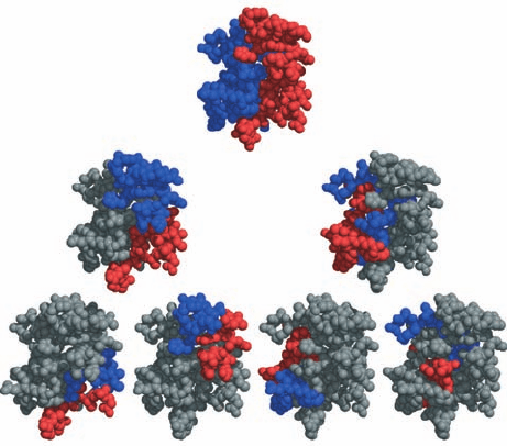
shown in Fig. 9-5 for high potential iron–sulfur protein
(HiPIP), it is clear that at every stage of the process, the red
Section 9-1. Protein Folding: Theory and Experiment 281
Figure 9-5 Hierarchical organization of globular proteins.
Here the X-ray structure of high potential iron–sulfur protein
(HiPIP) is represented by its C
␣
atoms shown as spheres. In the
top drawing, the first n/2 residues of this n-residue protein
(where n ⫽ 71) are colored red and the remaining n/2 residues
are colored blue. In the second row, the process is iterated such
that, on the right, for example, the first and last halves of the
second half of the protein are red and blue, with the remainder
of the chain gray. In the third row, the process is again iterated.
Note that at each stage of this hierarchy, the red and blue regions
do not intermingle. [Courtesy of George Rose, Johns Hopkins
University, and Robert Baldwin, Stanford University School of
Medicine.]
JWCL281_c09_278-322.qxd 2/24/10 1:17 PM Page 281
and blue regions do not interpenetrate. Evidently, protein
structures are organized hierarchically, that is, polypeptide
chains form locally compact structures that associate with
similar adjacent (in sequence) structures to form larger
compact structures, etc. This structural organization is, of
course, consistent with the observation that hydrogen
bonding interactions in proteins are mostly local (Section
8-4Bb). It also has important implications for how polypep-
tides fold to form native proteins (Section 9-1C).
d. Protein Structures Are Highly Adaptable
Globular proteins have packing densities comparable to
those of organic crystals (Section 8-3Bc) because the side
chains in a protein’s interior fit together with exquisite
complementarity.To ascertain whether this phenomenon is
an important determinant of protein structure, Eaton
Lattman and Rose analyzed 67 globular proteins of known
structure for the existence of preferred interactions be-
tween side chains. They found none, thereby indicating
that, at least in globular proteins, the native fold determines
the packing but packing does not determine the native fold.
This view is corroborated by the widespread occurrence of
protein families whose members assume the same fold
even though they may be so distantly related as to have no
recognizable sequence similarity (e.g., the ␣/ barrel pro-
teins; Section 8-3Bh).
The foregoing study indicates that there are a large num-
ber of ways in which a protein’s internal residues can pack
together efficiently.This was perhaps most clearly shown by
Brian Matthews in an extensive series of studies on
T4 lysozyme (a product of bacteriophage T4) in which the
X-ray structures of over 300 mutant varieties of this 164-
residue monomeric enzyme were compared. Replacements
of one or a few residues in T4 lysozyme’s hydrophobic core
were accommodated mainly by local shifts in the protein
backbone rather than by any global structural changes. In
many cases, T4 lysozyme could accommodate the insertion
of up to four residues without a major structural change or
even a loss of enzymatic activity. Moreover, assays of the
enzymatic activities of 2015 single-residue substitutions in
T4 lysozyme indicated that only 173 of these mutants had
significantly decreased enzymatic activity. Clearly protein
structures are highly resilient.
e. Secondary Structure Can Be Context-Dependent
The structure of a native protein is determined by its
amino acid sequence, but to what extent is the conforma-
tion of a given polypeptide segment influenced by the sur-
rounding protein? The NMR structure of protein GB1 (the
B1 domain of streptococcal protein G, which helps the bac-
terium evade the host’s immunological defenses by binding
to the antibody protein immunoglobulin G) reveals that
this 56-residue domain, which lacks disulfide bonds, con-
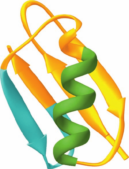
sists of a long ␣ helix lying across a 4-stranded mixed 
sheet (Fig. 9-6). In mutagenesis experiments by Peter Kim,
the 11-residue “chameleon” sequence AWTVEKAFKTF
was made to replace either residues 23 to 33 of GB1’s ␣ he-
lix (AATAEKFVFQY in GB1; a 7-residue change) to yield
Chm-␣, or residues 42 to 52 of its C-terminal  hairpin
(EWTYDDATKTF in GB1; a 5-residue change) to yield
Chm-.Both Chm-␣ and Chm- display reversible thermal
unfolding typical of compact single-domain globular pro-
teins, and their 2D NMR spectra indicate that each
assumes a structure similar to that of native GB1. Yet
NMR measurements also demonstrate that the isolated
chameleon peptide (Ac-AWTVEKAFKTF-NH
2
, where
Ac is acetyl) is unfolded in solution, which indicates that
this sequence has no strong preference for either an ␣ helix
or a  sheet conformation. This suggests that the informa-
tion specifying ␣ helix or  sheet secondary structures can
be nonlocal; that is, context-dependent effects may be im-
portant in protein folding (but see Section 9-1Ci).
f. Changing the Fold of a Protein
Proteins that share as little as ⬃20% sequence identity
may be structurally similar. To what degree must a pro-
tein’s sequence be changed in order to convert its fold to
that of another protein? This question was answered, at
least for the protein GB1, by the finding that changing 50%
of its 56 residues converted its fold to that of Rop protein
(Rop for repressor of primer; a transcriptional regulator).
Rop is a homodimer whose 63-residue subunits each form
an ␣␣ motif (Fig. 8-46c) that dimerizes with its 2-fold axis
perpendicular to the helix axes to form a 4-helix bundle
282 Chapter 9. Protein Folding, Dynamics, and Structural Evolution
Figure 9-6 NMR structure of protein GB1. Residues 23 to 33
are green and residues 42 to 53 are cyan.The 11-residue
chameleon sequence AWTVEKAFKTF can occupy either of
these positions without significantly altering the native protein’s
backbone conformation. [NMR structure by Angela Gronenborn
and Marius Clore, National Institutes of Health, Bethesda,
Maryland. PDBid 1GB1.]
JWCL281_c09_278-322.qxd 2/24/10 1:17 PM Page 282
(Fig. 9-7). Fifty percent of the residues of GB1 were
changed based largely on a secondary structure prediction
algorithm (Section 9-3Ad), energy minimization, and visual
modeling to yield a new polypeptide named Janus (after the
two-faced Roman god of new beginnings) that is 41% iden-
tical to Rop. In this manner, GB1 residues with high helix-
forming propensities were retained, whereas in regions
required to be ␣ helical, a number of residues with high
 sheet–forming propensities were replaced (helix- and
sheet–forming propensities are discussed in Section 9-3Aa);
hydrophobic residues were incorporated at the appropriate a
and d positions of a heptad repeat (Fig.8-26) to form the core
of Rop’s 4-helix bundle; and residue changes were made to
mimic Rop’s distribution of surface charges. Fluorescence
and NMR measurements reveal that Janus assumes a stable
Rop-like conformation. These studies indicate that not all
residues have equally important roles in specifying a par-
ticular fold. Indeed, the Janus sequence is more closely re-
lated to that of GB1 (50% identity) than to that of Rop
(41% identity), even though Janus structurally resembles
Rop but not GB1.
g. Many Proteins Are Natively Unfolded
In recent years it has become evident that many entire
native proteins and long protein segments (⬎30 residues)
are fully unfolded. Such intrinsically disordered proteins
lack specific tertiary structures and are therefore composed
of ensembles of conformations. They are characterized by
low sequence complexity, a low proportion of the bulky hy-
drophobic amino acids that form the cores of globular pro-
teins (Val, Leu, Ile, Met, Phe, Trp, and Tyr), and a high pro-
portion of certain polar and charged amino acids (Gln, Ser,
Pro, Glu, Lys, Gly, and Ala). Structure prediction techniques
based on amino acid sequences (Section 9-3) indicate that
an organism’s proportion of natively disordered proteins in-
creases with its complexity with ⬃2% of archeal proteins,
⬃4% of eubacterial proteins, and ⬃33% of eukaryotic pro-
teins predicted to contain long disordered regions.
Most natively disordered proteins specifically bind to
some other molecule such as a protein, a nucleic acid, or a
membrane component, and in doing so fold into stable sec-
ondary or tertiary structures. For example, the phosphory-
lated kinase-inducible domain (pKID) of the transcription
factor cyclic AMP response element-binding protein (CREB)
is disordered when free in solution, but folds to an ordered
conformation when it binds to the KID-binding domain of
CREB-binding protein (CBP, Fig. 9-8). Apparently, the in-
creased flexibility of natively disordered proteins enables
them to perform a relatively unhindered conformational
Section 9-1. Protein Folding: Theory and Experiment 283
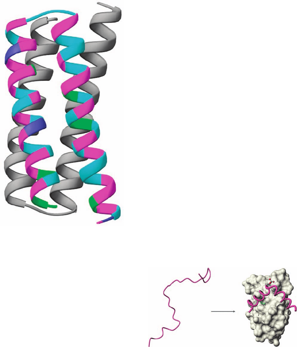
Figure 9-7 X-ray structure of Rop protein, a homodimer of ␣␣
motifs that associate to form a 4-helix bundle. On a change of
50% of its residues, protein GB1, whose structure is shown in Fig.
9-6, assumes the structure of Rop protein. One of the subunits of
the structure shown here is colored according to the sequence of
the GB1-derived polypeptide with purple residues identical in
both native proteins, magenta residues unchanged from native
GB1, cyan residues identical to those in native Rop, and green
residues different from those in either native protein.The
N-terminus of this subunit is at the lower right. [Based on an
X-ray structure by Demetrius Tsernoglou, Università di Roma,
Rome, Italy. PDBid 1ROP.]
Figure 9-8 The binding of the pKID domain of rat CREB to
the KID-binding domain of mouse CBP. The pKID, whose
backbone is drawn in worm form (pink), is unstructured when
free in solution (left) but forms two perpendicular helices when
bound to the KID-binding domain (right).The image on the right
shows the NMR structure of the pKID–KID-binding domain
complex with the side chains of pKID phosphoSer 133 and Leu
141 drawn in ball-and-stick form with C green, O red, and P
yellow and with the KID-binding domain (gray) represented by
its solvent-accessible surface. [Courtesy of Peter Wright, Scripps
Research Institute, La Jolla, California. PDBid 1KDX.]
JWCL281_c09_278-322.qxd 6/1/10 7:23 AM Page 283

a. Rapid Measurements Are Required to Monitor
Protein Folding
Folding studies on several small single-domain proteins,
including RNase A, cytochrome c, and apomyoglobin
(myoglobin that lacks its heme group), indicate that these
proteins fold to a significant degree within one millisecond
or less of being brought to native conditions. Hence, if the
earliest phases of the folding process are to be observed,
denatured proteins must be brought to native conditions in
significantly less time.This is most often done using a rapid
mixing device such as a stopped-flow apparatus (Fig. 9-9) in
which a protein solution at a pH that denatures the protein
or containing guanidinium chloride or urea at a concentra-
tion that does so is rapidly changed in pH or diluted to ini-
tiate folding.Most such instruments have “dead times” (the
interval between the times when mixing is initiated and
meaningful measurements can first be made) of ⬃0.5 ms.
However, recently developed ultrarapid mixing devices
have dead times of at little as 40 s.
An alternative technique involves the refolding of cold
denatured proteins. [For proteins whose folding has both
⌬H and ⌬S positive, a decrease in temperature is destabi-
lizing (Table 3-2). Since ⌬G ⫽⌬H ⫺ T ⌬S, these proteins
are unstable, that is denature, when T ⬍⌬H/⌬S. For many
of these proteins, solution conditions can be found for
which this temperature is ⬎0°C.] The refolding of the cold-
denatured protein is initiated by a so-called temperature-
jump in which the solution is heated with an infrared laser
pulse by 10 to 30°C in ⬍100 ns.
With either of the above methods, the folding protein
must be monitored by a technique that can report rapid
structural changes in a protein. The three such techniques
that have been most extensively used are (1) circular
dichroism (CD) spectroscopy, (2) pulsed HD exchange fol-
lowed by 2D-NMR spectroscopy or mass spectrometry,
and (3) fluorescence resonance energy transfer (FRET).
We discuss these methods below.
b. The Circular Dichroism Spectrum of a Protein Is
Indicative of Its Conformation
Polypeptides absorb strongly in the ultraviolet (UV) re-
gion of the spectrum (⫽100 to 400 nm) largely because their
aromatic side chains (those of Phe, Trp, and Tyr) have par-
ticularly large molar extinction coefficients (Section 5-3Ca)
search when binding to their target molecules. It has also
been suggested that a structured globular protein would
have to be two to three times larger than a disordered pro-
tein to provide the same size intermolecular interface and
hence the use of disordered proteins provides genetic
economy and reduces intracellular crowding. Disordered
regions may also aid in the transport of proteins across
membranes (Section 12-4Ea) and facilitate selective pro-
tein degradation (Section 32-6B).
The functions of natively disordered proteins are quite
varied.Their most common function appears to be binding
to specific DNA sequences to facilitate such processes as
replication, transcription, repair, and transposition (Chap-
ter 30). However, they have also been implicated in a vari-
ety of other functions including intracellular signal trans-
duction (Chapter 19), forming phosphorylation sites in
proteins whose activities are regulated by phosphorylation
(Section 18-3C), and in aiding other proteins and RNAs to
fold to their native conformations (Section 9-2C).
C. Folding Pathways
How does a protein fold to its native conformation? We, of
course, cannot hope to answer this question in detail until
we better understand why native protein structures are sta-
ble. Moreover, as one might guess, the folding process itself
is one of enormous complexity. Nevertheless, as we shall
see below, the broad outlines of how proteins fold to their
native conformations are beginning to come into focus.
The simplest folding mechanism one might envision is
that a protein randomly explores all of the conformations
available to it until it eventually “stumbles” onto its native
conformation. A back-of-the-envelope calculation first
made by Cyrus Levinthal, however, convincingly demon-
strates that this cannot possibly be the case: Assume that
the 2n backbone torsional angles, and , of an n-residue
protein each have three stable conformations.This yields 3
2n
⬇ 10
n
possible conformations for the protein, which is a
gross underestimate, if only because the side chains are ig-
nored. If a protein can explore new conformations at the
rate at which single bonds can reorient, it can find ⬃10
13
conformations per second, which is, no doubt, an overesti-
mate. We can then calculate the time t, in seconds, required
for a protein to explore all the conformations available to it:
[9.1]
For a small protein of n ⫽ 100 residues, t ⫽ 10
87
s, which is
immensely more than the apparent age of the universe
(⬃13.7 billion years ⫽ 4.3 ⫻ 10
17
s).
It would obviously take even the smallest protein an ab-
surdly long time fold to its native conformation by ran-
domly exploring all its possible conformations, an infer-
ence known as the Levinthal paradox. Yet several proteins
fold to their native conformations in microseconds. There-
fore, as Levinthal suggested, proteins must fold by some
sort of ordered pathway or set of pathways in which the ap-
proach to the native state is accompanied by sharply increas-
ing conformational stability (decreasing free energy).
t ⫽
10
n
10
13
s
⫺1
284 Chapter 9. Protein Folding, Dynamics, and Structural Evolution
Figure 9-9 A stopped-flow device. The reaction is initiated by
simultaneously and rapidly discharging the contents of both
syringes through the mixer. On hitting the stop switch, the
stopping syringe triggers the computer to commence optically
monitoring the reaction (via its UV/visible, fluorescence, or CD
spectrum).
Solution 1
Solution 2
Mixer
Light Source
Detector
Trigger
Stopping
syringe
Stop
switch
Computer
JWCL281_c09_278-322.qxd 2/24/10 1:17 PM Page 284
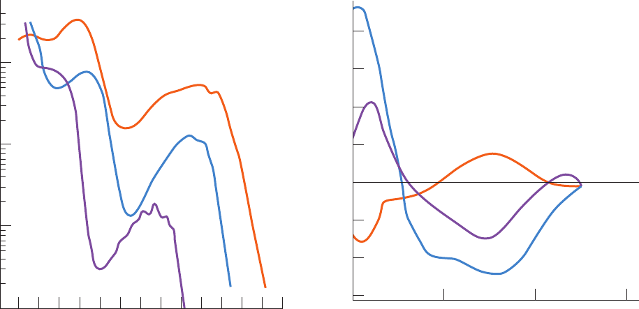
in this spectral region (ranging into the tens of thousands;
Fig. 9-10). However, polypeptides do not absorb visible
light (⫽400 to 800 nm), so that they are colorless.
For chiral molecules such as proteins, ε has different val-
ues for left and right circularly polarized light, ε
L
and ε
R
.
The variation with of the difference in these quantities,
⌬ε ⫽ ε
L
⫺ ε
R
, constitutes the circular dichroism (CD) spec-
trum of the solute of interest (for nonchiral molecules ε
L
⫽ e
R
and hence they have no CD spectrum). In proteins, ␣ he-
lices,  sheets, and random coils exhibit characteristic CD
spectra (Fig. 9-11). Hence the CD spectrum of a polypep-
tide provides a rough estimate of its secondary structure.
c. Pulsed H/D Exchange Provides Structural Details
on How Proteins Fold
Pulsed H/D exchange, a method devised by Walter Eng-
lander and Robert Baldwin, is the only known technique
that can follow the time course of individual residues in a
folding protein. Weakly acidic protons (
1
H), such as those
of amine and hydroxyl groups , exchange with
those of water, a process known as hydrogen exchange that
can be demonstrated with the use of deuterated water
[D
2
O; deuterium (D or
2
H) is a stable isotope of
1
H]:
Since
1
H has an NMR spectrum in a different frequency
range from that of D, the exchange of
1
H for D can be read-
ily followed by NMR spectroscopy. Under physiological
conditions, small organic molecules, such as amino acids
and dipeptides, completely exchange their weakly acidic
protons for D in times ranging from milliseconds to seconds.
X¬H ⫹ D
2
O Δ X¬D ⫹ HOD
(X¬H)
Proteins bear numerous exchangeable protons, particularly
those of its backbone amide groups. However, protons that
are engaged in hydrogen bonding do not exchange with
solvent and, moreover, groups in the interior of a native
protein are not in contact with solvent.
Through the use of 2D-NMR (Section 8-3Ac), pulsed
H/D exchange can be used to follow the time course of pro-
tein folding. The protein of interest, usually with its native
disulfide bonds intact, is denatured by guanidinium chlo-
ride or urea in D
2
O solution such that all of the protein’s
peptide nitrogen atoms become deuterated . Fold-
ing is then initiated in a stopped-flow apparatus by diluting
the denaturant solution with
1
H
2
O while the pH is simulta-
neously lowered so as to arrest hydrogen exchange (near
neutrality, hydrogen exchange reactions are catalyzed by
OH
⫺
and, therefore, their rates are highly pH dependent).
After a preset folding time, t
f
, the pH is rapidly increased
(using a third independently triggered syringe; the so-
called labeling pulse) to initiate hydrogen exchange. Pep-
tide nitrogen atoms whose D atoms have not formed hy-
drogen bonds by time t
f
exchange with
1
H, whereas those
that are hydrogen bonded at t
f
, and hence unavailable for
hydrogen exchange, remain deuterated. After a short time
(10 to 40 ms), the labeling pulse is terminated by rapidly
lowering the pH (with a fourth syringe). Folding is then al-
lowed to go to completion and the H/D ratio at each ex-
changeable site is determined by 2D-NMR (the peaks in the
2D proton NMR spectrum must have been previously as-
signed). By repeating the analysis for several values of t
f
, the
(N¬D)
Section 9-1. Protein Folding: Theory and Experiment 285
Figure 9-10 UV absorbance spectra of the three aromatic
amino acids, phenylalanine, tryptophan, and tyrosine. Note that
the molar absorbance, ε, is displayed on a log scale. [After
Wetlaufer, D.B., Adv. Prot. Chem. 7, 310 (1962).]
Figure 9-11 Circular dichroism (CD) spectra of polypeptides.
Polypeptides in the ␣ helix,  sheet, and random coil (rc)
conformations were determined from the CD spectra of proteins
of known X-ray structures. By comparing these spectra with the
absorption spectra in Fig. 9-10, it can be seen that ⌬ ε ⫽ ε
L
⫺ ε
R
is
a small difference of two large numbers. [After Saxena, V.P. and
Wetlaufer, D.B., Proc. Natl. Acad. Sci. 66, 971 (1971).]
λ (nm)
ε
320300280260240
Trp
Tyr
Phe
220200
20
10
50
200
100
500
1,000
2,000
5,000
10,000
20,000
40,000
190
λ (nm)
α
β
250230210
Δε
–15
–10
–5
0
5
10
15
20
rc
JWCL281_c09_278-322.qxd 2/24/10 1:17 PM Page 285
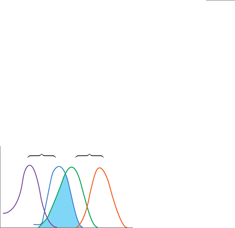
When two fluorescent molecules or groups, a donor (D)
and an acceptor (A), are within 100 Å of each other and D
is electronically excited (say, by a laser with a wavelength
within its absorption spectrum), some of the excitation en-
ergy will be transferred from D to A,
where the asterisk indicates an electronically excited state.
A will then fluoresce with its characteristic emission spec-
trum (Fig. 9-12), whose intensity can be measured. This
phenomenon is known as fluorescence resonance energy
transfer (FRET). Its efficiency E, the fraction of the energy
transferred to the acceptor per donor excitation event, is
given by
[9.2]
where r is the distance between D and A, and R
0
, their
Förster distance (named after Theodor Förster, who for-
mulated the theory for the mechanism of long-range
nonradiative energy transfer), is the value of r at which
the FRET efficiency is 50%. R
0
varies with the degree of
overlap between the donor’s emission spectrum and the
acceptor’s absorption spectrum (Fig. 9-12) as well as with
the relative orientation of the donor and acceptor.
Hence, the intensity of the acceptor’s fluoresence is in-
dicative of the distance between D and A as well as their
relative orientation.
In proteins, D and A can be the side chains of Trp or Tyr
residues. The number and placement of these residues in
the protein of interest can be manipulated by site-directed
mutagenesis (Section 5-5Gc). Alternatively, fluorescent
groups may be covalently linked to reactive side chains
such as Cys, which can also be placed via site-directed mu-
tagenasis. FRET measurements can then be used to track
how the distances between specific residues vary with time
in a folding protein.
e. The Earliest Protein Folding Events Are Initiated
by a Hydrophobic Collapse
Stopped-flow–CD measurements indicate that for many,
if not all, small single-domain proteins, much of the sec-
ondary structure that is present in native proteins forms
within a few milliseconds of when folding is initiated. This
is called the burst phase because subsequent folding
events occur over much longer time intervals. Pulsed H/D
exchange measurements of these small proteins show that
some protection against hydrogen exchange in some sec-
ondary structural elements develops by ⬃5 ms after fold-
ing initiation.
Since globular proteins contain a compact hydrophobic
core, it seems likely that the driving force in protein folding
is a so-called hydrophobic collapse, in which the protein’s
hydrophobic groups coalesce so as to expel most of their
surrounding water molecules. The polypeptide’s radius of
gyration is thereby dramatically reduced (from ⬃30 to ⬃15
Å for a 100-residue polypeptide), a phenomenon that is
E ⫽
1
1 ⫹ (r>R
0
)
6
D* ⫹ A S D ⫹ A*
286 Chapter 9. Protein Folding, Dynamics, and Structural Evolution
time course of hydrogen bond formation at each residue can
be determined.
Pulsed H/D exchange–NMR studies do not directly in-
dicate the structures of the folding intermediates. How-
ever, if the native structure of the protein under investiga-
tion is known (as it almost always is for proteins whose
folding is being investigated) and if it is assumed that the
protein folds without forming secondary structures not
present in the native protein, then the 2D-NMR spectra re-
veal the time course of the formation of the elements of the
native structure together with how fast they are excluded
from the bulk solvent.
The time course of protein folding may also be followed
by combining pulsed H/D exchange with mass spectrome-
try. In this method, a partially deuterated protein is frag-
mented by pepsin (a protease that functions under the
acidic conditions necessary to prevent further hydrogen
exchange; Table 7-2), the resulting fragments separated by
HPLC, and their degree of deuteration determined by
mass spectrometry.This method does not yield the residue-
level structural information that NMR provides. However,
unlike NMR, it can determine if a sample contains subpop-
ulations of protein fragments with different degrees of
deuteration and hence that have followed different folding
pathways.
d. Fluorescence Resonance Energy Transfer
Monitors Distances
Fluorescence is the phenomenon whereby an electroni-
cally excited molecule or group decays to its ground state by
emitting a photon. The initial excited state rapidly decays, via
nonradiative processes (e.g., heating; Section 24-2Aa), to an
excited state of lower energy before the photon is emitted.
Hence the molecule or group’s emission spectrum has a
longer wavelength than its absorption spectrum (Fig. 9-12).
Figure 9-12 Schematic diagram of the absorption and
emission spectra of a donor and an acceptor in fluorescence
resonance energy transfer (FRET). Note that the absorption
spectrum occurs at shorter wavelengths than the corresponding
emission spectrum and that the donor’s emission spectrum must
overlap the acceptor’s absorption spectrum (cyan) for FRET to
occur.
Wavelength
Intensity
Donor
Acceptor
Absorbance Emission Absorbance Emission
JWCL281_c09_278-322.qxd 2/24/10 1:17 PM Page 286
generally characteristic of polymers on being transferred
from a good to a poor solvent.
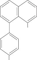
This hydrophobic collapse mechanism is consistent with
the observation that the hydrophobic dye 8-anilino-1-
naphthalenesulfonate (ANS)
binds to folding proteins. ANS undergoes a significant en-
hancement of its fluorescence when it occupies a nonpolar
environment, an enhancement that is observed within the
burst phase when ANS is present in a solution of a folding
protein. Since ANS is expected to preferentially bind to hy-
drophobic groups, this indicates that the hydrophobic core
of a protein rapidly forms once folding has been initiated.
The initial collapsed state of a folding protein is known
as a molten globule. Such a species has a radius of gyration
that is only 5 to 15% greater than that of the native protein
and has significant amounts of the native secondary struc-
ture and overall fold. However, a molten globule’s side
chains are extensively disordered, its structure fluctuates
far more than that of the native protein, and it has only
marginal thermodynamic stability. Nevertheless, to con-
tinue folding toward its native state, the polypeptide chain
need not undergo large rearrangements in the crowded
core of the partially folded protein.
f. Nativelike Tertiary Structure Appears During
Intermediate Folding Events
After the burst phase, small proteins exhibit increased
ANS binding, further changes in their CD spectrum, and
enhanced protection against H/D exchange. These inter-
mediate folding events typically occur over a time interval
of 5 to 1000 ms. This is the stage at which the protein’s sec-
ondary structure becomes stabilized and its tertiary struc-
ture begins to form. These nativelike elements are thought
to take the form of subdomains that are not yet properly
docked to each other. Side chains are probably still mobile,
so that, at this stage of folding,the protein can be described
as an ensemble of closely related and rapidly interconvert-
ing structures.
g. Final Folding Events Often Require
Several Seconds
In the final stage of folding, a protein achieves its native
structure.To do so, the polypeptide must undergo a series of
complex motions that permit the attainment of the relatively
NH
2
SO
3
2⫺
8-Anilino-1-naphthalene
sulfonate (ANS)
rigid native core packing and hydrogen bonding, while ex-
pelling the remaining water molecules from its hydropho-
bic core. For small single-domain proteins, this takes place
over a time interval of several seconds or less.
h. Landscape Theory of Protein Folding
The classic view of protein folding was that proteins fold
through a series of well-defined intermediates. The folding
of a random coil polypeptide was thought to begin with the
random formation of short stretches of 2° structure, such as
␣ helices and  turns, that acted as nuclei (scaffolding) for
the stabilization of additional ordered regions of the pro-
tein. Nuclei with the proper nativelike structure then grew
by the diffusion, random collision, and adhesion of two or
more such nuclei. The stabilities of these ordered regions
were thought to increase with size, so, after having ran-
domly reached a certain threshold size, they spontaneously
grew in a cooperative fashion until they formed a native-
like domain. Finally, through a series of relatively small
conformational adjustments, the domain rearranged to the
more compact 3° structure of the native conformation.
The advent of experimental methods that could observe
early events in protein folding led to a somewhat different
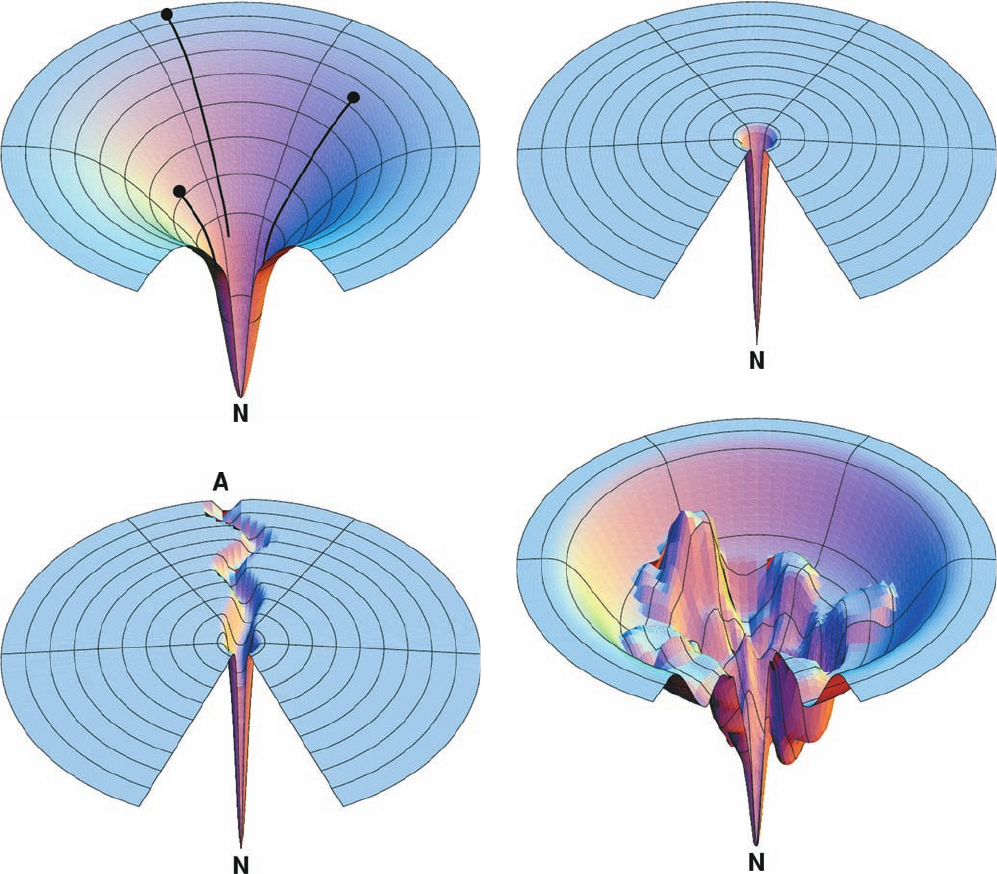
view of how proteins fold. In this so-called landscape theory,
which was formulated in large part by Peter Wolynes, Bald-
win, and Dill, folding is envisioned to occur on an energy
surface or landscape that represents the conformational
energy states available to a polypeptide under the prevail-
ing conditions.The horizontal coordinates of a point on this
surface represent a particular conformation of the
polypeptide, that is, the values of and for each of its
amino acid residues and the torsion angles for each of its
side chains (but here projected onto two dimensions from
its multidimensional space). The vertical coordinate of a
point on the energy surface represents the polypeptide’s
internal free energy in this conformation. The above-
described measurements indicate that the energy surface
of a folding polypeptide is funnel-shaped, with the native
state represented by the bottom of the funnel, the global
(overall) free energy minimum (Fig. 9-13a). The width of
the funnel at any particular height (free energy) above the
native state is indicative of the number of conformational
states with that free energy, that is, the entropy of the
polypeptide.
Polypeptides fold via a series of conformational adjust-
ments that reduce their free energy and entropy until the na-
tive state is reached. Since a collection of unfolded polypep-
tides all have different conformations (have different
positions on the folding funnel), they cannot follow pre-
cisely the same pathway in folding to the native state. If the
polypeptide actually folded to its native state via a random
conformational search, as Levinthal conjectured, its energy
surface would resemble a flat disk with a single small hole,
much like the surface of a golf course (Fig. 9-13b). Thus, it
would take an enormously long time for a polypeptide (a
golf ball) to achieve the native state (to fall in the hole) via
a random conformational search (by rolling about aim-
lessly on the surface of the golf course).
Section 9-1. Protein Folding: Theory and Experiment 287
JWCL281_c09_278-322.qxd 2/24/10 1:17 PM Page 287
The observation that many polypeptides acquire sig-
nificant nativelike structure within fractions of a millisec-
ond after folding commences indicates that their energy
surfaces are, in fact, funnel-shaped; that is, they tend to
slope toward the native conformation at all points. Thus,
the various pathways followed by initially unfolded
polypeptides in folding to their native state are analo-
gous to the various trajectories that could be taken by
skiers initially distributed around the top of a bowl-
shaped valley to reach the valley’s lowest point. Appar-
ently, there is no single pathway or closely related set of
288 Chapter 9. Protein Folding, Dynamics, and Structural Evolution
The energy surface of a protein that follows the classic
view of protein folding would have a deep radial groove in
its disklike surface that slopes toward the hole represent-
ing the native state (Fig. 9-13c). The extent of the confor-
mational search to randomly find this groove would be
much reduced relative to the Levinthal model, so that such
a polypeptide would readily fold to its native state. How-
ever, the conformational search for the pathway (groove)
leading to the native state would still take time, so that the
polypeptide would require perhaps several seconds to start
down the folding pathway.
Figure 9-13 Folding funnels. (a) An idealized funnel
landscape. As the chain forms increasing numbers of intrachain
contacts, its internal free energy (its height above the native
state, N) decreases together with its conformational freedom (the
width of the funnel). Polypeptides with differing conformations
(black dots) follow different pathways (black lines) in achieving
the native fold. (b) The Levinthal “golf course” landscape in
which the chain must search for the native fold (the hole)
randomly, that is, on a level energy surface. (c) The classic folding
landscape in which the chain must search at random on a level
energy surface until it encounters the canyon that leads it to the
native state. (d) A rugged energy surface containing local minima
in which a folding polypeptide can become transiently trapped.
The folding funnels of real proteins are thought to have such
topographies. [Courtesy of Ken Dill, University of California at
San Francisco.]
(a)
(b)
(c)
(d)
JWCL281_c09_278-322.qxd 2/24/10 1:17 PM Page 288
pathways that a polypeptide must follow in folding to its
native conformation.
The foregoing does not imply that the surface of the
folding funnel is necessarily smooth,as is drawn in Fig.9-13a.
Indeed, landscape theory suggests that this energy surface
has a relatively rugged topography, that is, has many local
energy minima and maxima (Fig. 9-13d). Consequently, in
following any particular folding pathway, a polypeptide is
likely to become trapped in a local minimum until it ran-
domly acquires sufficient thermal energy to surmount this
kinetic barrier and continue the folding process. Thus, in
landscape theory, the local energy maxima (transition
states; Section 14-1C) that govern the rate of folding are
not specific structures as the classic theory of protein fold-
ing suggests but, rather, are ensembles of structures.
i. Protein Folding Is Hierarchical
The observation that protein structures are hierarchi-
cally organized (Section 9-1B) suggests that they also fold
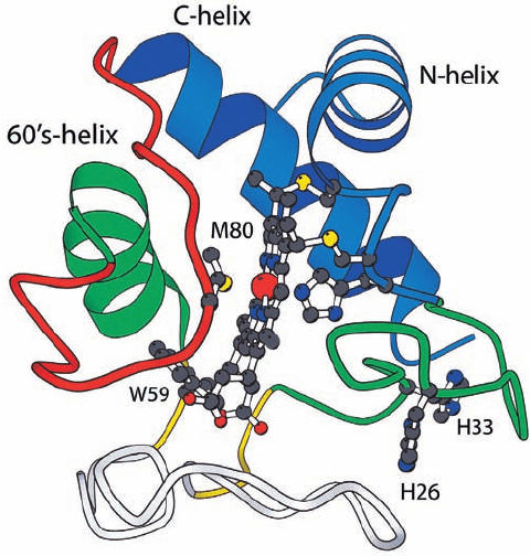
in a hierarchic manner. By this it is meant that folding be-
gins with the formation of marginally stable nativelike mi-
crostructures known as foldons (e.g., Fig.9-14) that are local
in sequence and that these foldons diffuse and collide with
nearby (in sequence) foldons to yield intermediates of in-
creasing complexity and stability that sequentially grow to
form the native protein. In contrast, in nonhierarchical fold-
ing, a protein’s tertiary structure would not only stabilize its
local structures but also determine them. Landscape theory
is consistent with hierarchical folding, whereas the classic
theory of protein folding is more in accord with nonhierar-
chical folding. Moreover, since a polypeptide in vivo begins
folding as it is being synthesized, that is, as it is extruded
from the ribosome, it would seem that it would most readily
achieve its native state if it folded in a hierarchical manner.
Several lines of evidence indicate that proteins, in fact,
fold in a hierarchical manner.
1. H/D exchange studies have established the existence
of foldons in numerous proteins. Indeed, it appears that
foldons rather than individual amino acid residues carry
out the unit steps in protein folding pathways.
2. Many peptide fragments excised from proteins either
form or exhibit a tendency to form foldons in the absence
of long-range (3°) interactions. Moreover, when proteins
such as cytochrome c and apomyoglobin are brought to a
pH sufficiently low to destabilize their native structures,
their foldons persist.
3. The boundaries of helices in native proteins are fixed
by their flanking sequences (Section 9-3) rather than by 3°
interactions.
4. The folding rates of proteins increase, on average,
with the degree to which their native contacts are local.
Thus fast folders tend to have a high proportion of helices
and tight turns, whereas slow folders tend to have a high
proportion of  sheets.
In Section 9-1B we saw that in protein GB1 (Fig. 9-6),
the 11-residue “chameleon” sequence assumed either an ␣
helix or a  hairpin, depending on its position in the protein.
Thus, its conformation appears to be determined by its con-
text rather than by local interactions. However, computer
simulations suggest that the conformation of the chame-
leon sequence is actually determined by local interactions
beyond its boundaries.
The folds of native proteins, as we have seen, are highly
resistant to sequence changes. Evidently, the sequence in-
formation specifying a particular fold is both distributed
throughout the polypeptide chain and highly overdeter-
mined. It is these characteristics that appear to be respon-
sible for hierarchical folding.
j. Primary Structures Determine Protein Folding
Pathways as Well as Structures
The above discussions suggest that protein primary struc-
tures evolved to specify efficient folding pathways as well as
stable native conformations. Evidence corroborating this hy-
pothesis has been obtained by Jonathan King in his study of
the renaturation of the tail spike protein of bacteriophage
P22. The tail spike protein is a trimer of identical 76-kD
polypeptides, whose T
m
⫽ 88°C. However, certain mutant
varieties of the protein fail to renature at 39°C.Nevertheless,
at 30°C, these mutant proteins fold to structures whose prop-
erties, including their T
m
’s, are indistinguishable from that of
the wild-type tail spike protein.The amino acid changes caus-
ing these temperature-sensitive folding mutations apparently
act to destabilize intermediate states in the folding process but
do not affect the native protein’s stability. This observation
suggests that a protein’s amino acid sequence dictates its native
structure by specifying how it folds to its native conformation.
Section 9-1. Protein Folding: Theory and Experiment 289
Figure 9-14 Ribbon diagram of cytochrome c. Its several foldon
units are shown in different colors. Its heme group and several of its
functionally important side chains are drawn in ball-and-stick form
with C black, N blue, O red, S yellow, and Fe a large red sphere.
[Courtesy of Walter Englander, University of Pennsylvania.]
JWCL281_c09_278-322.qxd 2/24/10 1:17 PM Page 289
that is, with a large fraction of the polypeptide chains as-
suming quasi-stable non-native conformations and/or
forming nonspecific aggregates. In vivo, however, poly-
peptides efficiently fold to their native conformations as
they are being synthesized, a process that normally re-
quires a few minutes or less. This is because all cells con-
tain three types of accessory proteins that function to as-
sist polypeptides in folding to their native conformations
and in assembling to their 4° structures: protein disulfide
isomerases, peptidyl prolyl cis–trans isomerases, and mo-
lecular chaperones. We discuss these essential proteins in
this section.
290 Chapter 9. Protein Folding, Dynamics, and Structural Evolution
This hypothesis is supported by the observation that, in native
proteins, a greater number of polar residues than would be
randomly expected occupy helix-capping positions (Section
8-4Bb) even though they do not make helix-capping hydro-
gen bonds. This suggests that they do so as the helix forms so
as to facilitate the protein’s proper folding.
2 FOLDING ACCESSORY PROTEINS
Most unfolded proteins renature in vitro over periods rang-
ing from minutes to days and, quite often, with low efficiency,
S
S
S
S
S
S
SS
S
S
S
S
S
S
HS
S
S
S
S
SH
PDI PDI
PDI
PDI
SH
SH
SH
SH
PDI
SH
SH
PDI
S
S
SH
SH
PDI
S
S
HS
HS
SH
Reduced
PDI
Oxidized
PDI
Reduced
PDI
1
1
1
1
2
2
2
2
4
4
4
4
3
3
3
3
..
..
..
..
Native S–S bondsMixed disulfide
Mixed disulfide nietorp )evitan( dezidixOnietorp decudeR
Non-native S–S bonds
(a)
(b)
S
S
SH
1
2
4
3
SH
..
PDI
S
S
Oxidized
PDI
SH
..
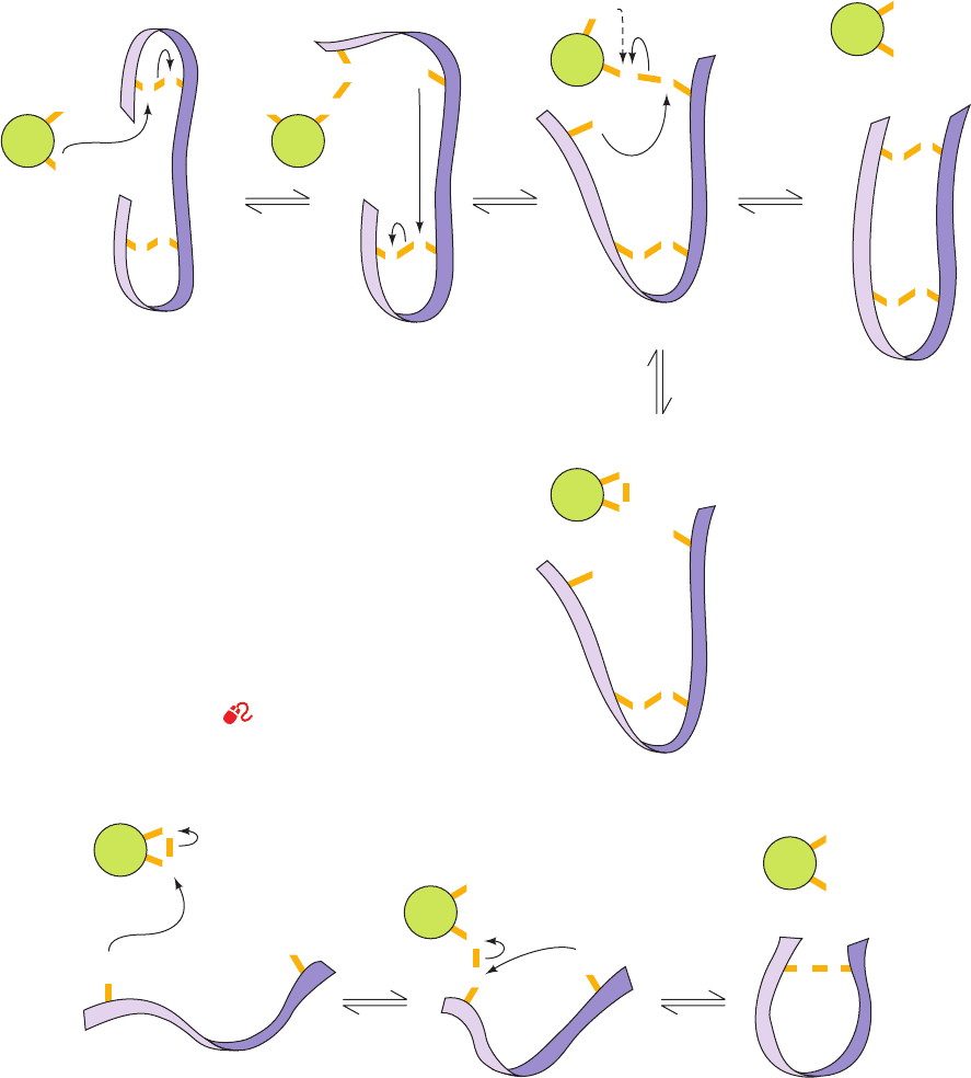
Figure 9-15 Reactions catalyzed by protein disulfide
isomerase (PDI). (a) Reduced PDI catalyzes the rearrangement
of the non-native disulfide bonds in a substrate protein (purple
ribbon) via disulfide interchange to yield the native disulfide
bonds (horizontal reactions). If a disulfide bond between PDI
and the substrate protein is resistant to disulfide interchange, it is
reduced by PDI’s second SH group to yield reduced substrate
protein and oxidized PDI (vertical reaction and dashed curved
arrow). (b) The oxidized PDI-dependent synthesis of disulfide
bonds in proteins.The reaction occurs with the intermediate
formation of a mixed disulfide between PDI and the protein.The
reduced PDI reaction product reacts with cellular oxidizing
agents to regenerate oxidized PDI.
See the Animated Figures
JWCL281_c09_278-322.qxd 2/24/10 1:17 PM Page 290
