Vlak J.M., de Gooijer C.D., Tramper J., Miltenburger H.G. (Eds.) Insect Cell Cultures: Fundamental and Applied Aspects
Подождите немного. Документ загружается.

This page intentionally left blank.

Cytotechnology 20: 73–93, 1996. 73
© 1996 Kluwer Academic Publishers. Printed in the Netherlands.
Baculovirus–insect cell interactions
Gary W. Blissard
Boyce Thompson Institute, Cornell University, Tower Road, Ithaca, NY 14853–1801, U.S.A.
Key words: Baculoviridae, viral interactions
Introduction
The Baculoviridae are a family of large, enveloped
DNA viruses that are characterized by rod-shaped
nucleocapsids and relatively large double stranded
DNA genomes. Baculoviruses are infectious only to
arthropods, with the vast majority of permissive species
falling within one Order (Lepidoptera) of the Class
Insecta. Genomes of different baculoviruses range
from approximately 80–180 kbp. The Baculoviridae
contain two Genera, the Nucleopolyhedroviruses (or
NPVs) and Granuloviruses (or GVs) (Volkman et al.,
1995). Because it has been difficult to develop con-
venient cell culture systems for the propagation of
GVs (Winstanley & Crook, 1993), most molecular,
biochemical, and genetic studies have focused on the
NPVs. Baculoviruses interact at many levels with the
host cell; yet much remains to be discovered about
these interactions. Few studies have examined the
participation of host proteins in baculovirus infection
processes. However, information on viral proteins, and
their structures and roles in infection is accruing rapid-
ly. The sequence of the genome of the Autographa
californica Multicapsid Nuclear Polyhedrosis Virus
(AcMNPV) was recently reported along with an exten-
sive analysis of known and predicted baculovirus genes
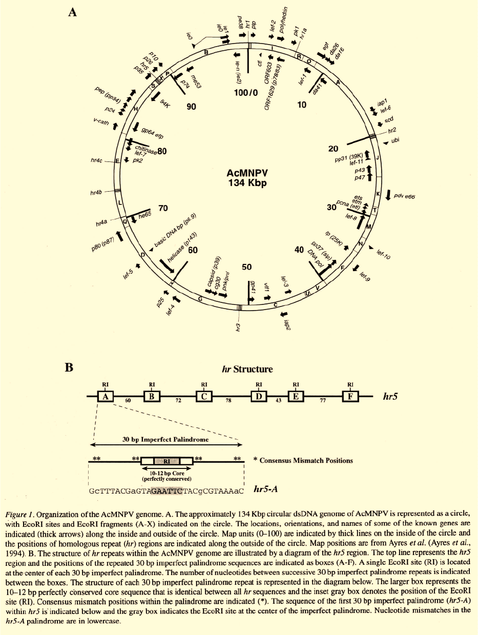
(Ayres et al., 1994). The AcMNPV genome contains
154 open reading frames (ORFs) encoding potential
proteins of amino acids. A map of the approx-
imately 134 kbp AcMNPV genome and some genes
which have been identified are shown in Figure 1.
In addition to reviews included in the present vol-
ume, previous reviews have also described aspects of
baculovirus structure and molecular biology (Blissard
& Rohrmann, 1990; Friesen & Miller, 1986; Grana-
dos & Federici, 1986; King & Possee, 1992; Miller,
1995; O’Reilly et al., 1992b; Rohrmann, 1992). This
review will examine selected topics with an emphasis
on known and likely viral interactions at the cell and
molecular level.
Baculovirus virions
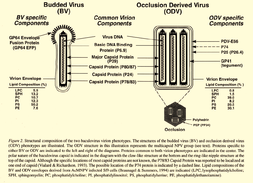
A hallmark of the Baculoviridae is the production of
two virion phenotypes, termed budded virions (BV)
and occlusion derived virions (ODV) (Figure 2). These
two virion phenotypes are produced at different loca-
tions in the cell, and at different times in the infec-
tion cycle. They serve distinctly different functional
roles. In addition, BV and ODV enter cells by differ-
ent mechanisms. BV are produced in the late phase
of the infection cycle, when nucleocapsids bud from
the surface of infected cells. Thus, the BV envelope
is derived from the modified plasma membrane of the
host cell. Very late in infection, nucleocapsids become
enveloped within the nucleus to form the ODV. ODV
are subsequently occluded within an occlusion matrix
protein (“Polyhedrin” in the NPVs; “Granulin” in the
GVs). Since nucleocapsids of both BV and ODV are
produced in the nucleus, the nucleocapsids and viral
DNA of the BV and ODV appear to be identical (Fig-
ure 2). Thus, BV and ODV differ primarily in compo-
sition of the envelopes and associated structures, and
these differences result in different functional roles of
the BV and
ODV.

74

75
Biology and structure of ODV
In nature, baculovirus occlusion bodies are released
into the environment after cell lysis and death of an
infected insect host. New hosts acquire baculoviruses
by feeding on foliage contaminated with viral occlu-
sion bodies. After ingestion, occlusion bodies are
exposed to the alkaline pH of the lepidopteran midgut,
and this results in the dissolution of occlusion bodies
and release of the occlusion derived virions (ODV).
The ODV are highly infectious to epithelial cells of
the insect midgut and appear to be less infectious in
other tissues (Volkman & Summers, 1977). The num-
ber of nucleocapsids within an ODV envelope varies
between different subgroups of baculoviruses. ODV
may contain one or many nucleocapsids per envelope.
GVs typically contain a single nucleocapsid per enve-
lope and a single (ODV) virion per occlusion body (or
capsule). In contrast, each NPV occlusion body con-
tains many (ODV) virions. Historically, two subgroups
have been described within the NPVs: Those typi-
cally containing a single nucleocapsid per virion are
referred to as “single-capsid NPVs” (SNPVs), whereas
those containing multiple nucleocapsids per virion are
known as “multicapsid NPVs” (MNPVs). It is not clear
whether the phenotypic differences observed between
SNPVs and MNPVs represent significant phylogenet-
ic differences between these groups since preliminary
phylogenetic analyses of NPVs suggest that SNPVs
and MNPVs do not segregate into separate groupings
(Rohrmann, 1986; Zanotto et al., 1993).
Do ODV contain viral encoded envelope proteins
that interact with cellular receptors on insect midgut
epithelial cells? Little direct data exist to address this
question, and no specific virus-host cell receptor inter-
action has been identified for ODV. Electron micro-
graphs of ODV do not reveal distinct spike, or peplom-
er structures on the surface of the envelope, as can
be observed on some other enveloped viruses and on
the BV of baculoviruses (see below). A recent study
76
of ODV (Horton & Burand, 1993), suggests that spe-
cific saturable virion binding sites exist on the brush
border of midgut epithelial cells. However, specific
interactions between viral encoded proteins and spe-
cific cellular proteins or other ligands have not yet
been demonstrated.
After successful binding of the virion at the cell
surface, nucleocapsids must enter the cytoplasm.
Enveloped viruses normally enter cells by either direct
membrane fusion at the cell surface, or by receptor
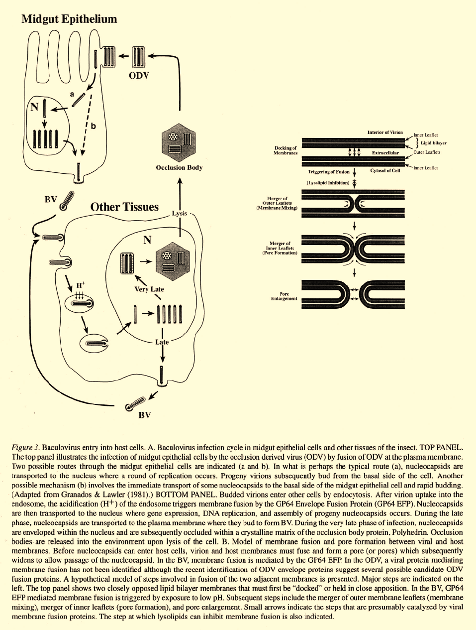
mediated endocytosis. ODV enter cells by fusion of
the virion envelope with the plasma membrane at the
cell surface (Figure 3). Evidence for this mechanism
of entry is largely from electron micrographic observa-
tions of midgut epithelial cells (Granados, 1978; Sum-
mers, 1971), and the finding that ODV entry is not
inhibited by treatment with chloroquine, an agent that
buffers the pH of the endosome and thus inhibits the
entry of viruses by endocytosis (Horton & Burand,
1993). In the GVs, ODV infectivity is aided by a high
molecular weight protein named “Enhancin” which is
found in occlusion bodies (granules) (Gijzen et al.,
1995; Hashimoto et al., 1991). The Enhancin protein
has structural and functional characteristics of metallo-
proteases (R. Granados, pers. comm.) and the primary
mode of action of Enhancin appears to be proteolysis
of the peritrophic membrane, a structure that lines the
insect midgut (Derksen & Granados, 1988; Wang et
al., 1994).
Information on the structural composition of the
ODV is rapidly emerging. The ODV envelope, per-
haps the most important component in the initial inter-
action of ODV with the host cell, contains a number of
structural proteins. Viral encoded ODV envelope pro-
teins include P25, PDV-E66, ODV-E56 (ODVP-6E)
and possibly P74 (Braunagel & Summers, 1994; Brau-
nagel et al., 1996; Hong et al., 1994; Kuzio et al., 1989;
Roberts, 1989; Russell & Rohrmann, 1993a; Theil-
mann et al., 1996) (Figure 2). An additional protein
associated with ODV virions, GP41 (Liu & Maruniak,
1995; Ma et al., 1993; Whitford & Faulkner, 1992a;
Whitford & Faulkner, 1992b), is believed to local-
ize in the “tegument” region. The tegument is a dis-
tinct region between the nucleocapsid and ODV enve-
lope that has been observed in electron micrographs
(Kawamoto
et al
., 1977) (see Figure 2, tegument).
The functional roles of ODV-specific structural pro-
teins are largely unknown. However, occlusion bodies
from an AcMNPV virus containing an inactivated p74
gene are not infectious, suggesting that P74 plays an
important role in ODV infectivity (Kuzio et al., 1989).
Neutralizing antibodies directed against specific ODV
structural proteins have not been reported. In addition
to the protein composition of the ODV envelope, the
lipid composition is also likely to be a critical factor
in virion infectivity and function. The mechanism of
ODV envelope assembly in the nucleus is not known.
However, the ODV envelope appears to be a typical
lipid bilayer membrane and it has been suggested that
the ODV envelope may be derived from invaginations
of the inner nuclear membrane, forming microvesi-
cles within the infected cell nucleus (Fraser, 1986b;
Hong et al., 1994). One ODV envelope protein, PDV-
E66, was shown to localize to nuclear microvesicles,
suggesting that nuclear microvesicles are the likely
precursors of the ODV envelope (Hong et al., 1994).
A recent comparison of membranes from Spodoptera
frugiperda Sf9 cell nuclei and envelopes from ODV
and BV (of the AcMNPV baculovirus) showed signifi-
cant differences in membrane lipid profiles (Braunagel
& Summers, 1994). The composition of phospholipids
from ODV and Sf9 nuclei differed quantitatively, for
all classes of phospholipids examined except phos-
phatidylethanolamine. Whilephosphatidylcholine and
phosphatidylethanolamine were the predominant phos-
pholipids in the ODV, phosphatidylserine was the
major phospholipid in Sf9 nuclei. Thus, lipid profiles
of Sf9 cell nuclei and ODV differ significantly indicat-
ing that the ODV envelope, if derived from the nuclear
envelope, appears to contain significant modifications.
Biology and structure of BV
Budded virions observed in electron micrographs typi-
cally contain a single rodshaped nucleocapsid which is
surrounded by an envelope that has been described as
a “loosely fitting” lipid bilayer membrane. Prominent
spike-like structures or peplomers are often observed in
the envelope, at one end of the mature virion (Figure 2).
In addition, similar structures have been observed con-
centrated in the cellular plasma membrane at sites
where budding occurs (Volkman, 1986). The major
envelope protein of the BV is the GP64 Envelope
Fusion Protein (GP64 EFP) (Blissard & Rohrmann,
1989; Whitford et al., 1989), and this protein is not
found in ODV. Immunoelectron microscopic studies of
budding and mature virions indicate that the peplomers
are composed of GP64 EFP (Volkman, 1986; Volkman
et al., 1984). Recently, the baculovirus transcriptional
activator, IE1, was identified in BV but not ODV viri-
ons of the Orgyia pseudotsugata MNPV (OpMNPV)
(Theilmann & Stewart, 1993). However, the location

77
78
of IE1 in the virion is not known and it is current-
ly unclear whether IE1 is present in the BV of all
baculoviruses. A modified form of ubiquitin was also
recently identified on the inner surface of the BV enve-
lope (Guarino et al., 1995). This modified ubiquitin
contains an unusual phospholipid anchor that likely
results in the membrane association. Because mod-
ified ubiquitin in the BV is composed of both viral
and host-encoded ubiquitins (Guarino, 1990; Guari-
no et al., 1995), further study will be necessary to
determine whether ubiquitin plays a role in virion pro-
duction or infectivity. The lipid composition of the BV
envelope differs significantly from that of the ODV
envelope (Figure 2) (Braunagel & Summers, 1994). In
BV envelopes from AcMNPV propagated in Sf9 cells,
phosphatidylserine is the major phospholipid, com-
prising approximately 50% of the total phospholipid
content. In contrast phosphatidylcholine and phos-
phatidylethanolamine are the predominant phospho-
lipids of the ODV envelope. Because lipid composi-
tion can dramatically influence membrane fluidity and
perhaps the mobility of proteins, lipid composition is
likely to be an important factor in the function of the
two baculovirus virion phenotypes. Analysis of lipid
compositions of the respective envelopes suggests that
the BV envelope is more fluid than that of the ODV
(Braunagel & Summers, 1994). In addition, the ODV
envelope appears to contain higher concentrations of
protein than the BV envelope.
Following ODV infection of midgut epithelial cells,
infection is extended to other tissues within the insect
by the BV (Figure 3A). This was recently demon-
strated by following the progression of disease from
a GP64-null AcMNPV baculovirus, a virus that does
not produce infectious BV (Monsma et al., 1996). The
GP64-null virus is also defective for cell-to-cell move-
ment in cell culture. Thus, the role of the BV is the
dissemination of infection from cell to cell and from
tissue to tissue within the infected animal. The typical
infection is believed to initiate by a round of viral repli-
cation in the midgut epithelium, and the subsequent
production of progeny BV by budding from the basal
side of midgut epithelial cells. In addition, an alter-
native mechanism for production of BV has also been
demonstrated. When lepidopteran larvae are experi-
mentally infected by feeding high doses of ODV, BV
can be observed budding from the basal side of MG
epithelial cells and infectious virus can be detected
in the hemolymph within a few hours (Granados &
Lawler, 1981). Thus, Granados & Lawler (1981) pro-
posed that an alternative pathway of viral pathogenesis
may involve the rapid conversion of ODV to BV, prior
to viral replication in the midgut. The observation that
BV can be produced prior to viral DNA replication is
consistent with the early expression of the major enve-
lope glycoprotein, GP64 EFP. Unlike most structural
protein genes which are regulated as late transcrip-
tion units, GP64 EFP is expressed both early and late
in the infection cycle (Blissard & Rohrmann, 1989;
Blissard & Rohrmann, 1991; Bradford et al., 1990;
Jarvis & Garcia, 1994; Whitford et al., 1989). Because
infected midgut epithelial cells may be rapidly shed
in some larvae, a mechanism for rapidly traversing
this tissue may provide a selective advantage. In addi-
tion, such a mechanism would require that multiple
nucleocapsids enter a single cell so that some nucleo-
capsids may uncoat and express GP64 EFP (and any
other necessary gene products), while other nucleo-
capsids migrate to the basal side of the cell for subse-
quent budding. Thus, two mechanisms for traversing
or penetrating the midgut epithelial cell layer appear
to be possible in baculovirus infections: One, per-
haps the typical mechanism, requires DNA replica-
tion for amplification of virus in the midgut cell. The
second mechanism (direct budding without viral repli-
cation) would not provide the benefit of viral amplifi-
cation in the midgut but would rapidly move the virus
through the midgut epithelial cell. How does the BV
traverses the basal lamina of the midgut epithelium
to infect tissues within the hemocoel? One possibili-
ty is that BV may directly traverse the basal lamina
of the midgut epithelium during budding (Granados &
Lawler, 1981). However, recent studies (Engelhard et
al., 1994; Flipsen, 1995) using recombinant “mark-
er” viruses to follow the sequence of tissues infected,
suggest that the BV may also use the tracheal system
as a conduit to cross the basal lamina of the midgut
epithelium, since tracheoblasts are among the first cells
infected after midgut epithelial cells. Thus, in the ani-
mal, infection begins by ODV infection of the midgut
and production of infectious BV, which disseminates
the infection first through the tracheal cells and per-
haps hemocytes, and then to other tissues within the
hemocoel.
Cellular entry by BV
In contrast to the ODV, which enter cells by direct
fusion at the plasma membrane, BV enter cells by
endocytosis (Figure 3). Entry by endocytosis was orig-
inally demonstrated by showing that BV infectivity

79
was neutralized by chloroquine and ammonium chlo-
ride (reagents that buffer the endosomal pH) (Volkman
& Goldsmith, 1985). Viral entry by endocytosis is a
multistep process that usually includes: 1) virion bind-
ing to a host cell receptor, 2) invagination of the host
plasma membrane, 3) formation of an endocytic vesicle
containing the enveloped virion, 4) acidification of the
endosome, 5) activation of the viral envelope fusion
protein, 6) fusion of the viral and endosomal mem-
branes, and 7) release of the viral nucleocapsid into
the cytoplasm (Figure 3). While a specific virus-cell
receptor interaction has not been characterized for the
baculovirus BV, scatchard analysis of BV interactions
with insect cells indicate that specific binding between
the BV and a host cell ligand occurs (Wickham et al.,
1990). Data from the envelope protein of an unrelat-
ed virus may provide some insight into the identity of
the baculovirus protein involved. Morse and coworkers
(Morse et at., 1992) found that the envelope glycopro-
teins of the Thogoto (THO) and Dhori (DHO) virus-
es (orthomyxovirus-like arboviruses that are vectored
by ticks) contain a remarkable degree of amino acid
sequence identity with the baculovirus GP64 EFP pro-
tein, indicating a clear but unexplained ancestral rela-
tionship between the envelope protein genes of these
two unrelated virus groups. Studies of the THO enve-
lope protein have demonstrated both fusion and hemag-
glutinating activities (Portela et al., 1992). Hemag-
glutination serves as an indicator of receptor binding
activity. Because of the high level (and colinearity) of
sequence identity between the baculovirus GP64 EFP
and the THO envelope protein, similarities in function
are likely. Indeed, membrane fusion activity has been
demonstrated for the baculovirus GP64 EFP Blissard
& Wenz, 1992; Monsma & Blissard, 1995) but host
receptor binding activity has not. Thus, while the bac-
ulovirus GP64 EFP protein appears to be a likely can-
didate BV “attachment protein,” data to demonstrate
this function are lacking, and the possibility that BV
attachment or binding activity may reside in a different
BV envelope protein cannot be excluded.
After BV binding at the plasma membrane and
uptake of the virion into an endocytic vesicle, mem-
brane fusion must occur before the nucleocapsid can be
released into the cytoplasm. The fusion of biological
membranes is a multistep process that is not well under-
stood, even in the most intensively studied systems.
[For discussions of membrane fusion models, the read-
er is referred to the following reviews: (Benz, 1993;
White, 1992; Zimmerberg et al., 1993)]. In the case
of the baculovirus BV, membrane fusion and nucleo-
capsid release likely involves the following processes:
a) docking of the BV envelope and host endosome
membrane (possibly mediated by the same interaction
required for BV binding to the cell), b) triggering of
fusion activity by low pH, c) merger of the outer mem-
brane leaflet and membrane mixing, d) merger of the
inner leaflet to form a fusion pore, and e) expansion
of the fusion pore (Figure 3B). Membrane fusion of
the BV envelope and endosome membrane is mediat-
ed by GP64 EFP. Initial studies utilizing a neutralizing
monoclonal antibody (AcVl) directed against GP64
EFP showed that the GP64 EFP protein was necessary
for the acid-induced fusion activity of the purified BV
(Hohmann & Faulkner, 1983; Volkman & Goldsmith,
1985). More recent studies showed that GP64 EFP
expressed on the surface of uninfected insect cells (in
the absence of other viral proteins) was sufficient to
mediate acid-induced membrane fusion activity (Blis-
sard & Wenz, 1992; Monsma & Blissard, 1995). Thus,
the GP64 EFP is necessary and sufficient for the acid-
induced membrane fusion activity that is required for
fusion of the BV envelope and endosome membrane.
GP64 EFP is extensively processed and is one of the
best characterized of baculovirus proteins. GP64 EFP
is glycosylated, phosphorylated, acylated, and contains
intra- and inter-molecular disulfide bonds (Hohmann
& Faulkner, 1983; Jarvis & Finn, 1995; Jarvis & Gar-
cia, 1994; Oomens et al., 1995; Roberts & Manning,
1993; Roberts & Faulkner, 1989; Volkman, 1986;
Volkman & Goldsmith, 1984). The native protein is
present on the cell surface and in the BV envelope
as an oligomer. Recent mass spectrometry analysis,
using a soluble form of GP64 EFP, indicates that the
oligomeric form of GP64 EFP is trimeric (Oomens et
al., 1995). In addition, a recent mutagenesis study of
GP64 EFP showed that a predicted amphipathic alpha
helical domain containing a leucine zipper motif, is
necessary for oligomerization (Monsma & Blissard,
1995).
How does GP64 EFP participate in the fusion
process? In the best characterized membrane fusion
protein, hemagglutinin (HA) of influenza virus, struc-
tural studies indicate that exposure to acid pH in the
endosome results in a conformational change that pro-
pels the hydrophobic N-terminal domain of (the
fusion peptide) into the adjacent (endosomal) mem-
brane (Carr & Kim, 1993). Although the baculovirus
GP64 EFP bears no apparent similarity to the influen-
za HA protein, a similar displacement or exposure of
a hydrophobic domain may be necessary. Unlike HA,
which is proteolytically processed to produce a rela-
80
lively large (approximately 20 amino acids) hydropho-
bic N-terminal fusion peptide, the baculovirus GP64
EFP is not similarly processed and contains no termi-
nal hydrophobic domain on the mature protein (Mons-
ma & Blissard, unpublished). In the highly conserved
GP64 EFP proteins of AcMNPV and OpMNPV, the
most highly hydrophobic portion of the ectodomain
consists of a relatively small, 6 amino acid region near
the center of the protein. Using amino acid substitu-
tion mutations in the OpMNPV GP64 EFP protein, it
was recently demonstrated that this small hydropho-
bic domain was required for membrane fusion activ-
ity (Monsma & Blissard, 1995). Whether this fusion
domain is involved in the triggering of fusion, mem-
brane mixing, or pore formation remains to be deter-
mined.
The use of cell-to-cell fusion, mediated by GP64
EFP expressed on the surface of infected cells, has
served as a valuable technique for studying mecha-
nisms by which GP64 EFP mediates membrane fusion
within the endosome. Using AcMNPV infected Sf9
cells for cell-to-cell fusion studies, it has been shown
that two of the steps in membrane fusion, triggering
and membrane mixing, can be experimentally sepa-
rated. By performing membrane fusion studies in the
presence or absence of a lysolipid (lysophosphatidyl-
choline), it was demonstrated that lysophosphatidyl-
choline inhibits GP64 EFP mediated fusion of infected
cells at a step after triggering, but prior to membrane
merger (Chernomordik et al., 1995b; Chernomordik et
al., 1993; Vogel et al., 1993) (see Figure 3B). Although
the mechanism of lysolipid inhibition is not clear, it is
believed that when inserted into membranes, the mole-
cular shape of lysophosphatidylcholine (an inverted
cone) may alter the ability of membranes to bend into
the highly curved membrane intermediates that are nec-
essary for membrane fusion to proceed (Chernomordik
et al., 1995a; Chernomordik et al., 1995b;Zimmerberg
et al., 1993). It is clear that this specific inhibitor will be
useful for dissecting the steps in GP64 EFP-mediated
membrane fusion and will provide a powerful tool for
understanding the molecular and biophysical interac-
tions between viral and host membranes.
Nucleocapsid transport and uncoating
After baculovirus nucleocapsids enter the cytoplasm,
they are transported to the nucleus. Although little is
known about this process, host cell actin cables may
play a role in these early stages of infection. Actin is an
abundant cellular protein that is involved in numerous
cellular activities (cell movement, phagocytosis, secre-
tion, etc.) and cell structure. In studies of BV entry
into cultured Sf21 cells, Charlton & Volkman (1993)
observed that filamentous actin (F-actin) aggregates
within 30 minutes post infection. The formation of F-
actin aggregates can be inhibited by treatments that
prevent virion entry but not by cyclohexamide, sug-
gesting that nucleocapsid entry, rather than production
of early viral proteins results in F-actin cable forma-
tion. Surprisingly, cytochalasin D, a fungal toxin that
binds to actin and prevents polymerization, does not
appear to affect the efficiency of infection by BV (Volk-
man et al., 1987). Thus, although significant changes
in the localization of F-actin result from viral entry into
host cells, the role of actin during the early stages of
infection is unknown.
Uncoating of baculovirus DNA occurs at or within
the nucleus. Electron microscopic studies indicate that
the two baculovirus genera, the NPVs and GVs, differ
in the location of viral DNA uncoating. While nucle-
ocapsids of NPVs enter host cell nuclei and uncoat
within the nucleus (Granados, 1978), GV nucleocap-
sids appear to remain in the cytoplasm and line up
at the nuclear pore, releasing viral DNA directly into
the nucleus through the pore (Summers, 1971). Bac-
ulovirus nucleocapsids are cylindrical structures com-
posed of helically wound subunits of the capsid protein.
Optical diffraction studies suggested that each turn of
the helix may consist of 12 copies of the capsid protein
(Burley et al., 1982). The ends of the capsid are mor-
phologically dissimilar to the cylindrical portion of the
capsid and have been described as “nipple and claw”
structures (Figure 2). One end, the “nipple” end, has
the appearance of stacked rings of decreasing diameter
(Federici, 1986; Teakle, 1969). One might speculate
that specific structures or proteins at the end of the
baculovirus nucleocapsid may interact with compo-
nents of the nuclear pore complex. One viral protein,
the P78/83 phosphorylated capsid protein, appears to
be localized at one end of the nucleocapsid (Pham &
Sivasubramanian, 1992; Possee et al., 1991; Vialard
& Richardson, 1993) and thus may be a candidate for
possible interactions with nuclear pores.
The extrusion of viral DNA from the intact nucle-
ocapsid is believed to involve the phosphorylation of
the basic DNA binding protein (known also as Basic
Protein, P6.9, or VP12) (Russell & Rohrmann, 1990;
Wilson et al., 1987). The basic DNA binding pro-
tein is associated with viral DNA in the nucleocap-
sid. In addition, a protein kinase activity capable of
81
phosphorylating the Basic DNA Binding Protein has
been identified from nucleocapsids of the Plodia inter-
punctella GV (PiGV) (Wilson & Consigli, 1985a; Wil-
son & Consigli, 1985b). Although the Basic DNA
Binding Protein is phosphorylated in infected cells,
it is not phosphorylated in mature nucleocapsids and is
associated with zinc in the nucleocapsid. Interestingly,
Zn
2+
was found to inhibit the nucleocapsid-associated
kinase activity. In vitro studies have shown that activa-
tion of the nucleocapsid associated kinase (by a diva-
lent cation, Mn
2+
or Mg
2+
) results in extrusion of
viral DNA, similar to the uncoating observed in natural
infections (Wilson & Consigli, 1985b). It has been not-
ed (Funk & Consigli, 1993) that the basic DNA bind-
ing protein has similarities to cellular protamines, the
simple and highly basic proteins that substitute for his-
tones in the packaging of DNA within sperm of many
species. Similarities between the basic DNA binding
protein and protamines include the highly basic charge
that results from a high arginine content, the abili-
ty to bind zinc and the cycling of phosphates. Funk
& Consigli (1993) proposed the following model for
uncoating of baculovirus DNA. The stable nucleocap-
sid contains the unphosphorylated form of the Basic
DNA Binding Protein which is also complexed with
zinc. At the time of uncoating, the Zn
2+
may be chelat-
ed, activating the nucleocapsid associated kinase which
then phosphorylates the Basic DNA Binding Protein.
As with protamines, the phosphorylated form of the
Basic DNA Binding Protein may have a lower affinity
for DNA, resulting in unwrapping and the extrusion
of DNA from the capsid. While a number of aspects
of this model are speculative, the structural and func-
tional similarities between eukaryotic protamines and
the basic DNA binding protein make this an attractive
model for the packaging and uncoating of viral DNA.
Early gene expression
Upon uncoating in the nucleus, unreplicated viral DNA
is transcribed by a host RNA polymerase. Because ear-
ly viral transcription is inhibited by alpha amanitin, a
fungal toxin that specifically inhibits eukaryotic RNA
polymerase II, host RNA polymerase II is believed
to mediate most, if not all, early transcription from
the viral genome (Fuchs et al., 1983). Although tran-
scription from only a few baculovirus early genes has
been examined in detail, promoter sequences from bac-
ulovirus early genes resemble insect RNA polymerase
II promoters.
Core promoter elements
In many early promoters, a canonical TATA box is
found approximately 30 bp upstream of the transcrip-
tion start site. The role of the TATA box in eukaryotic
RNA polymerase II promoters is well defined as a site
for recognition and binding of the “TATA binding pro-
tein” (TBP). TBP binding to TATA sequences nucleates
the assembly of an RNA polymerase II complex that
directs transcription initiation to a site approximately
30 nt downstream. Experiments in which TATA boxes
from baculovirus early promoters have been deleted
or mutagenized confirm the function of these basal
elements in baculovirus early promoters (Blissard et
al., 1992; Blissard & Rohrmann, 1991; Dickson &
Friesen, 1991; Guarino & Smith, 1992; Kogan et al.,
1995; Pullen & Friesen, 1995b; Theilmann & Stewart,
1991). In addition, basal promoter elements such as the
TATA box are functionally reiterated in some (perhaps
many) baculovirus early genes. In one case, this basal
promoter redundancy takes the form of dual TATA
boxes (Guarino & Smith, 1992), while in other cases,
overlapping TATA-dependent and TATA-independent
basal promoter activities may provide basal promot-
er redundancy (Kogan
et al
., 1995; Pullen & Friesen,
1995b). TATA-less baculovirus early promoters have
also been identified but have not been studied as exten-
sively as TATA-containing promoters.
Baculovirus early genes frequently contain a con-
served “CAGT” sequence at the transcription start site
and are similar in this regard to insect RNA poly-
merase II genes (Blissard & Rohrmann, 1989; Blis-
sard & Rohrmann, 1990; Bucher, 1990; Cherbas &
Cherbas, 1993; Hultmark et al., 1986). Conservation
of sequences at the transcription start site is not simi-
larly observed in vertebrate RNA polymerase II genes
(Bucher, 1990). In the majority of cases where bac-
ulovirus early transcription has been mapped within or
near a CAGT sequence, a TATA box is also located
upstream. Although a survey of the AcMNPV genome
indicates that the conserved CAGT motif is located
upstream of many of the predicted ORFs (Ayres et
al., 1994), an understanding of the overall distribution
of these motifs and associations with early promoters
will require extensive transcriptional mapping of the
genome. Functionally, the conserved CAGT start site
sequence has been shown to play an important role in
the efficiency of transcription initiation in several bac-
ulovirus early promoters (Blissard et al., 1992; Guarino
& Smith, 1992; Kogan et al., 1995; Pullen & Friesen,
1995b). In some cases, sequences at or near the start
