Nielsen M. Atlas of Human Anatomy
Подождите немного. Документ загружается.

35
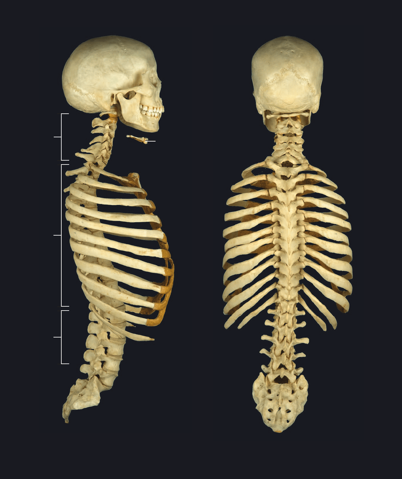
Axial skeleton
Lateral view
Axial skeleton
Posterior view
2
1
1
4
4
5
5
3
6
6
7
8
8
9
9
10
11
11
12
12
13
13
2
3
7
10
Atlas_AxialSkel.indd Page 35 15/03/11 8:42 PM user-F391Atlas_AxialSkel.indd Page 35 15/03/11 8:42 PM user-F391 /Users/user-F391/Desktop/Users/user-F391/Desktop
36
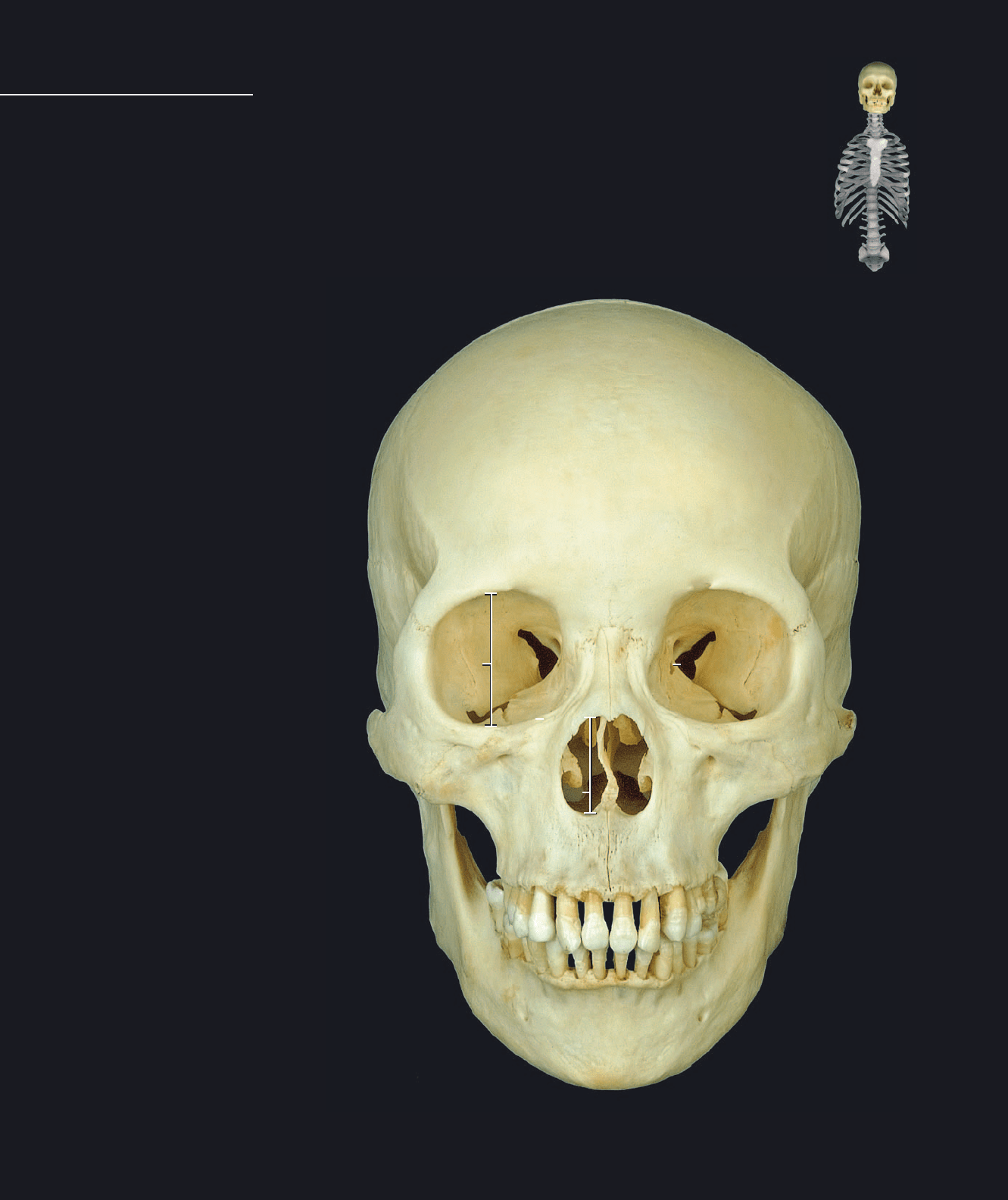
1 Frontal bone
2 Parietal bone
3 Occipital bone
4 Sphenoid bone
5 Temporal bone
6 Ethmoid bone
7 Inferior nasal concha
8 Lacrimal bone
9 Nasal bone
10 Vomer
11 Maxilla
12 Palatine bone
13 Zygomatic bone
14 Mandible
15 Bony nasal cavity
16 Piriform aperture
17 Inferior nasal meatus
18 Middle nasal meatu
19 Orbit
Cranium
Anterior view
functions, that include protecting the delicate brain tissue, fi xing the vestibular apparatus of the inner ear in
three-dimensional space, maintaining open air passageways for respiration, and acquiring and processing
food, to name a few. There are two main subdivisions of the cranium — the neurocranium or brain box is the
region that surrounds and encases the brain, and the viscerocranium or facial skeleton is the area contributing
to the orbits, nasal cavity, and oral cavity. This page and the facing page, and the four page spreads that follow,
depict the fi ve normas, or views, of the cranium in both articulated and disarticulated cranial images. The
bones of the skull are labeled on these views, along with key landmarks that can only be labeled on the articu-
lated cranium. Individual landmarks of the bones are labeled on the individual pictures of the cranial bones on
the pages that follow. This spread is of the norma facialis or facial aspect of the cranium.
The cranium is the composite skeleton of the head and is composed of 29 bones.
The bones of the cranium range from simple, non-descript plates of bone to the
most intricate bones of the skeleton. The cranial bones have a range of important
Cranium
1
2
4
5
6
6
7
8
9
10
11
12
13
14
16
15
18
17
19
12
16
15
18
17
Atlas_AxialSkel.indd Page 36 15/03/11 8:42 PM user-F391Atlas_AxialSkel.indd Page 36 15/03/11 8:42 PM user-F391 /Users/user-F391/Desktop/Users/user-F391/Desktop
37
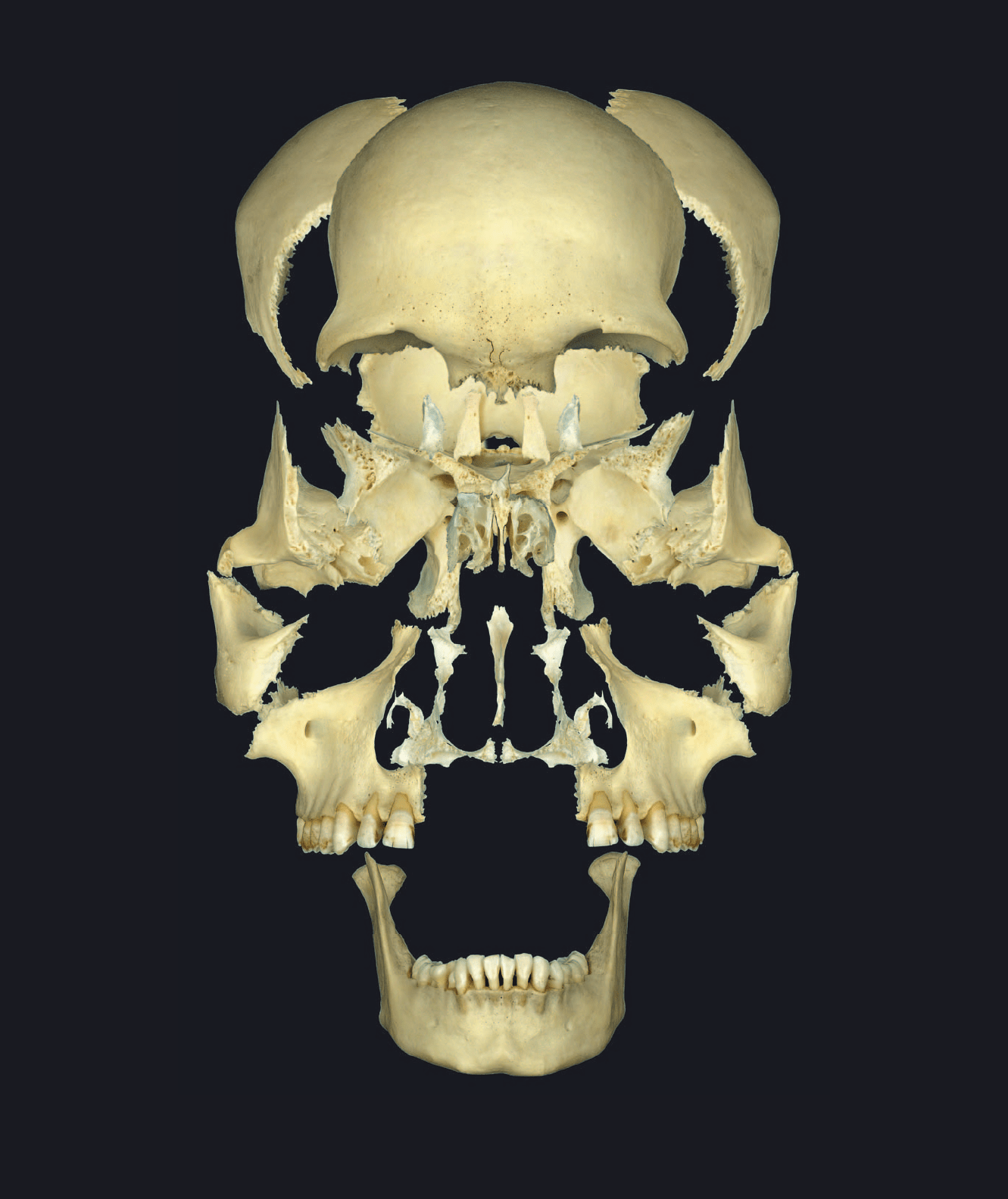
Bones of the cranium disarticulated
Anterior view
1
2
3
4
4
5
6
7
89
10
11
12
13
14
Atlas_AxialSkel.indd Page 37 15/03/11 8:42 PM user-F391Atlas_AxialSkel.indd Page 37 15/03/11 8:42 PM user-F391 /Users/user-F391/Desktop/Users/user-F391/Desktop

38
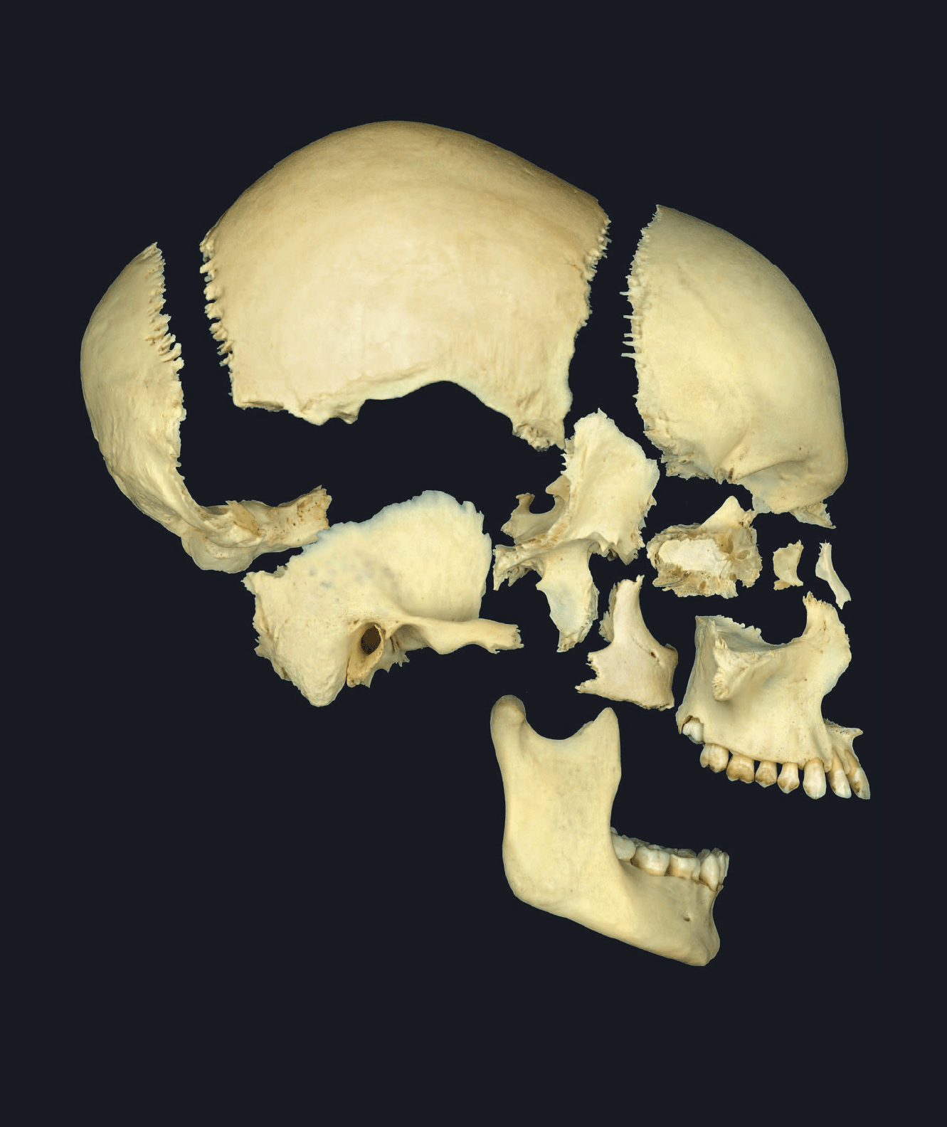
1 Frontal bone
2 Parietal bone
3 Occipital bone
4 Sphenoid bone
5 Temporal bone
6 Ethmoid bone
7 Lacrimal bone
8 Nasal bone
9 Maxilla
10 Zygomatic bone
11 Mandible
12 Zygomatic arch
13 Pterygopalatine fossa
Cranium
Cranium
Lateral view
This page spread depicts the norma lateralis, or lateral aspect of the cranium. In this view both
the brain box and facial skeleton are clearly visible and the relative proportions of the two cranial
regions are evident. In the disarticulated view, only those bones that are visible in the lateral as-
pect are shown.
12
3
4
5
6
7
8
9
10
11
12
13
Atlas_AxialSkel.indd Page 38 15/03/11 8:42 PM user-F391Atlas_AxialSkel.indd Page 38 15/03/11 8:42 PM user-F391 /Users/user-F391/Desktop/Users/user-F391/Desktop
39
Bones of the cranium disarticulated
Lateral view
1
2
3
4
5
6
7
8
9
10
11
Atlas_AxialSkel.indd Page 39 15/03/11 8:42 PM user-F391Atlas_AxialSkel.indd Page 39 15/03/11 8:42 PM user-F391 /Users/user-F391/Desktop/Users/user-F391/Desktop
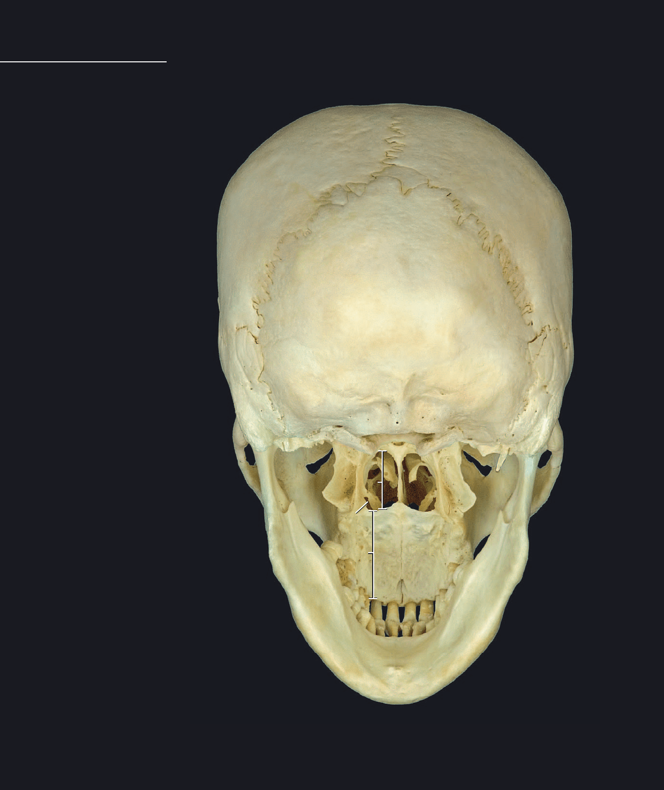
40
Cranium
1 Parietal bone
2 Occipital bone
3 Sphenoid bone
4 Temporal bone
5 Ethmoid bone
6 Inferior nasal concha
7 Vomer
8 Maxilla
9 Palatine bone
10 Zygomatic bone
11 Mandible
12 Choana or posterior nasal aperture
13 Inferior orbital fissure
14 Bony nasal cavity
15 Middle nasal meatus
16 Inferior nasal meatus
17 Bony palate
18 Sutural bone
Cranium
Posterior view
This page spread depicts the norma occipitalis, or occipital aspect of the cranium. From this pos-
terior view the internal aspects of the bones of the oral and nasal cavities are clearly visible. In the
disarticulated view only those bones that are visible in the occipital aspect of the cranium are
depicted.
1
2
3
4
5
6
7
8
9
10
11
12
13
14
15
16
17
18
5
12
13
14
15
16
Atlas_AxialSkel.indd Page 40 15/03/11 8:43 PM user-F391Atlas_AxialSkel.indd Page 40 15/03/11 8:43 PM user-F391 /Users/user-F391/Desktop/Users/user-F391/Desktop
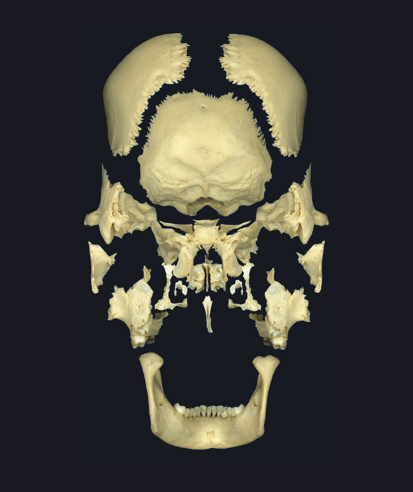
41
Bones of the cranium disarticulated
Posterior view
1
2
3
4
5
6
7
8
9
10
11
Atlas_AxialSkel.indd Page 41 15/03/11 8:43 PM user-F391Atlas_AxialSkel.indd Page 41 15/03/11 8:43 PM user-F391 /Users/user-F391/Desktop/Users/user-F391/Desktop
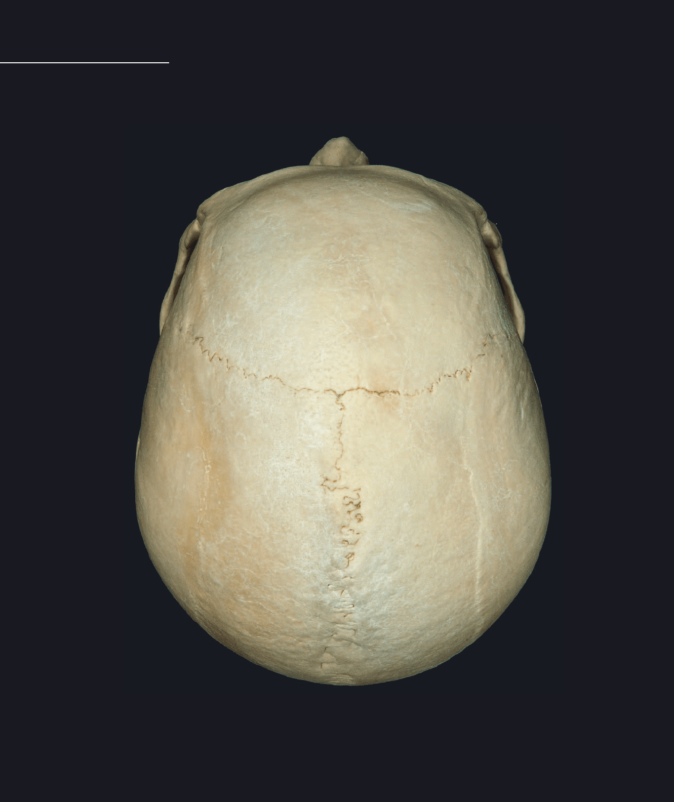
42
Cranium
1 Frontal bone
2 Parietal bone
3 Occipital bone
4 Temporal bone
5 Nasal bone
6 Maxilla
7 Zygomatic bone
Cranium
Superior view
This page spread depicts the norma superior, or superior aspect of the cranium. This view clearly
depicts the neurocranium or brain box, while the facial skeleton is almost completely hidden from
view. In the disarticulated view only those bones that are visible in the superior aspect of the cranium
are depicted.
1
2
4
5
6
7
5
6
7
Atlas_AxialSkel.indd Page 42 15/03/11 8:43 PM user-F391Atlas_AxialSkel.indd Page 42 15/03/11 8:43 PM user-F391 /Users/user-F391/Desktop/Users/user-F391/Desktop
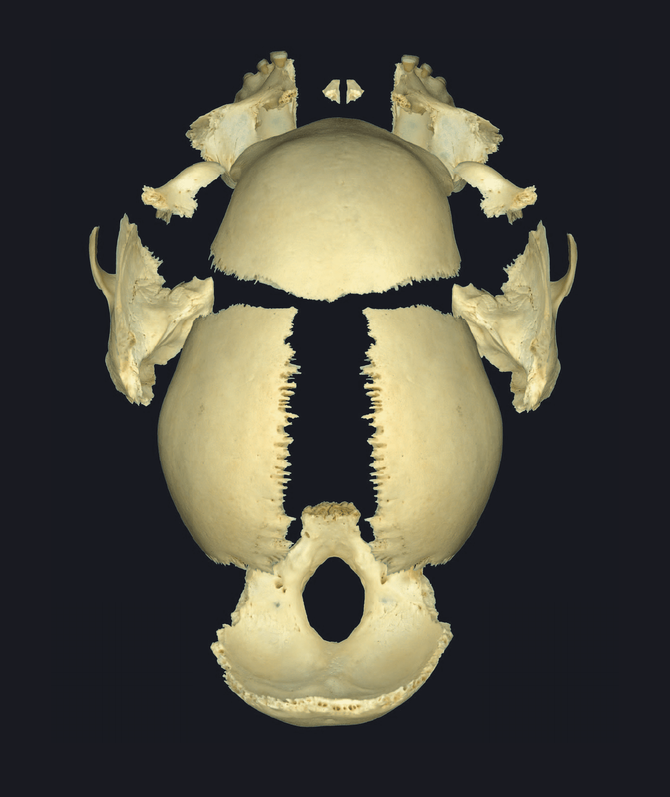
43
Bones of the cranium disarticulated
Superior view
1
2
3
4
5
6
7
Atlas_AxialSkel.indd Page 43 15/03/11 8:43 PM user-F391Atlas_AxialSkel.indd Page 43 15/03/11 8:43 PM user-F391 /Users/user-F391/Desktop/Users/user-F391/Desktop
44
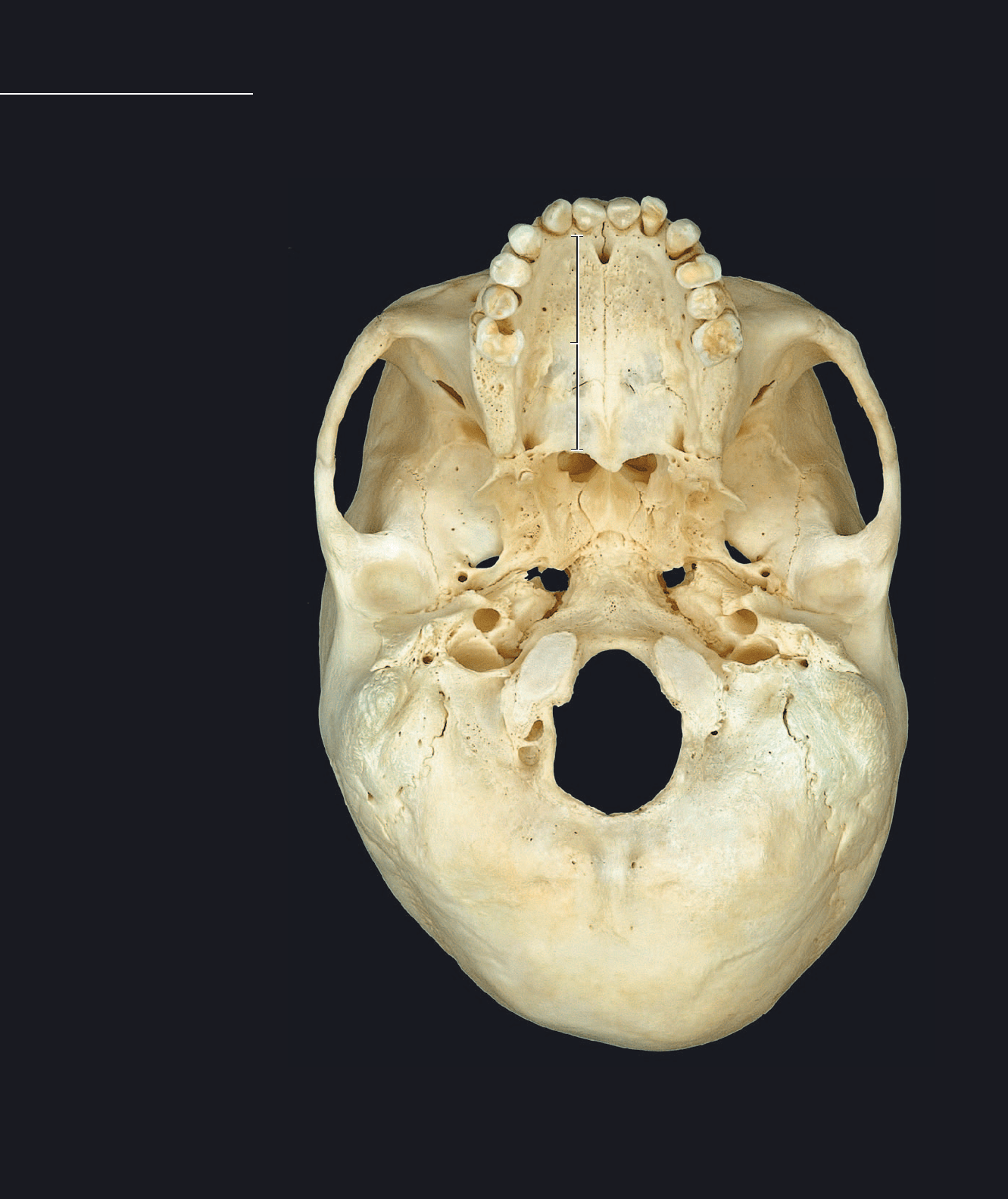
Cranium
1 Occipital bone
2 Sphenoid bone
3 Temporal bone
4 Vomer
5 Maxilla
6 Palatine bone
7 Zygomatic bone
8 Bony palate
9 Choana or posterior nasal aperture
10 Zygomatic arch
11 Jugular foramen
12 Foramen lacerum
13 Greater palatine foramen
14 Incisive fossa
Cranium
Inferior view
This page spread depicts the norma inferior (basalis), or inferior aspect of the cranium. The man-
dible has been removed to more clearly reveal the basicranium. This view clearly depicts the fl oor
of the brain box, the bony palate forming the roof of the oral cavity, and mandibular tooth row. In
the disarticulated view only those bones that are visible in the inferior aspect of the cranium are
depicted.
1
2
3
4
5
6
7
9
10
11
12
13
8
14
14
9
12
13
14
Atlas_AxialSkel.indd Page 44 15/03/11 8:43 PM user-F391Atlas_AxialSkel.indd Page 44 15/03/11 8:43 PM user-F391 /Users/user-F391/Desktop/Users/user-F391/Desktop
