Moss Tom. DNA-protein interactions: principles and protocols
Подождите немного. Документ загружается.

170 Geiselmann and Boccard
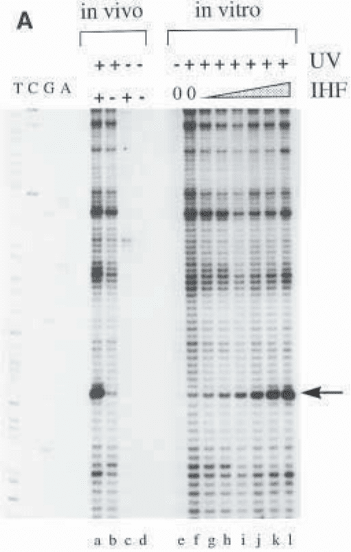
Fig. 2. UV-laser footprinting. (A) Primer extension profile of a UV-laser foot-
printing experiment showing the binding of IHF to a specific binding site. The
four lanes on the left, labeled TCGA, are a sequencing reaction using the same
primer as the one used for the primer extension of the UV-laser footprinting reac-
tion (only the T lane is clearly visible on the picture). Increasing amounts of IHF
(0–200 nM, lanes f to l) are incubated with 5 nM plasmid and footprinted in vitro,
as described in Subheading 3. The arrow points to the major footprinting signal.
Lane e is identical to lane f, but the DNA has not been irradiated. The in vivo
reactions are carried out as described in the protocol. Lane a is derived from wt
cells expressing IHF, lane b is a footprinting reaction from a strain lacking IHF.
UV-Laser Footprinting 171
The control lanes c and d show that the preparation of the plasmid DNA is suffi-
ciently clean for an efficient primer extension and that the bands seen in lanes a
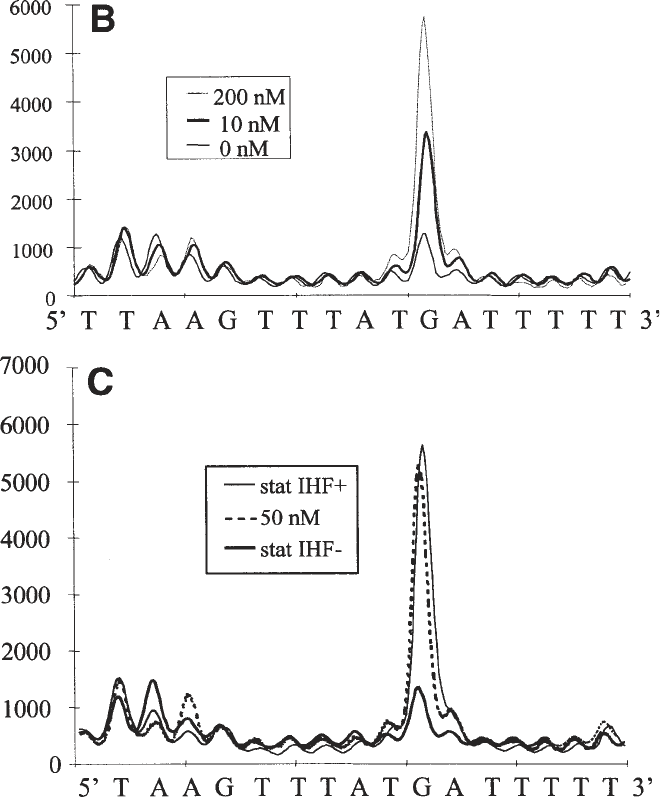
and b are due to the UV irradiation. (B) Superposition of line profiles from lanes f,
h, and l, corresponding to the indicated concentrations of IHF. The intensities of
the bands are in arbitrary units. (C) An equivalent superposition of the in vivo
profiles and the in vitro profile corresponding to the 50-nM IHF lane shows that
E. coli contains roughly the same amount of free IHF in stationary-phase cells as
was present in the in vitro sample using 50 nM (total) IHF. As expected, the foot-
print in a strain lacking functional IHF shows the same profile as DNA alone in
the in vitro reaction.
172 Geiselmann and Boccard
13. In vivo footprinting. Primer extension of the irradiated template can only be per-
formed in vitro. It is therefore necessary to extract the irradiated DNA from the
bacteria. Our current technology allows the measurement of protein binding to
specific binding sites carried on a multicopy plasmid. In principle, a primer
extension reaction on chromosomal DNA should work as well. In practice the
signals obtained from chromosomal DNA are too weak. Increasing the number
of samples irradiated does not remedy the problem. Because of the large excess
of chromosomal DNA with respect to the primer extension product, we observe
abnormal migration of the band in the sequencing gel.
The sample for in vivo UV-laser footprinting must be prepared such that a
single pulse of the laser (typically about 30 mJ per pulse, corresponding to
4 × 10
16
photons [i.e., 67 nmol of photons]) delivers more photons than there are
absorbing molecules in the sample. For an in vivo experiment, the absorbing
molecules are mostly made up of cellular DNA and RNA, as well as free nucleo-
side phosphates. An upper estimate of the concentration of absorbing molecules
within an Escherichia coli cell is about 100 mM, corresponding to 6 × 10
8
absorbers per cell. Because a single pulse delivers 4 × 10
16
photons and because
we want an excess of photons over absorbers, we want to irradiate less than about
10
8
E. coli cells per pulse. This numbers corresponds to about 100 µL of a sus-
pension at 1 OD
600
. A large number of 50-µL samples are therefore irradiated
and the cells are frozen immediately after irradiation.
References
1. von Hippel, P. H. and Berg, O. G. (1986) On the specificity of DNA–protein
interactions. Proc. Natl. Acad. Sci. USA 83(6), 1608–1612.
2. Sauer, R. T. (1991) Protein–DNA interactions, in Methods in Enzymology, vol.
208, Academic Press, San Diego, CA.
3. Jost, J.-P. and Saluz, H. P. (1991) A laboratory guide to in vitro studies of protein-
DNA interactions, in Biomethods, vol. 5, Birkhäuser Verlag, Basel.
4. Hockensmith, J. W., Kubasek, W. L., Vorachek, W. R., and von Hippel, P. H.
(1993) Laser cross-linking of proteins to nucleic acids. I. Examining physical
parameters of protein–nucleic acid complexes. J. Biol. Chem. 268, 15,712–15,720.
5. Pashev, I. G., Dimitrov, S. I., and Angelov, D. (1991) Crosslinking proteins to
nucleic acids by ultraviolet laser irradiation. Trends Biochem. Sci. 16, 323–326.
6. Panyutin, I. G., Kovalsky, O. I., and Budowsky, E. I. (1989) Irradiation of the
template with high-intensity (pulse-laser) ultraviolet light results in DNA–poly-
merase termination events at deoxyguanosine residues. FEBS Lett. 258, 274–276.
7. Menshonkova, T. N., Simukova, N. A., Budowsky, E. I., and Rubin, L. B. (1980)
The effect of high intensity ultraviolet irradiation on nucleic acids and their com-
ponents. Cleavage of N-glycosidic bond in thymidine, adenosine and 2'-deoxy-
adenosine. FEBS Lett. 112, 299–301.
8. Matsunaga, T., Hieda, K., and Nikaido, O. (1991) Wavelength dependent formation
of thymine dimers and (6–4) photoproducts in DNA by monochromatic ultraviolet
light ranging from 150 to 365 nm. Photochem. Photobiol. 54, 403–410.
UV-Laser Footprinting 173
9. Hockensmith, J. W., Kubasek, W. L., Vorachek, W. R., Evertsz, E. M., and von
Hippel, P. H. (1991) Laser cross-linking of protein-nucleic acid complexes. Meth-
ods Enzymol. 208, 211–236.
10. Eichenberger, P., Dethiollaz, S., Buc, H., and Geiselmann, J. (1997) Structural
kinetics of transcription activation at the malT promoter of Escherichia coli by
UV laser footprinting. Proc. Natl. Acad. Sci. USA 94, 9022–9027.
11. Buckle, M., Pemberton, I. K., Jacquet, M. A., and Buc, H. (1999) The kinetics of
sigma subunit directed promoter recognition by E. coli RNA polymerase. J. Mol.
Biol. 285, 955–964.
12. Murtin, C., Engelhorn, M., Geiselmann, J., and Boccard, F. (1998) A quantitative
UV laser footprinting analysis of the interaction of IHF with specific binding
sites: re-evaluation of the effective concentration of IHF in the cell. J. Mol. Biol.
284, 949–961.
13. Nash, H. A. (1996) The HU and IHF proteins: accessory factors for complex pro-
tein-DNA assemblies, in Regulation of Gene Expression in Escherichia coli (Lin,
E. E. C. and Lynch, A. S., eds.), R.G. Landes Company, Austin, TX, pp. 149–179.
14. Rice, P. A., Yang, S., Mizuuchi, K., and Nash, H. A. (1996) Crystal structure of
an IHF-DNA complex: a protein-induced DNA U-turn. Cell 87, 1295–1306.
15. Engelhorn, M., Boccard, F., Murtin, C., Prentki, P., and Geiselmann, J. (1995) In
vivo interaction of the Escherichia coli integration host factor with its specific
binding sites. Nucleic Acids Res. 23, 2959–2965.
16. Miller, J. H. (1992) A Short Course in Bacterial Genetics. Cold Spring Harbor
Laboratory, Cold Spring Harbor, NY.
17. Buckle, M., Buc, H., and Travers, A. (1992) DNA deformation in nucleoprotein
complexes between RNA polymerase, cAMP receptor protein and the lac UV5
promoter probed by singlet oxygen. EMBO J. 11, 2619–2625.

In Vivo DNA Analysis 175
175
From:
Methods in Molecular Biology, vol. 148: DNA–Protein Interactions: Principles and Protocols, 2nd ed.
Edited by: T. Moss © Humana Press Inc., Totowa, NJ
13
In Vivo DNA Analysis
Régen Drouin, Jean-Philippe Therrien, Martin Angers,
and Stéphane Ouellet
1. Introduction
The in vivo analysis of DNA–protein interactions and chromatin structure
can provide several kinds of critical information regarding regulation of gene
expression and gene function. For example, DNA sequences spanned by
nuclease-hypersensitive sites or bound by transcription factors often corre-
spond to genetic regulatory elements. Using the ligation-mediated polymerase
chain reaction (LMPCR) technology it is possible to map such DNA sequences
and to demonstrate the existence of unusual DNA structures directly in living
cells. LMPCR analyses can thus be used as a primary investigative tool to
identify the regulatory sequences involved in gene expression. Once specific
promoter sequence sites are shown to be bound by transcription factors in liv-
ing cells, it is often possible to establish the identity of these factors simply by
comparison with the consensus binding sites of known factors such as Sp1,
AP-1, NF-1, and so forth. The identity of each factor can then be confirmed
using in vitro gel shift (electrophoretic mobility shift assay [EMSA]) or
footprinting assays.
Clearly, gene promoters are best studied in their natural state in the living
cell and, thus, it is not surprising that in vivo DNA footprinting is one of the
most accurate predictors of the state of transcriptional activity of genes (1–3).
The native state of a gene and most of the special DNA structures are unavoid-
ably lost when DNA is cloned or purified (1–4). Hence, the commonly used in
vitro methods, such as in vitro footprinting and EMSAs, cannot demonstrate
that a given DNA–protein interaction actually occurs within the cells of inter-
est. With the advent of in vivo DNA footprinting, in vitro studies have been
extended to the situation in living cells, revealing the cellular processes impli-

176 Drouin et al.
cated in the regulation of gene expression. LMPCR is the method of choice for
in vivo footprinting and DNA structure studies because it can be used to inves-
tigate complex animal genomes, including that of human. The quality and use-
fulness of the information obtained from any in vivo DNA analysis, however,
depends on three parameters: (1) the integrity of the native chromatin substrate
used in the experiment, (2) the structural specificity of the chromatin probe,
and (3) the sensitivity of the assay. The ideal chromatin substrate is, of course,
that found inside intact cells. However, a near-ideal chromatin substrate is still
to be found in permeabilized cells, allowing the application of a wider range of
DNA cleavage agents, including DNase I.
In vivo footprinting assesses the local reactivity of modifying agents on the
DNA of living cells as compared to that on purified DNA (see Figs. 1–4). Two
steps characterize an in vivo footprinting analysis: (1) the treatment of purified
DNA and of cells with a given DNA modifying agent and (2) the visualization
of nucleotide modifications on a DNA sequencing gel. The latter step requires
that the modifying agent either directly induces DNA strand breaks or modi-
fies DNA nucleotides such that strand breaks can subsequently be induced in
vitro. A comparison is then made between the modification frequency on puri-
fied DNA and that on the DNA in living cells. For example, each guanine
residue of purified DNA has a near-equivalent probability of being methylated
by dimethylsulfate (DMS) and, thus, the cleavage pattern of in vitro modified
DNA appears on a sequencing gel as a ladder of bands of roughly equal inten-
sity. However, as a result of the presence of DNA-binding proteins, all guanine
residues do not show the same accessibility to DMS in living cells (Fig. 1).
Thus, differences between banding patterns obtained from in vitro and in vivo
modified DNA can be used to infer the sites of protein binding in living cells.
As will be seen, it is always advisable to validate such interpretations using
more than one footprinting agent.
The step of visualizing in vivo footprints has historically been problematic
because of the dilute nature of the sequences of interest and the complexity of
the genomes of higher eukaryotes. The development of an extremely sensitive
and specific technique, such as LMPCR, was thus necessary. The LMPCR tech-
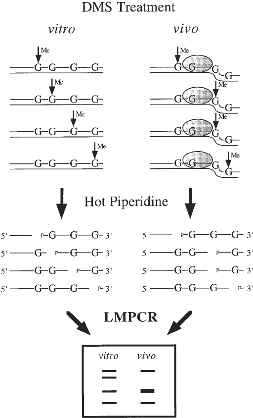
Fig. 1. (opposite page) Overall scheme for in vivo DNA analysis using DMS. The
methylation of guanine residues following DMS treatment of purified DNA (in vitro)
and cells (in vivo) is shown by vertical arrows and methylated residues (Me). When
purified DNA is treated with DMS, every guanine residue has a similar probability of
being methylated. However, the guanine residue in intimate contact with a sequence-
specific DNA-binding protein illustrated by the dotted oval is protected from DMS
methylation, whereas the guanine residues localized close to the boundary of a DNA–
protein contact that modifies DNA structure, allowing a better accessibility to DMS, is
In Vivo DNA Analysis 177
methylated more frequently. The methylated guanine residues are cleaved by hot pip-
eridine leaving phosphorylated 5' ends. On the sequencing ladder following LMPCR,
guanine residues that are protected from methylation appear as missing or less intense
bands when compared with the sequencing ladder from the same DNA sequence
obtained after DMS treatment of purified DNA. On the other hand, guanine residues
that undergo enhanced DMS methylation appear as darker bands in the sequencing
ladder relative to the purified DNA control.
178 Drouin et al.
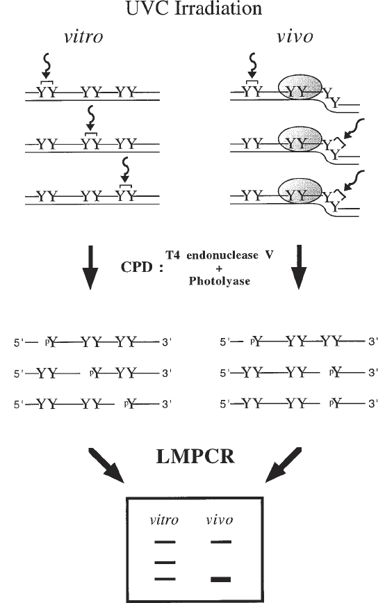
Fig. 2. Overall scheme for in vivo DNA analysis using UVC and CPD formation.
The CPD formation following UVC exposure of purified DNA (in vitro) and cells (in
vivo) is shown with curved arrows and brackets linking two adjacent pyrimidines (Y).
When purified DNA is irradiated with UVC, the frequency of CPD formation at
dipyrimidine sites is determined by the DNA sequence. However, the presence of a
sequence-specific DNA-binding protein illustrated by the dotted oval as well as DNA
structure can prevent (negative photofootprint) or enhance (positive photofootprint)

In Vivo DNA Analysis 179
nique quantitatively maps single-strand DNA breaks having phosphorylated 5'
ends within single-copy DNA sequences. It was first developed by Mueller
and Wold (5) for DMS footprinting, and, subsequently, Pfeifer and colleagues
adapted it to DNA sequencing (6), methylation analyses (1,6,7), DNase I
footprinting (2), nucleosome positioning (2), and UV photofootprinting (4,8).
LMPCR can be combined with a variety of DNA-modifying agents used to
probe the chromatin structure in vivo. It is our opinion that no single technique
can provide as much information on the DNA–protein interactions and DNA
structures existing within living cells as can LMPCR.
1.1. General Overview of LMPCR
Genomic sequencing techniques such as that developed by Church and Gil-
bert (9) can be used to map strand breaks in mammalian genes at nucleotide
resolution. However, by incorporating an exponential amplification step,
LMPCR (outlined in Fig. 5) constitutes a genomic sequencing method orders
of magnitude more sensitive than the direct technique of Church and Gilbert. It
uses 20 times less DNA than this latter technique to obtain a nucleotide-resolu-
tion banding pattern and allows short autoradiographic exposure times. The
unique aspect of LMPCR is the blunt-end ligation of an asymmetric double-
stranded linker (5' overhanging to avoid self-ligation or ligation in the wrong
direction) onto the 5' end of each cleaved blunt-ended DNA molecule (5,6).
The blunt end is created by the extension of a gene-specific primer (primer 1 in
Fig. 5) until a footprinting strand break is reached. Because the generated
breaks will be randomly distributed along the genomic DNA and thus have 5'
ends of unknown sequence, the asymmetric linker adds a common and known
sequence to all 5' ends. This then allows exponential PCR amplification from
an adjacent genomic sequence to that of the generated breaks using the longer
oligonucleotide of the linker (linker-primer) and a second nested gene-specific
primer (primer 2, see Fig. 5). After 20–22 cycles of PCR, the DNA fragments
are size-fractionated on a sequencing gel. LMPCR preserves the quantitative
representation of each fragment in the original population of cleaved molecules
(10–13), allowing quantification on a phosphorimager (14–17). Thus, the band
intensity pattern obtained by LMPCR directly reflects the frequency distribu-
CPD formation. The CPDs are cleaved by T
4
endonuclease V digestion and photolyase
photoreactivation leaving phosphorylated 5' ends. On the sequencing ladder following
LMPCR, the negative photofootprints appear as missing or less intense bands when
compared with the sequencing ladder from the same DNA sequence obtained after
UVC irradiation of purified DNA. On the other hand, positive photofootprints appear
as darker bands in the sequencing ladder relative to the purified DNA control.
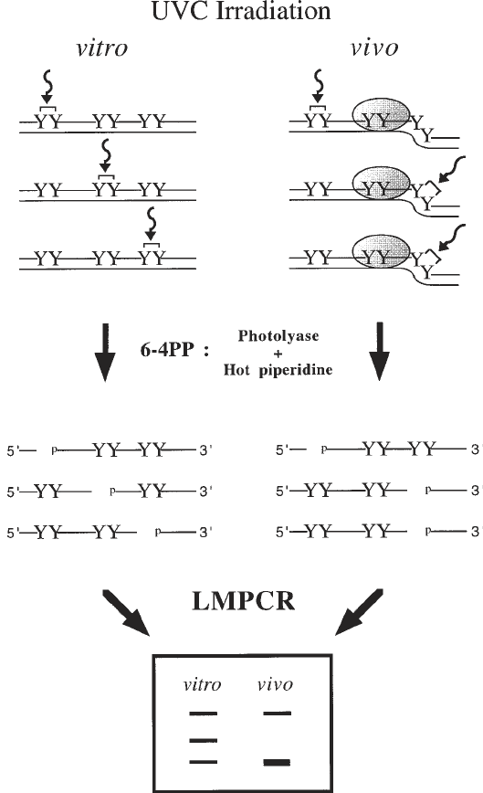
180 Drouin et al.
Fig. 3. Overall scheme for in vivo DNA analysis using UVC and 6–4PP formation.
The 6–4PP formation following UVC exposure of purified DNA (in vitro) and cells
(in vivo) is shown with curved arrows and brackets linking two adjacent pyrimidines
(Y). When purified DNA is irradiated with UVC, the frequency of 6–4PP formation at
dipyrimidine sites is determined by the DNA sequence. However, the presence of a
sequence-specific DNA-binding protein illustrated by the dotted oval as well as DNA
structure can prevent (negative photofootprint) or enhance (positive photofootprint)
