Moss Tom. DNA-protein interactions: principles and protocols
Подождите немного. Документ загружается.


In Vivo DNA Analysis 181
tion of 5'-phosphoryl DNA breaks along a 200-bp sequence adjacent to the
nested primer.
Two methods exist to reveal the sequence and footprinting ladders created
by LMPCR. Pfeifer and colleagues (6) took advantage of electroblotting DNA
onto a nylon membrane followed by hybridization with a gene-specific probe
to reveal sequence ladders, otherwise known as “indirect end labeling”. On the
other hand, Mueller and Wold (5) used a nested third radiolabeled primer for
the last one or two cycles of the PCR amplification step. We find Pfeifer’s
method much more sensitive than Mueller and Wold’s (unpublished data). In
this chapter, we will describe our LMPCR protocol as modified from the pro-
tocol of Pfeifer and colleagues.
In summary, LMPCR is the method of choice to study the in vivo structure
of promoters with respect to the positions of DNA–protein interactions, of
special DNA structures and of chromatin structures such as nucleosomes. To
perform in vivo DNA analysis, three probing agents are regularly combined
with LMPCR: DMS, ultraviolet (UV) and DNase I (Figs. 1–4, Table 1). These
probing agents provide complementary information and each has its associated
advantages and drawbacks (Table 2). To best characterize DNA–protein
interactions, it is often necessary to use two or even all three of these methods.
Treatments with any probing agents must produce either strand breaks
or modified nucleotides that can be converted to DNA strand breaks with a
5'-phosphate in vitro (Figs. 1–4, Table 3). In this chapter, we describe protocols
routinely used in our laboratory for DMS, UV, and DNase I in vivo treatments
as well as the associated LMPCR technology. These protocols may also be
adapted to footprinting with other probing agents, such as KMnO
4
and OsO
4
(see Chapters 6 and 9), although a detailed description is beyond the scope of
the present chapter.
1.2. In Vivo Dimethylsulfate (DMS) Footprint Analysis (Fig. 1)
Dimethylsulfate is a small, highly reactive molecule that easily diffuses
through the outer cell membrane and into the nucleus. It preferentially methy-
lates not only the N7 position of guanine residues via the major groove but, to
a lesser extent, also the N3 position of adenine residues via the minor groove.
6–4PP formation. First, CPDs are photoreactivated by photolyase and then 6–4PPs are
cleaved by hot piperidine treatment leaving phosphorylated 5' ends. On the sequenc-
ing ladder following LMPCR, the negative photofootprints appear as missing or less
intense bands when compared with the sequencing ladder from the same DNA
sequence obtained after UVC irradiation of purified DNA. On the other hand, positive
photofootprints appear as darker bands in the sequencing ladder relative to the puri-
fied DNA control.
182 Drouin et al.
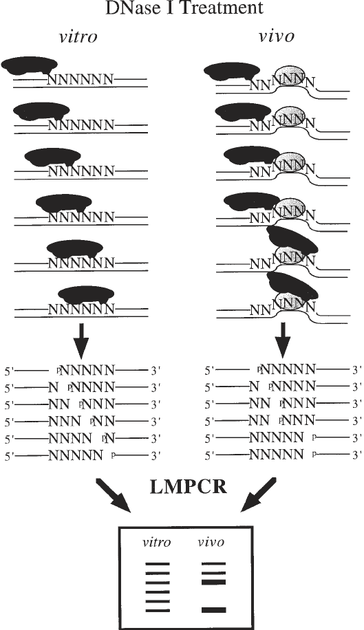
Fig. 4. Overall scheme for in vivo DNA analysis using DNase I. The DNase I
enzyme (the solid black) digestion of purified DNA (in vitro) and cells (in vivo) is
shown. When purified DNA is digested with DNase I, the cleavage pattern shows that
sites of the nucleotide sequence have similar probabilities of being cleaved. However,
the presence of a sequence-specific DNA-binding protein illustrated by the dotted oval

In Vivo DNA Analysis 183
The most significant technical advantage of in vivo DMS footprinting is that
DMS can be simply added to the cell culture medium, requiring no cell
manipulation (see Table 2 for advantages and drawbacks). Each guanine resi-
due of purified DNA displays about the same probability of being methylated
by DMS. Because DNA inside living cells forms chromatin and is often found
associated with a number of proteins, it is expected that its reactivity toward
DMS will differ from purified DNA. Figures 6 and 7 show in vivo DMS treat-
ment patterns compared to the treatment of purified genomic DNA. Proteins in
contact with DNA either decrease accessibility of specific guanines to DMS
(protection) or, as frequently observed at the edges of a footprint, increase
reactivity (hyperreactivity) (1). Hyperreactivity can also indicate a greater
DMS accessibility of special in vivo DNA structures (19). Hot piperidine
cleaves the glycosylic bond of methylated guanines and adenines, leaving a
ligatable 5'-phosphate (20).
Genomic footprinting using DMS reveals DNA–protein contacts located in
the major groove of the DNA double helix (Table 1). However, it should be
noted that in vivo DNA studies using DMS alone may not detect some DNA–
protein interactions (21). First, no DNA–protein interaction will be detected in
the absence of guanine residues. Second, some proteins do not affect DNA
accessibility to DMS. Third, certain weak DNA–protein contacts could actually
be disrupted because of the high reactivity of the DMS. Thus when using DMS,
it is often important to also apply alternative footprinting approaches (21,22).
1.3. Photofootprint Analysis (Figs. 2 and 3)
Ultraviolet light (UVC: 200–280 nm; UVB: 280–320 nm) can also be used
as a modifying agent for in vivo footprinting (4,8,23–25). When cells are sub-
jected to UV light (UVC or UVB), two major classes of lesions may be intro-
duced into the DNA at dipyrimidine sequences (CT, TT, TC, and CC): the
cyclobutane pyrimidine dimer (CPD) and the pyrimidine (6–4) pyrimidone
photoproduct (6–4PP) (26). CPDs are formed between the 5,6 bonds of any
two adjacent pyrimidines, whereas a stable bond between positions 6 and 4
of two adjacent pyrimidines characterizes 6–4PPs. 6–4PP are formed at a rate
15–30% of that of CPDs (27) and are largely converted to their Dewar valence
as well as DNA structure can prevent (protection) or enhance (hypersensitive) DNase
I cleavage. The DNase I cleavage leaves phosphorylated 5' ends. On the sequencing
ladder following LMPCR, DNA sequences that are protected from DNase I cleavage
appear as missing or less intense bands when compared with the sequencing ladder
from the same DNA sequence obtained after DNase I digestion of purified DNA. On
the other hand, hypersensitive sites that undergo enhanced DNase I cleavage appear as
darker bands in the sequencing ladder relative to the purified DNA control.
184 Drouin et al.

In Vivo DNA Analysis 185
isomers by direct secondary photolysis (photoisomerization) (27). In living
cells, the photoproduct distribution is determined both by sequence context
and chromatin structure (28). In general, CPDs and 6–4PPs appear to form
preferentially in longer pyrimidine runs. Because UVB and UVC radiation are
primarily absorbed in the cell by the DNA, there are relatively few perturba-
tions of other cellular processes, and secondary events that could modify the
chromatin structure or release DNA–protein interactions. Furthermore, intact
cells are exposed for a short period of time only to a high-intensity UV irradia-
tion. Thus, UV irradiation is probably one of the least disruptive footprinting
method and, hence, truly reflects the in vivo situation (Table 2). As for DMS,
DNA-binding proteins influence the distribution of UV photoproducts in a sig-
nificant way (23). When the photoproduct spectrum of irradiated purified DNA
is compared with that obtained after irradiation of living cells, some striking
differences become apparent. These are referred to as “photofootprints” (23).
The photoproduct frequency within sequences bound by sequence-specific
DNA-binding proteins (transcription factors) is suppressed or enhanced in com-
parison to purified DNA (4,8,29). Effects of chromatin structure may be sig-
nificant in regulatory gene regions that bind transcription factors (Fig. 6).
Mapping of CPDs at the single-copy gene level can reveal positioned nucleo-
somes because CPDs are modulated in a 10-bp periodicity within nucleosome
core DNA (30,31). 6–4PPs form more frequently in linker DNA than in core
DNA (32).
Photofootprints reveal variations in DNA structure associated with the pres-
ence of transcription factors or other proteins bound to the DNA. UV light has
the potential to reveal all DNA–protein interactions provided there is a
dipyrimidine sequence on either DNA strand within a putative protein-binding
sequence. Because photofootprints can be seen outside protein-binding sites,
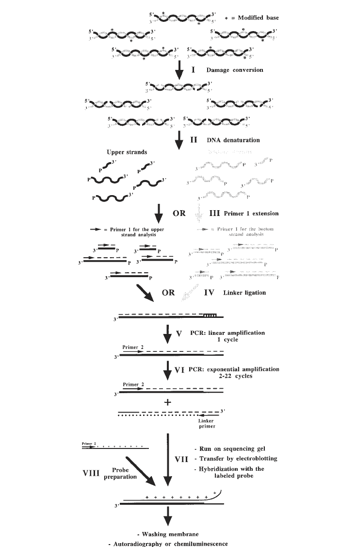
Fig. 5. (previous page) Outline of the LMPCR procedure. Step I: specific conver-
sion of modified bases to phosphorylated 5' single-strand breaks; Step II: denaturation
of genomic DNA; Step III: annealing and extension of primer 1 (although both strands
can be studied, each LMPCR protocol only involves the analysis of either the
nontranscribed strand or the transcribed strand); Step IV: ligation of the linker;
Step V: first cycle of PCR amplification, this cycle is a linear amplification because
only the gene-specific primer 2 can anneal; Step VI: cycles 2 to 22 of exponential PCR
amplification of gene-specific fragments with primer 2 and the linker primer (the
longer oligonucleotide of the linker); Step VII: separation of the DNA fragments on a
sequencing gel, transfer of the sequence ladder to a nylon membrane by electroblotting,
and visualization of the sequence ladder by hybridization with a labeled single-
stranded probe; Step VIII: preparation and isotopic or nonisotopic labeling of single-
stranded probe.

186 Drouin et al.
UV light should not be used as the only in vivo footprinting agent. The precise
delimitations of the DNA–protein contact are difficult to determine with the
simple in vivo UV probing method.
The distribution of UV-induced CPDs and 6–4PPs along genomic DNA can
be mapped at the sequence level by LMPCR following conversion of these
photoproducts into ligatable 5'-phosphorylated single-strand breaks. CPD are
enzymatically converted by cleavage with T
4
endonuclease V followed by
UVA (320–400 nm) photoreactivation of the overhanging pyrimidine using
photolyase (Fig. 2) (8). Because the 6–4PPs and their Dewar isomers are hot
alkali-labile sites, they can be cleaved by hot piperidine (Fig. 3) (29).
1.4. In Vivo DNase I Footprint Analysis (Fig. 4)
DNase I treatment of permeabilized cells gives clear footprints when the
DNase I-induced breaks are mapped by LMPCR (2). Both living cells (in vivo)
and purified DNA (in vitro) are treated with DNase I. As with DMS and UV,
footprint analyses are obtained by comparing in vivo DNase I digestion
patterns to patterns obtained from the digestion of purified genomic DNA
(Fig. 7). When compared to purified DNA, permeabilized cells show protected
bands at DNA–protein interaction sequences and DNase I hypersensitive bands
in regions of higher-order nucleoprotein structure (2). Compared to DMS,
DNase I is less base selective, is more efficient at detecting minor groove
DNA–protein contacts, provides more information on chromatin structure, dis-
plays larger and clearer footprints, and better delimits the boundaries of DNA–
protein interactions (Fig. 7). The nucleotides covered by a protein are almost
completely protected on both strands from DNase I nicking, allowing a better
Table 1
Purposes of the Three Main In Vivo Footprinting Approaches
Approaches Activities
1. Dimethylsulfate i. Localizes in vivo DNA–protein contacts located in the major
(DMS) groove of the DNA double helix
ii. Can detect special DNA structures
2. UV irradiation i. Localizes in vivo DNA–protein interactions and shows how
(UVB or UVC) DNA structure is affected in the presence of transcription factors
ii. Can detect special DNA structures
iii. Can show evidence of positioned nucleosomes
3. DNase I i. Localizes in vivo DNA–protein contacts
ii. Precisely maps in vivo DNase I hypersensitive sites
iii. Shows evidence of nucleosomes and their positions;
can differentiate core DNA from linker DNA

In Vivo DNA Analysis 187
187
Table 2
Advantages and Drawbacks of the Three Main In Vivo Footprinting Approaches
Approaches Advantages Drawbacks
DMS Treatment is technically easy to carry out; the 1. Requires guanines, therefore is sequence dependent.
DMS is a small molecule that penetrates very 2. Does not detect all DNA–protein interactions.
easily into living cells with little disruption.
UV irradiation 1. Treatment is technically easy to carry out; 1. Requires two adjacent pyrimidines, therefore is
(UVB or UVC) UV light penetrates through the outer sequence dependent.
membrane of living cells without disruption. 2. The interpretation of the results is sometime difficult;
2. Detects many DNA–protein interactions. to differentiate between DNA–protein interactions.
3. Very sensitive to particular and DNA structures. and special DNA structures can be very difficult.
DNase I 1. Little sequence dependency. 1. Technically difficult to carry out; reproducibility is
2. No conversion of modified bases required. often a problem.
3. Detects all DNA-protein contacts. 2. DNase I is a protein that can penetrate in living cells
4. Very sensitive to particular DNA structures. only following membrane permeabilization, thus
causing some cell disruption.

188 Drouin et al.
delimitation of the boundaries of DNA–protein contacts. However, it should
be underlined that the relatively bulky DNase I molecule cannot cleave the
DNA in the immediate vicinity of a bound protein because of steric hindrance.
Consequently, the regions protected from cutting can extend beyond the actual
DNA–protein contact site. On the other hand, when DNA is wrapped around a
nucleosome-size particle, DNase I cutting activity is increased at 10-bp inter-
vals and no footprint is observed (Tables 1 and 2).
DNase I, a relatively large 31-kDa protein, cannot penetrate cells without
previous cell-membrane permeabilization. Cells can be efficiently permeabi-
lized by lysolecithin (2) or Nonidet P40 (33). It has been shown that cells
permeabilized by lysolecithin remain intact, replicate their DNA very
efficiently, and show normal transcriptional activities (34,35). There are
numerous studies showing that lysolecithin-permeabilized cells maintain a nor-
mal nuclear structure to a greater extent than isolated nuclei, because the
chromatin structure can be significantly altered during the nuclear isolation
procedures (2). Indeed, DNase I footprinting studies using isolated nuclei
can be flawed because transcription factors are lost during the isolation of
nuclei in polyamine containing buffers (2). Even though other buffers may be
less disruptive, factors can still be lost during the isolation procedure, leading
to the loss of footprints or partial loss of footprints.
DNase I digestion of DNA leaves ligatable 5’-phosphorylated breaks, but
the 3’-ends are free hydroxyl groups. Pfeifer and colleagues (2,36) observed
that these genomic 3'-OH ends can be used as primers and be extended by the
DNA polymerases during the initial extension and/or PCR steps of LMPCR,
thereby reducing significantly the overall efficiency of LMPCR and giving a
Table 3
Mapping Schemes Used with the Three Main In Vivo Footprinting Approaches
Conversion of modified
Strand bases to DNA
Approaches breaks Modified bases single-strand breaks
DMS Few Guanine: methylated guanines Hot piperidine
at N7 position
Adenine: to a much lesser
extent, methylated adenines
at N3 position
UV irradiation Very (i) Cyclobutane pyrimidine (i) T
4
endonuclease V
(UVB or UVC) few dimers followed by photolyase
(ii) 6–4 Photoproducts (ii) Photolyase followed
by hot piperidine
DNase I Yes None No conversion required
In Vivo DNA Analysis 189
background smear on sequencing gels. To avoid the nonspecific priming of
these 3'-OH ends, three alternative solutions have been applied: (1) blocking
these ends by the addition of a dideoxynucleotide (2,36); (2) enrichment of
fragments of interest by extension product capture using biotinylated gene-
specific primers and magnetic streptavidin-coated beads (18,37–39); and (3)
performing primer 1 hybridization and primer 1 extension at a higher tempera-
ture (52–60°C vs 48°C, and 75°C vs 48°C, respectively) using a thermostable
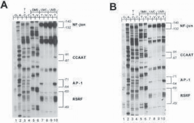
Fig. 6. LMPCR analysis of methylated guanines and CPD along the nontranscribed
strand of the c-jun promoter following DMS treatment, and UVB and UVC irradiation
respectively. (A) The membrane was hybridized with an isotopic [
32
P]-dCTP-labeled
probe. The membrane was exposed on film between two intensifying screens for 25 min
at –70°C. (B) The membrane was hybridized with a digoxigenin-labeled probe and
exposed on film for 40 min at room temperature. For this experiment, one LMPCR
protocol was carried out and only one gel was run on which all the samples (20 in
total) were loaded symmetrically in duplicate. Each symmetrical well of each set of
samples was loaded with exactly the same amount of DNA. Lanes 1–4: LMPCR of
DNA-treated with chemical cleavage reactions. These lanes represent the sequence
of the c-jun promoter analyzed with JD primer set (18). Lanes 5–6: LMPCR of DMS-
treated naked DNA (T: in vitro) and fibroblasts (V: in vivo) followed by hot piperidine
treatment. Lanes 7–10: LMPCR of UVC- and UVB-irradiated naked DNA (T) and
fibroblasts (V) followed by T
4
endonuclease V/photolyase digestion. On the right, the
consensus sequences of transcription factor binding sites are delimited by brackets.
The numbers indicate their positions relative to the major transcription initiation site.
190 Drouin et al.
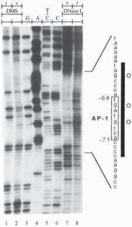
Fig. 7. LMPCR analysis of methylated guanines and DNA strand breaks along the
transcribed strand of the c-jun promoter following DMS treatment and DNase I diges-
tion, respectively. The membrane was hybridized with an isotopic [
32
P]-dCTP-labeled
probe. Lanes 1–2: LMPCR of DMS-treated purified DNA (t: in vitro) and fibroblasts
(v: in vivo) followed by hot piperidine treatment. Lanes 3–6: LMPCR of DNA-treated
with chemical cleavage reactions. These lanes represent the sequence of the c-jun pro-
moter analyzed with JC primer set (18). Lanes 7–8: LMPCR of DNase I-digested
permeabilized fibroblasts (v) and purified DNA (t). As a reference, a small portion
of the chemically derived sequence is shown on the right of the autoradiogram, the
AP-1-like binding sequence is enclosed by a box, and the numbers indicate its posi-
tion relative to the major transcription initiation site. Open circles represent guanines