Martienssen W., Warlimont H. (Eds.). Handbook of Condensed Matter and Materials Data
Подождите немного. Документ загружается.


32 Part 1 General Tables
Table 1.3-4 Crystal families, crystal systems, crystallographic point groups, conventional coordinate systems, and Bravais lattices
in three dimensions. Lattice point symmetries (holohedries) are given in bold
Crystal Symbol Crystal Crystallographic No. of Conventional Bravais lattice
family
system point groups space coordinate system (Pearson symbol)
groups Restrictions on Parameters
cell parameters to be determined
Triclinic a Triclinic 1,
¯
1 2 None a, b, c, aP
(anorthic)
α, β, γ
monoclinic m Monoclinic 2, m, 2/m 13 Setting with b unique: a, b, c, mP, mS
α = γ = 90
◦
β (mC, mA, mI)
Setting with c unique: a, b, c, mP, mS
α = β = 90
◦
γ (mA, mB, mI)
orthorhombic o Orthorhombic 222, mm2, 59 α = β = γ = 90
◦
a, b, c oP,oS,
mmm (oC, oA, oB)
oI, oF
tetragonal t Tetragonal 4,
¯
4, 4/m, 68 a = b a, c tP, tI
422, 4mm, α = β = γ = 90
◦
¯
42m, 4/mmm
hexagonal h Trigonal 3,
¯
3, 18 a =b, a, c hP
32, 3m,
¯
3m α = β = 90
◦
γ = 120
◦
(hexagonal
axes)
7 a = b =c a,α hR
α = β = γ =90
◦
(rhombohedral
axes)
Hexagonal 6,
¯
6, 6/m, 27 a = b, a, c hP
622, 6mm, α = β = 90
◦
¯
62m, 6/mmm γ = 120
◦
cubic c Cubic 23, m
¯
3, 36 a = b = c a cP, cI,cF
432,
¯
43m, α = β = γ = 90
◦
m
¯
3m
ing of the symbols is the following: P denotes a primitive
Bravais lattice. It belongs to the tetragonal crystal sys-
tem indicated by 4. Along the first standard blickrichtung
[001] there is a 4
2
screw axis with a perpendicular mir-
ror plane m. Along [100]there is a twofold rotation axis,
named 2, with a perpendicular glide plane c parallel to c.
Third, along [110]there is a twofold rotation axis 2, with
a perpendicular mirror plane m.
Decoration of the Lattice with the Basis
At this point we have to recall that in a real crys-
tal structure we have not only the lattice, but also the
basis. In [3.8], there are standardized sets of general
and special positions (i. e. coordinates x, y, z) within
the unit cell (Wyckoff positions). An atom placed in
a general position is transformed into more than one
atom by the action of all symmetry operators of the
respective space group. Special positions are located
on special points which are mapped onto themselves
by one or more symmetry operations – for example
a position in a mirror plane or exactly on a rotational
axis. Reference [3.8] also provides information about
symmetry relations between individual space groups
(group–subgroup relations). These are often useful for
describing relationships between crystal structures and
for describing phase transitions of materials.
The use of the space group allows us to further re-
duce the basis to the asymmetric unit: this is the minimal
set of atoms that needs to be given so that the whole crys-
tal structure can be generated via the symmetry of the
space group. This represents the main power of a crystal-
lographically correct description of a material: just some
10 parameters are sufficient to describe an ensemble of
some 10
23
atoms.
Part 1 3.1

Rudiments of Crystallography 3.1 Crystalline Materials 33
Thus, a crystallographically periodic structure of
a material is unambiguously characterized by
•
the cell parameters;
•
the space group;
•
the coordinates of the atoms (and their chemical
type) in the asymmetric unit;
•
the occupation and thermal displacement factors of
the atoms in the asymmetric unit.
For an example, the reader is referred to the crystal-
lographic description of the spinel structure of MgAl
2
O
4
given below under the heading “Structure Types”.
To complete the information on space group sym-
metries given here, periodic magnetic materials should
also be mentioned. Magnetic materials contain magnetic
moments carried by atoms in certain positions in the unit
cell. If we take into account the magnetic moments in the
description of the structure, the classification by space
groups (the 230 “gray” groups, described above) has to
be extended to 1651 the “black and white”, or Shub-
nikov, groups [3.9]. A magnetic periodic structure is
then characterized by
•
the crystallographic structure;
•
the Shubnikov group;
•
the cell parameters of the magnetic unit cell;
•
the coordinates of the atoms carrying magnetic mo-
ments (the asymmetric unit in the magnetic unit
cell);
•
the magnitude and direction of the magnetic mo-
ments on these atoms.
Structure Types
It is useful to classify the crystal structures of materials
by the assignment of structure types. The structure type
is based on a representative crystal structure, the param-
Table 1.3-5 Complete crystallographic parameter set for MgAl
2
O
4
, spinel structure type
Material MgAl
2
O
4
Structure type MgAl
2
O
4
, spinel
Pearson symbol cF56
Space group Fd
¯
3m (No. 227)
a (Å) 8.174(1)
Atom Wyckoff position xyzOccupancy
Mg 8a 0001.0
Al 16d 5/85/85/81.0
O32e 0.3863(2) xx1.0
eters of which describe the essential crystallographic
features of other materials of the same type. As an ex-
ample, we consider the structure of the spinel oxides
AB
2
O
4
. The generic structure type is MgAl
2
O
4
, cF56.
The Pearson symbol, here cF56, denotes the cubic crys-
tal family and a face-centered Bravais lattice with 56
atoms per unit cell (see Table 1.3-5 and also the last
column in Table 1.3-4).
Regarding the free parameters, for example a,the
notation 8.174(1) in Table 1.3-5 means 8.174 ±0.001.
The chemical formula and the unit cell contents can
easily be calculated from the site multiplicities (given by
the Wyckoff positions) and the occupancies. So can the
(crystallographic) density, using the appropriate atomic
masses.
There is a huge variety of other materials belonging
to the same structure type as in this example. The only
parameters that differ (slightly) are the numerical value
of a, the types of atoms in the positions, the numerical
value of the parameter x for Wyckoff position 32e,and
the occupancies. Thus, for example, the crystal structure
of the iron sulfide Fe
3
S
4
can be characterized in its
essential features via the information that it belongs to
the same structure type.
1.3.1.2 Aperiodic Materials
In addition to the crystalline periodic state of matter,
a class of materials exists that lacks 3-D translational
symmetry and is called aperiodic. Aperiodic materials
cannot be described by any of the 230 space groups
mentioned above. Nevertheless, they show another type
of long-range order and are therefore included in the
term “crystal”. This notion of long-range order is the
major feature that distinguishes crystals from amor-
phous materials. Three types of aperiodic order may
Part 1 3.1
34 Part 1 General Tables
be distinguished, namely modulated structures, com-
posite structures, and quasicrystals. All aperiodic solids
exhibit an essentially discrete diffraction pattern and
can be described as atomic structures obtained from
a 3-D section of a n-dimensional (n-D) (n > 3) periodic
structure.
Modulated Structures
In a modulated structure, periodic deviations of the
atomic parameters from a reference or basic structure are
present. The basic structure can be understood as a pe-
riodic structure as described above. Periodic deviations
of one or several of the following atomic parameters are
superimposed on this basic structure:
•
atomic coordinates;
•
occupancy factors;
•
thermal displacement factors;
•
orientations of magnetic moments.
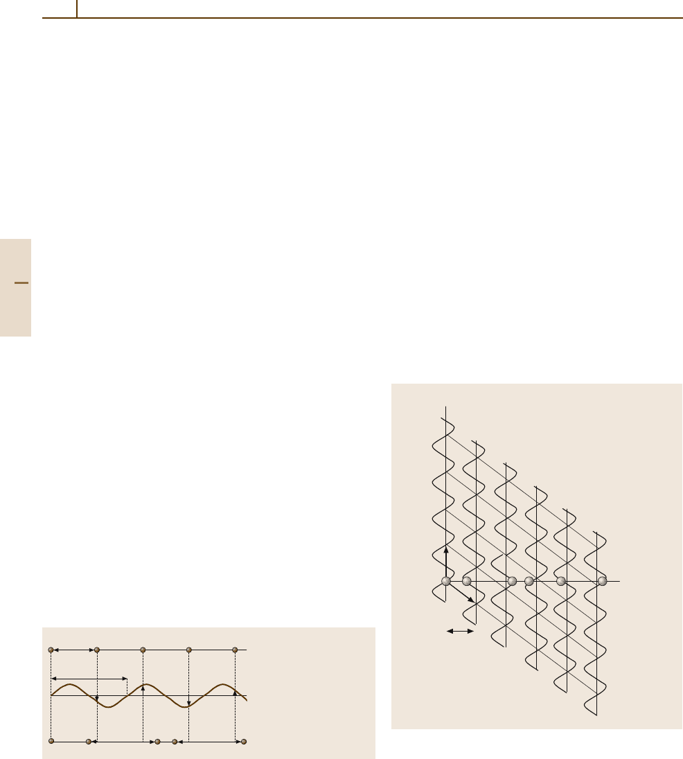
Let the period of the basic structure be a and the
modulation wavelength be λ; the ratio a/λ may be
(1) a rational or (2) an irrational number (Fig. 1.3-7).
In case (1), the structure is commensurately modulated;
we observe a qa superstructure, where q = 1/λ.This
superstructure is periodic. In case (2), the structure is
incommensurately modulated. Of course, the experi-
mental distinction between the two cases is limited by
the finite experimental resolution. q maybeafunc-
tion of external variables such as temperature, pressure,
or chemical composition, i. e. q = f(T, p, X),andmay
adopt a rational value to result in a commensurate
“lock-in” structure. On the other hand, an incommensu-
rate charge-density wave may exist; this can be moved
through a basic crystal without changing the internal
energy U of the crystal.
When a 1-D basic structure and its modulation func-
tion are combined in a 2-D hyperspace R = R
parallel
⊕
Basic structure s(r)
f(r) = sin(q r)
Modulated structure s
m
(r)
a
λ =1/q
+
=
Fig. 1.3-7 A 1-D modulated structure s
m
(r) can be described as
a sum of a basic structure s(r) and a modulation function f(r) of its
atomic coordinates. If a/λ is irrational, the structure is incommen-
surately modulated. Circles denote atoms
R
perpendicular
, periodicity on a 2-D lattice results. The
real atoms are generated by the intersection of the
1-D physical (external, parallel) space R
parallel
with the
hyperatoms in the complementary 1-D internal space
R
perpendicular
. In the case of a modulated structure, the
hyperatoms have the shape of the sinusoidal modulation
function in R
perpendicular
.
Figure 1.3-8 illustrates this construction. We have
to choose a basis (a
1
, a
2
) in R where the slope of a
1
with respect to R
parallel
corresponds to the length of the
modulation λ.
It is clear that real atomic structures are always mani-
festations of matter in 3-D real, physical space. The
cutting of the 2-D hyperspace to obtain real 1-D atoms
illustrated in Fig. 1.3-8 may serve as an instructive ba-
sic example of the concept of higher-dimensional (n-D,
n > 3) crystallography. The concept is also called a su-
perspace description; it applies to all aperiodic structures
and provides a convenient finite set of variables that can
be used to compute the positions of all atoms in the real
3-D structure.
R
perpendicular
R
parallel
,
s
m
(r)
a
2
a
a
1
Fig. 1.3-8 2-D hyperspace description of the example of
Fig. 1.3-7. The basis of the hyperspace R = R
parallel
⊕
R
perpendicular
is (a
1
, a
2
); the slope of a
1
with respect to
R
parallel
is proportional to λ. Atoms of the modulated struc-
ture s
m
(r) occur in the physical space R
parallel
and are
represented by circles
Part 1 3.1

Rudiments of Crystallography 3.1 Crystalline Materials 35
The modulation may occur in one, two, or
three directions of the basic structure, yielding
1-D, 2-D, or 3-D modulated structures. If we in-
troduce one additional dimension per modulation
vector (the direction r that the modulation corres-
ponding to λ runs along), these structures can be
described as periodic in 4-D, 5-D, or 6-D superspace,
respectively.
Composite Structures
Composite crystals are crystalline structures that consist
of two or more periodic substructures, each one hav-
ing its own 3-D periodicity to a first approximation.
The symmetry of each of these subsystems is character-
ized by one of the 230 space groups. However, owing
to their mutual interaction, the true structure consists
of a collection of incommensurately modulated subsys-
tems. All known composite structures to date have at
least one lattice direction in common and consist of
a maximum of three substructures. There are three main
classes:
•
channel structures;
•
columnar packings;
•
layer packings.
These composite structures are also known as inter-
growth or host–guest structures. Figure 1.3-9 illustrates
an example of a host with channels along a,inwhich
atoms of the substructure with a periodicity λa reside as
a guest.
a
b
λa
c
Fig. 1.3-9 Host–guest channel structure. The guest atoms
reside in channels parallel to a, with a periodicity λa
The higher-dimensional n-D formalism (n > 3) used
to describe composite structures is essentially the same
as that which applies to modulated structures.
Quasicrystals
Quasicrystals represent the third type of aperiodic ma-
terials. Quasiperiodicity may occur in one, two, or three
dimensions of physical space and is associated with
special irrational numbers such as the golden mean
τ =(1+
√
5)/2, and ξ = 2 +
√
3. The most remarkable
feature of quasicrystals is the appearance of noncrystal-
lographic point group symmetries in their diffraction
patterns, such as 8/mmm,10/mmm,12/mmm,and
2/m
¯
3
¯
5. The golden mean is related to fivefold symme-
try via the relation τ =2cos(π/5); τ can be considered
as the “most irrational” number, since it is the irrational
number that has the worst approximation by a truncated
continued fraction,
τ =1 +
1
1+
1
1+
1
1+
1
1+
1
1+...
.
This might be a reason for the stability of quasiperi-
odic systems where τ plays a role. A prominent 1-D
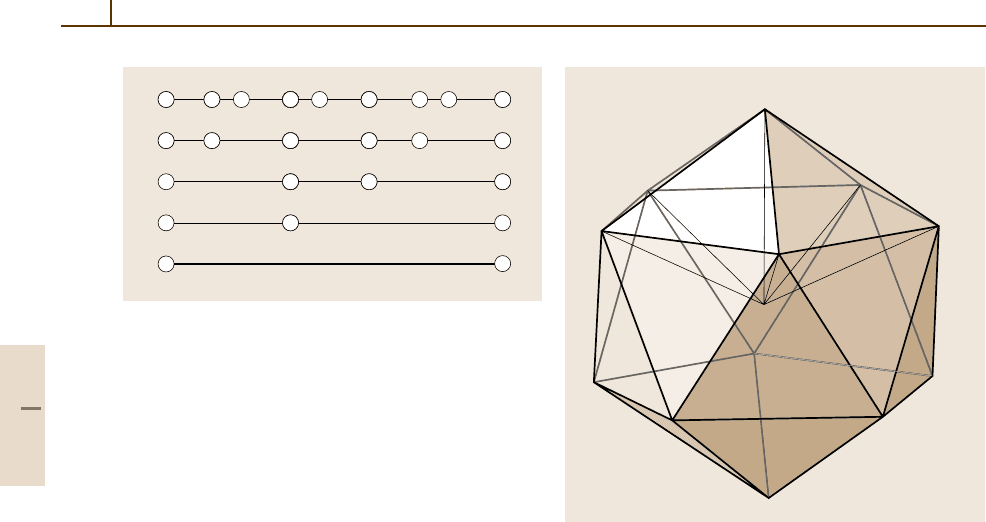
example is the Fibonacci sequence, an aperiodic chain
of short and long segments S and L with lengths
S and L, where the relations L/S = τ and L +S = τL
hold. A Fibonacci chain can be constructed by the sim-
ple substitution or inflation rule L → LS and S → L
(Table 1.3-6, Fig. 1.3-10). Materials quasiperiodically
modulated in 1-D along one direction may occur. Again,
their structures are readily described using the super-
space formalism as above.
The Fibonacci sequence can be used to explain the
idea of a periodic rational approximant. If the sequence
...LSLLSLSLS... represents a quasicrystal, then the
Table 1.3-6 Generation of the Fibonacci sequence using
the inflation rule L → LS and S → L. The ratio F
n+1
/F
n
tends towards τ for n →∞. F
n
is a Fibonacci num-
ber; F
n+1
= F
n
+F
n−1
. The sequence starts with F
0
= 0,
F
1
= 1
Sequence n F
n+1
/F
n
L 1 1/1 = 1
LS 2 2/1 =2
LSL 3 3/2 = 1.5
LSLLS 4 5/3 = 1.66666...
LSLLSLSL 5 8/5 =1.6
...
... LSLLSLSLS ... ∞ τ = 1.61803...
Part 1 3.1
36 Part 1 General Tables
L
LL
L
L
L
L
L
L
L
S
L
L
S
SS
S
S
S
Fig. 1.3-10 1-D Fibonacci sequence. Moving downwards
corresponds to an inflation of the self-similar chains, and
moving upwards corresponds to a deflation
periodic sequence ...LSLSLSLSLS..., consisting only
of the word LS, is its 2/1 approximant (Table 1.3-6).
In real systems, such approximants often exist as large-
unit-cell (periodic!) structures with atomicarrangements
locally very similar to those in the corresponding qua-
sicrystal. When described in terms of superspace, they
would result via cutting with a rational slope, in the
above example 2/1 = 2, instead of τ =1.6180....
To date, all known 2-D quasiperiodic materials
exhibit noncrystallographic diffraction symmetries of
8/mmm, 10/mmm,or12/mmm. The structures of these
materials are called octagonal, decagonal, and do-
decagonal structures, respectively. Quasiperiodicity is
present only in planes stacked along a perpendicular pe-
riodic direction. To index the lattice points in a plane,
four basis vectors a
1
, a
2
, a
3
, a
4
are needed; a fifth one,
a
5
, describes the periodic direction. Thus a 5-D hyper-
crystal is appropriate for describing the solid periodic-
ally. In an analogous way to the 230 3-D space groups,
the 5-D superspace groups (e.g. P10
5
/mmc) provide
•
the multiplicity and Wyckoff positions;
•
the site symmetry;
•
the coordinates of the hyperatoms.
Again, the quasiperiodic structure in 3-Dcan be obtained
from an intersection with the external space.
On the atomic scale, these quasicrystals consist of
units of some 100 atoms, called clusters. These clus-
ters, of point symmetry 8/mmm, 10/mmm,or12/mmm
a
5
a
4
a
1
a
3
a
2
a
6
Fig. 1.3-11 Unit vectors a
1
,... ,a
6
of an icosahedral lattice
(or less), are fused, may interpenetrate partially, and
can be considered to decorate quasiperiodic tilings.
In a diffraction experiment, their superposition leads
to an overall noncrystallographic symmetry. There are
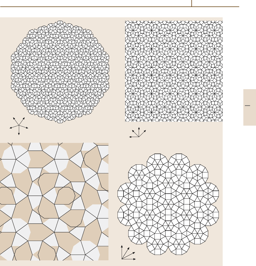
a number of different tilings that show such noncrys-
tallographic symmetries. Figure 1.3-12 depicts four of
them, as examples of the octagonal, decagonal, and
dodecagonal cases.
Icosahedral quasicrystals are also known. In 3-D,
the icosahedral diffraction symmetry 2/m
¯
3
¯
5 can be ob-
served for these quasicrystals. Their diffraction patterns
can be indexed using six integers, leading to a 6-D super-
space description (see Fig. 1.3-11). On the atomic scale
in 3-D, in physical space, clusters of some 100 atoms
are arranged on the nodes of 3-D icosahedral tilings;
the clusters have an icosahedral point group symmetry
or less, partially interpenetrate, and generate an overall
symmetry 2/m
¯
3
¯
5. Many of their structures are still wait-
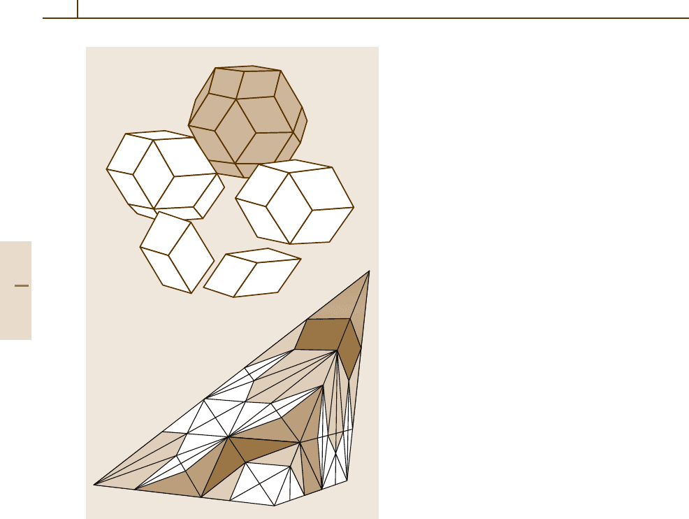
ing to be determined completely. Figure 1.3-13 shows
the two golden rhombohedra and the four Danzer tetra-
hedra that can be used to tile 3-D space icosahedrally.
Part 1 3.1
Rudiments of Crystallography 3.1 Crystalline Materials 37
a
2
a
1
a
3
a
4
a) b)
d)
a
2
a
1
a
3
a
4
–(a
1
+ a
2
+ a
3
+ a
4
)
a
2
a
1
a
3
a
4
c)
Fig. 1.3-12a–d Some 2-D quasiperiodic tilings; the corresponding four basis vectors a
1
,... ,a
4
are shown. Linear
combinations of r =
i
u
i
a
i
reach all lattice points. (a) Penrose tiling with local symmetry 5mm and diffraction symmetry
10mm,
(b) octagonal tiling with diffraction symmetry 8mm, (c) Gummelt tiling with diffraction symmetry 10mm,and
(d) dodecagonal Stampfli-type tiling with diffraction symmetry 12mm
Part 1 3.1
38 Part 1 General Tables
a)
b)
Fig. 1.3-13 Icosahedral tilings. (a) The two golden rhombo-
hedra (bottom) can be used to form icosahedral objects (the
rhombic triacontahedron with point symmetry m
¯
3
¯
5shown
in gray).
(b) Danzer’s {ABCK} tiling: three inflation steps
for prototile A
1.3.2 Disorder
In between the ideal crystalline and the purely amor-
phous states, most real crystals contain degrees of
disorder. Two types of statistical disorder have to be
distinguished: chemical disorder and displacive disor-
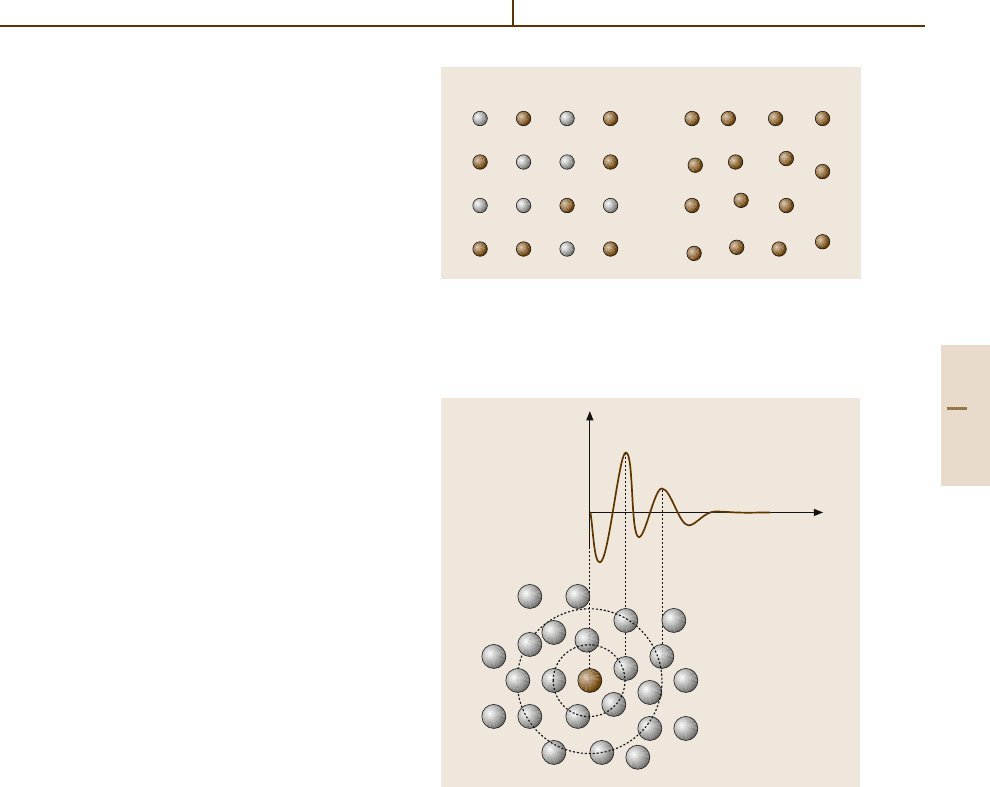
der (Fig. 1.3-14). Statistical disorder contributes to the
entropy S of the solid and is manifested by diffuse scat-
tering in diffraction experiments. It may occur in both
periodic and aperiodic materials.
Chemical Disorder
Chemical disorder is observed, for example, in the case
of solid solutions, say of B in A,orA
1−x
B
x
for short.
Here, an average crystal structure exists. On the crys-
tallographic atomic positions, different atomic species
(the chemical elements A and B) are distributed ran-
domly. Generally, the cell parameter a varies with x.
For x = 0 or 1, the pure end member is present. A lin-
ear variation of a(x) is predicted by Vegard’s law. On
the atomic scale, however, differences in the local struc-
ture, are present owing to the different contacts A–A,
B–B,andA–B. These differences are usually repre-
sented by enlarged displacement factors, but can be
investigated by analyzing the pair distribution function
G(r). G(r) represents the probability of finding any
atom at a distance r from any other atom relative to
an average density. Chemical disorder can also occur
on only one or a few of the crystallographically dif-
ferent atomic positions (e.g. A(X
1−x
Y
x
)
2
). This type
of disorder is often intrinsic to a material and may be
temperature-dependent.
Part 1 3.2
Rudiments of Crystallography 3.4 Methods for Investigating Crystallographic Structure 39
Displacive Disorder
The displacive type of disorder can be introduced by
the presence of voids or vacancies in the structure or
may exist for other reasons. Vacancies can be an im-
portant feature of a material: for example, they may
leading to ionic conductivity or influence the mechanical
properties.
Fig. 1.3-14 Schematic sketch of (a) chemical and (b) dis-
placive disorder
a) b)
A
B
1.3.3 Amorphous Materials
The second large group of condensed matter is classified
as the amorphous or glassy state. No long-range order
is observed. The atoms are more or less statistically
distributed in space, but a certain short-range order is
present.
This short-range order is reflected in the certain
average coordination numbers or average coordination
geometries. If there are strong (covalent) interactions
between neighboring atoms, similar basic units may
occur, which are in turn oriented randomly with re-
spect to each other. The SiO
4
tetrahedron in silicate
glasses is a well-known example. In an X-ray diffrac-
tion experiment on an amorphous solid, only isotropic
diffuse scattering is observed. From this information,
the radial atomic pair distribution function (Fig. 1.3-15)
can be obtained. This function G(r) can be interpreted
as the probability of finding any atom at a dis-
tance r from any other atom relative to an average
density.
r
G(r)
0
Fig. 1.3-15 Radial atomic pair distribution function G(r)
of an amorphous material. Its shape can be deduced from
diffuse scattering
1.3.4 Methods for Investigating Crystallographic Structure
So far, we have been dealing with the formal descrip-
tion of solids. To conclude this chapter, the tool kit that
an experimentalist needs to obtain structural informa-
tion about a material in front of him/her will be briefly
described.
The major technique used to derive the atomic
structure of solids is the diffraction method. To ob-
tain the most comprehensive information about a solid,
other techniques besides may be used to complement
a model based on diffraction data. These tech-
niques include scanning electron microscopy (SEM),
wavelength-dispersive analysis of X-rays (WDX),
energy-dispersive analysis of X-rays (EDX), extended
X-ray atomic fine-structure analysis (EXAFS), trans-
mission electron microscopy (TEM), high-resolution
transmission electron microscopy (HRTEM), differen-
tial thermal analysis (DTA), and a number of other
methods.
For diffraction experiments, three types of radia-
tion with a wavelength λ of the order of magnitude of
Part 1 3.4

40 Part 1 General Tables
interatomic distances are used: X-rays, electrons, and
neutrons. The shortest interatomic distances in solids
are a few times 10
−10
m. Therefore the non-SI unit the
angstrom (1 Å = 10
−10
m) is often used in crystallogra-
phy. In the case of electrons and neutrons, their energies
have to be converted to de Broglie wavelengths:
λ = h/mv,
λ(Å) = 0.28/
E(eV).
Figure 1.3-16 compares the energies and wavelengths of
the three types of radiation.
From wave optics, it is known that radiation of wave-
length λ is diffracted by a grid of spacing d.Ifwetake
a 3-D crystal lattice as such a grid, we expect diffrac-
tion maxima to occur at angles 2θ, given by the Bragg
equation (Fig. 1.3-17)
λ = 2d
hkl
sin θ
hkl
.
For the aperiodic (n-D periodic crystal) case, d
hkl
has to be replaced by d
h
1
h
2
...h
i
...h
n
.Togiveasimple3-D
example, for the determination of the cell parameter a
in the cubic case, the Bragg equation can be rewritten in
the form
(Q/2π)
2
= 4sin
2
θ
hkl
/λ
2
= (h
2
+k
2
+l
2
)/a
2
.
10
5
1.0
0.5
0.1
1 5 10 50 100
X-ray
Neutron
Electron
λ(Å)
E/ keV (X-ray); 0.01eV (neutron); 100eV (electron)
Fig. 1.3-16 Wavelengths λ in Å and particle energies E
for X-ray photons (energies in keV), neutrons (energies in
0.01 eV), and electrons (energies in 100 eV)
θ
θ
d
d sin θ
Fig. 1.3-17 Geometrical derivation of the Bragg equation
nλ = 2d sin θ. n can be set to 1 when it is included in
a higher-order hkl
Thus the crystal lattice is determined by a set of θ
hkl
.
In the case of X-rays and neutrons, information about
the atomic structure is contained in the set of diffraction
intensities I
hkl
. Here we have I
hkl
= F
2
hkl
where F
hkl
are
the structure factors.
To reconstruct the matter distribution ρ(xyz) inside
a unit cell of volume V, the crystallographic phase prob-
lem has to be solved. Once the phase factor φ for each
hkl is known, the crystal structure is solved.
ρ(xyz) = 1/V
×
all h,k,l
|
F
|
cos[2π(hx +ky+lz) −φ] .
Non-Bragg diffraction intensities I(Q) and therefore
a normalized structure function S(Q) can be obtained,
for example, from an X-ray or neutron powder diffrac-
togram. The sine Fourier transform of S(Q) yields
a normalized radial atomic pair distribution function
G(r):
G(r) = (2/π)
∞
0
Q[S(Q) −1]sin(Qr) dQ .
For measurements at high Q, the 1-D function G(r)
contains detailed information about the local structure.
This function therefore resolves, for example, disorder
or vacancy distributions in a material. The method can be
applied to 3-D diffuse scattering distributions as well and
thus can include angular information with respect to r.
Part 1 3.4

Rudiments of Crystallography References 41
X-rays
X-rays can be produced in the laboratory using a con-
ventional X-ray tube. Depending on the anode material,
wavelengths λ from 0.56 Å (Ag Kα)to2.29 Å (Cr Kα)
can be generated. Filtered or monochromatized radi-
ation is usually used to collect diffraction data, from
either single crystals or polycrystalline fine powders.
A continuous X-ray spectrum, obtained from a tungsten
anode, for example, is used to obtain Laue images to
check the quality, orientation, and symmetry of single
crystals.
X-rays with a higher intensity, a tunable energy,
a narrower distribution, and higher brilliance are pro-
vided by synchrotron radiation facilities.
X-rays interact with the electrons in a structure and
therefore provide information about the electron density
distribution – mainly about the electrons near the atomic
cores.
Neutron Diffraction
Neutrons, generated in a nuclear reactor, are useful for
complementing X-ray diffraction information. They in-
teract with the atomic nuclei, and with the magnetic
moments of unpaired electrons if they are present in
a structure. Hydrogen atoms, which are difficult to lo-
cate using X-rays (the contain one electron, if at all, near
the proton), give a far better contrast in neutron diffrac-
tion experiments. The exact positions of atomic nuclei
permit “X minus N” structure determinations, so that
the location of valence electrons can be made observ-
able. Furthermore, the magnetic structure of a material
can be determined.
Electron Diffraction
The third type of radiation which can be used for diffrac-
tion purposes is an electron beam; this is usually done in
combination with TEM or HRTEM. Because electrons
have only a short penetration distance – electrons, being
charged particles, interact strongly with the material –
electron diffraction is mainly used for thin crystallites,
surfaces, and thin films. In the TEM mode, domains and
other features on the nanometer scale are visible. Nev-
ertheless, crystallographic parameters such as unit cell
dimensions, and symmetry and space group information
can be obtained from selected areas.
In some cases, information about, for example,
stacking faults or superstructures obtained from an
electron diffraction experiment may lead to a revised,
detailed crystal structure model that is “truer” than
the model which was originally deduced from X-ray
diffraction data. If only small crystals of a material are
available, crystallographic models obtained from unit
cell and symmetry information can be simulated and
then adapted to fit HRTEM results.
The descriptions above provide the equipment
needed to understand the structure of solid matter on
the atomic scale. The concepts of crystallography, the
technical terms, and the language used in this frame-
work have been presented. The complementarities of the
various experimental methods used to extract coherent,
comprehensive information from a sample of material
have been outlined. The “rudiments” presented here,
however, should be understood only as a first step into
the fascinating field of the atomic structure of condensed
matter.
References
3.1 L. V. Azaroff: Elements of X-Ray Crystallography
(McGraw-Hill, New York 1968)
3.2 J. Pickworth Glusker, K. N. Trueblood: Crystal Struc-
ture Analysis – A Primer (Oxford Univ. Press, Oxford
1985)
3.3 E. R. Wölfel: Theorie und Praxis der Röntgenstruk-
turanalyse (Vieweg, Braunschweig 1987)
3.4 W. Kleber, H.-J. Bautsch, J. Bohm: Einführung in die
Kristallographie (Verlag Technik, Berlin 1998)
3.5 C. Giacovazzo (Ed.): Fundamentals of Crystallog-
raphy, IUCr Texts on Crystallography (Oxford Univ.
Press., Oxford 1992)
3.6 C. Janot: Quasicrystals – A Primer (Oxford Univ. Press,
Oxford 1992)
3.7 S. J. L. Billinge, T. Egami: Underneath the Bragg
Peaks: Structural Analysis of Complex Materials
(Elsevier, Amsterdam 2003)
3.8 T. Hahn (Ed.): International Tables for Crystal-
lography, Vol. A (Kluwer, Dordrecht 1992)
3.9 A. V. Shubnikov, N. V. Belov: Colored Symmetry
(Pergamon Press, Oxford 1964)
Part 1 3
