Lima J.J.Pedroso, de (ed.). Nuclear Medicine Physics
Подождите немного. Документ загружается.


468 Nuclear Medicine Physics
Considering two spheres with radius and r
1
and r
2
, and using Equation
8.69, the fluence ratio for the two spheres is given by
Φ
1
Φ
2
=
r
2
2
r
2
1
(8.70)
As previouslymentioned, the absorbed dose can be calculated from fluence,
leadingto the replacementofΦ by Din Equation 8.70.Making r
1
= 1m,r
2
= r,
and D
2
= D and if the dose rate at one meter,
˙
D
1
, is known, the relationship
becomes
D =
˙
D
1
r
2
×t. (8.71)
The dose is proportional to the inverse of the square distance from the point
source to the point of interest.
Given the dose rate at one meter per unit activity, Equation 8.71 allows
simple calculations, for example, on radiation protection around radioactive
sources.
If we take into account scattered radiation by the buildup factor, this
becomes
D(r) =
˙
D
1
r
2
×t ×B(μr) (8.72)
where r is the distance from source to the point of interest.
If there is a homogeneous medium between the radiation source and the
point of interest, the exponential attenuation must be included, leading to
D(r) =
˙
D
1
r
2
×t ×B(μr) × e
−μ(E,Z)r
. (8.73)
For air, we often use B(μ(air)r) ×e
−μ(air)r
≈ 1, which simplifies some radia-
tion shielding calculations. If the air attenuation is neglected and there exists
a slab shield of thickness x, then Equation 8.73 becomes
D(r) =
˙
D
1
r
2
×t ×B(μx) ×e
−μ(E,Z)x
. (8.74)
To determine the value of
˙
D
1
for radionuclides, we can use the air kerma
rate constant, Γ
δ
. This factor is also called the k factor, specific γ-ray constant,
or exposure rate constant in the last case used, obviously, to calculate the
exposure instead of dose. It is defined for each radionuclide.
Assuming the kerma approximation, then the dose rate
˙
D
1
can be calculated
by the expression
˙
D =
Γ
δ
A
l
2
, (8.75)
Dosimetry and Biological Effects of Radiation 469
where A is the activity of the source and l is the distance from a point source.
The index δ means that only the photons with energy greater than δ are
considered. If A = 1 Bq and l = 1 m, then
˙
D
1
= Γ
δ
. (8.76)
Therefore, Γ
δ
is the kerma rate at 1 m from a gamma source of activity 1 Bq.
The constant Γ
δ
has to have units (m
2
Gy Bq
−1
s
−1
) in order to give
˙
D
1
in
units (Gy s
−1
).
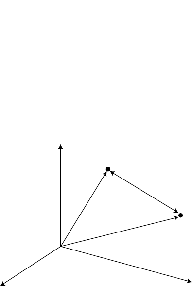
Let us consider the geometry outlined in Figure 8.19.
To calculate the dose at the target point, t, due to the source at point s,we
can define the photon dose point kernel. The dose point kernel is a function
that represents the dose at a given distance from an isotropic point source, in
a homogeneous and infinite medium, which can be written as
P(r, E) =
˙
S
4πr
2
μ
en
ρ
tB(μr)e
−μr
(8.77)
where r =|
r
s
−
r
1
| and
˙
s is the source strength in photons per second. The
coefficients in Equation 8.77, already earlier mentioned, are functions of the
energy and the atomic number of the medium. In general, a gamma source
emits photons of several energies so that it must be considered as an integral
in terms of energy:
P(r) =
E
P(r, E)dE. (8.78)
r
s
|r
s
– r
t
|
r
t
→ →
→
→
t
s
z
xy
FIGURE 8.19
Isotropic point source at point s and the point of interest t, within a homogeneous and infinite
medium.
470 Nuclear Medicine Physics
After this integration, the point kernel can be defined as a function of the
distance from the point source,
P(r) = P
|
r
s
−
r
1
|
. (8.79)
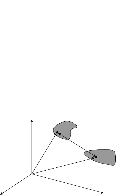
The point kernel is used in more general and complex geometry, such as
volume source and volume target. This is the case for NM, where dosimetric
calculations are made at the organ level or, in some cases, at the cellular level.
For the case of the organ level, the organ source and the organ target must be
considered. This geometry is outlined in Figure 8.20.
The concept of point kernel was introduced for use in the volume integra-
tion:
D(T ← S) =
1
V
1
V
s
dV
s
V
t
dV
t
P
|
r
s
−
r
1
|
(8.80)
where D(T ← S) means the dose in the target volume T, due to a source in
volume S.
In suitable models such as those involving spherical geometry, for example,
spherical source and spherical target, theoretically the integration (Equation
8.80) can be analytically solved.
The point kernel can be evaluated for any type of emission (photons,
electrons, α-particles, etc.) and any homogeneous medium.
The calculation of Equation 8.80 at an organ or tissue level, where the
geometry is rather more complex than the spherical case and the medium
is non homogeneous, is only possible by Monte Carlo simulation, which was
briefly reviewed in this chapter.
Due to the importance of absorbed dose calculations during the admin-
istration of radionuclides in the human body, several years ago, a scheme
S
T
s
t
z
x y
r
s
→
|r
s
– r
t
|
→ →
→
r
t
FIGURE 8.20
Volume source and volume target illustrating the general geometry for dose calculations, for
example, in nuclear medicine.

Dosimetry and Biological Effects of Radiation 471
known as MIRD (medical internal radiation dose) scheme was developed by
the MIRD committee.
The advantage of this scheme is that the integration (Equation 8.80) is solved
for several pairs of human organs, and the results are presented in tabu-
lar form or implemented in software packages. The integral equation 8.80 is
replaced by the equation
D(O
t
← O
s
) =
˜
A
s
S(O
t
← O
s
). (8.81)
In this equation, the absorbed dose in the target organ,O
t
, due to cumulative
activity,
˜
A
s
, in the source region O
s
is simply obtained by multiplying the
cumulated activity by an S-value, which is the absorbed dose at the target
organ per unit cumulative activity in the source region.
All the complexity of the integration (Equation 8.80) is hidden in the
simplicity of the S-value. All we have to do is calculate the cumulated
activity,
˜
A
s
:
˜
A
s
=
A(t)dt (8.82)
The S-value is defined as
S(O
t
← O
s
) =
i
Δ
i
φ
i
(O
t
← O
s
)
m
t
(8.83)
where m
t
is the mass of the target region, Δ
i
is the mean energy emitted per
nuclear transition, and φ
i
(O
t
← O
s
) is the fraction of energy emitted from
the organ source that is absorbed in the target region for the ith radiation
component.
Next there are defined radiation protection quantities for application in
internal dosimetry.
The committed equivalent dose for an organ or tissue, T, is represent by
H
T
(τ).
Since the radionuclide irradiates the body organs after its incorporation,
this quantity is defined as the integral in time τ of the equivalent dose rate in
the tissue of interest. It is defined as the total committed equivalent dose in
the organ or tissue for the time τ after the incorporation:
H
T
(τ) =
t
0
+τ
t
0
˙
H
T
(t)dt (8.84)
When the time of integration is not mentioned, a time of 50 years for adults
or 70 years for children is assumed.

472 Nuclear Medicine Physics
The committed effective dose, E(τ), is obtained, by multiplying the commit-
ted equivalent dose for each organ or tissue by the respective tissue weighting
factor. This quantity is defined as the sum:
E(τ) =
t
H
T
(τ)w
T
(8.85)
To end this short review of the radiation protection quantities, it is often
useful to have a measure of the exposure of a population, which is made by
the collective effective dose.
The effective collective S, is defined by the next integral:
S =
∞
0
E
dN
dE
dE (8.86)
or by the sum
S =
i
¯
E
i
N
i
, (8.87)
where
¯
E
i
is the average effective dose for the subgroup i of a population. The
time interval is not explicitly specified during which the population were
exposed, resulting in the corresponding effective dose. Therefore, that time
must be expressly specified in the presentation of effective dose values.
The requirement of homogeneous medium and point isotropic kernel has
already been mentioned. However, the MIRD scheme is not limited to organ
or suborgan dosimetry. In fact, a lot of useful applications have more recently
appeared in cellular dosimetry, for example, in targeted radiotherapy, where
dose at the cellular level is a major concern.
Adaptation of the formalism shortly summarized in Equations 8.81 through
8.83 for organs can be straightforwardly applied to cells or subcellular regions,
by simply replacing the reference of organs to a referenceof cells and adapting
the formalism for absorbed dose calculation to multiple sourceregions, so that
Equation 8.81 becomes
D(C
t
← C
s
) =
s
˜
A
s
S(C
t
← C
s
). (8.88)
At the cellular level, the concepts of self-dose and cross-dose are often used,
so that the mean dose of a cell cluster is a sum of the self-dose and the cross-
dose. Similarly, we can define S-values for self-dose and cross-dose, S
self
and
S
cross
, respectively.
Differences between organ dosimetry, often called conventional dosimetry,
and cellular dosimetry are that in conventional dosimetry, no distinction is
made between the cells and the medium. The radioactivity is assumed to be
Dosimetry and Biological Effects of Radiation 473
homogeneously distributed throughout the volume considered. This implies
that the dose to the target cell nucleus is the same as that to the overall organ or
to any given subvolume. In cellular dosimetry, the approach of Equation 8.88
can be further extended to subcellular regions, such as cell membrane, cyto-
plasm, and cell nucleus. Conventional dosimetry predicts poorly absorbed
dose at the cell nucleus, which can be of great importance for small range
particles, such as Auger electrons.
A drawback of this cellular approach lies in the validation of cross-dose
due to difficulties in available data for comparison of results. In fact, calcu-
lations are made defining a given cell cluster. Cell clusters can differ in the
number of cells, the cell and nucleus size, and the packaging of cells within
the cluster. Sometimes, cells are assumed to be spheres. A typical approach
at the cellular level assumes the last geometric details for a given cell cluster
[26], allowing the evaluation of S
cross
, S
selfNuc
, S
selfCyt
, and S
selfMb
, where the
index refers to cross, nucleus, cytoplasm, and cell membrane. Other authors
[27] calculate intracellular S-values for cell–cell, cell–cell surface, nucleus–
nucleus, nucleus–cytoplasm, and nucleus–cell surface, where the first region
is the target and the second region is the source.
Different number of cells, cell size, or cell arrangement (package) make any
comparison difficult. It seems apparentthat there is a need to define some fun-
damental quantities at the cellular level in order to allow comparison between
different calculation methods and clusters. With this scope, and considering
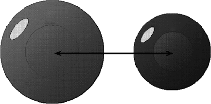
that the cell is the basic unit of life, the basic unit of cellular dosimetry can be
defined as a pair of cells, one of them with radionuclide incorporated and the
other clean (Figure 8.21).
For the basic unit of cellular dosimetry, we can define several S-values:
In the earlier definitions of all the S-values, only the cell and the nucleus are
considered as targets. The source can be the cell (C1), the cytoplasm (Cy1),
the cell surface (CS1), and the cell nucleus (N1). Additional factors affect-
ing the S-values are RC1, RN1, RC2, RN2, and R12, which are the radius of
cell 1, radius of cell nucleus 1, radius of cell 2, radius of cell nucleus 2, and
Source
Ã
s
Tar ge t
R
FIGURE 8.21
The basic unit of cellular dosimetry can be defined as a pair of cells. Acontaminated one as source
and a clean one as target.

474 Nuclear Medicine Physics
TABLE 8.1
S-Values between Two Cells Including
Subcellular Regions
Self-dose Cross-dose
S(C1 ← C1) S(C2 ← C1)
S(C1 ← Cy1) S(C2 ← Cy1)
S(C1 ← CS1) S(C2 ← CS1)
S(C1 ← N1) S(C2 ← N1)
S(N1 ← C1) S(N2 ← C1)
S(N1 ← Cy1) S(N2 ← Cy1)
S(N1 ← CS1) S(N2 ← CS1)
S(N1 ← N1) S(N2 ← N1)
intercellular distance, respectively. The intercellular distance can be defined
as the distance between the cell centers. As can be seen, cellular dosimetry
involves a lot of parameters that contribute to the difficulties already men-
tioned in comparisons between different methods and cell clusters. In Table
8.1, the need appears for a rigorous definition of all the parameters involved.
8.10 Physics of the Biological Effects of Radiation
Radiationdosimetry quantifiesthe energydeposited byradiation inbiological
tissue through the main quantity in radiation protection: the absorbed dose.
Deterministic or stochastic effects appear, depending on its value. From the
physical point of view, the absorbed dose can be determined independently
of the biological situation. From the biological point of view, the studies of the
effects in the living matter, for a given localization and absorbed dose value,
are made independently of the physical action.
Very often, the biological effects of ionizing radiation are correlated with
absorbed dose values in tissues or cells at risk. In spite of that, other physical
factors are also related, such as the radiation type, the energy spectrum, the
spatial and time distribution of the energy deposited, the total number of cells
irradiated, or different biological responses to radiation qualities.
In the attempt to estimate biological effects, for example, carcinogenesis,
it is often assumed that the risk is simply proportional to the dose received.
However, it is well known that biological experiments reveal a much more
complex relationship [20].
In a simplified approach, we can consider that radiation protection studies
the results of the interaction between a physical agent (radiation) and man
(biological medium) or the environment. If we seek a field of knowledge
that is multidisciplinary, embracing a vast set of areas, we can arrive at, for
example, the characterization of the biological effects of radiation.

Dosimetry and Biological Effects of Radiation 475
It is considered that two distinct types of biological effects exist: deter-
ministic and stochastic. Edwards and Lloyd [28] and Doll [29] publish two
review papers about deterministic and stochastic effects, respectively. From
a pragmatic point of view, the effects that can be diagnosed in a medical
examinationwithin the few weeks after radiation exposurearecertainly deter-
ministic effects, such as skin burns or reduction in the levels of the blood cells,
which, in extreme situations, can be fatal within weeks or months.
Perhaps, the best distinction between deterministic and stochastic effect
is to mention the mechanisms of their production. In general, the stochastic
effects (mainly cancer) are caused by nonlethal mutations in cells, whereas
deterministic effects are caused by cell death.
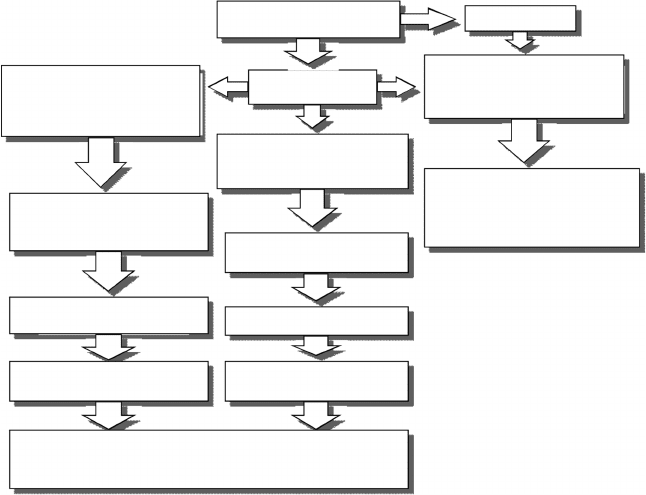
The scheme shown in Figure 8.22 describes the processes related to the
effects of ionizing radiation in living tissues.
The occurrence and proliferation of modified cells can have influences from
modifications through other agents that act beforethe irradiation. These influ-
ences are common and can include exposure to other carcinogenic agents.
As already indicated, in general, there are two types of radiation effects:
stochastic and deterministic (Figure 8.23).
The replacement of dead cells is a natural process of maintenance of tissues.
Through sophisticated feedback mechanisms, the rates of production of new
cells are changed to compensate the rate of loss of mature cells. For example,
Radiation
Ionization
Modification of the structure of molecules
Cellular damage
DNA damage
(most serious damage)
The cell don’t survive
of doesn’t reproduce
The cell repairs the damage
Imperfect repair
(modified viable cell)
Perfect repair
(without consequences)
Chemical modification in molecules
which are not critical for the working of the cell
(production of free radicals)
Chemical modification in molecules
which are critical for the working of the cell
FIGURE 8.22
Radiation effect in living tissues.
476 Nuclear Medicine Physics
Radiation
Modification of cells in gonads,
with function of transmission
of information to the descendants
of the exposed individual
Transmission of incorrect
hereditary information
Severe damages to
some of the descendants
Eventually cancer
Somatic effect
Hereditary effect
Stochastic effect
Modified viable cell
Modification of a somatic cell.
Maintain the
reproductive capacity
It can originate
clones of modified cells
Deterministic effect
Loss of the function of the organ.
This loss is greater for
a high number of cells affected
Dead cell
FIGURE 8.23
Stochastic and deterministic effects of radiation.
the external layer of the skin is replaced approximately every 6 weeks; and
blood cells, depending on the type, are replaced with half-lives varying from
some days to some years. The body has the capacity to tolerate the death
of some additional cells due to external agents, such as radiation or chemi-
cals, because it is able to replace cells at a higher velocity than under normal
conditions.
The deterministic effects occur when the number of dead cells exceeds the
capacity for replacement, that is, when the equilibrium state between produc-
tionand death isperturbed byexcess ofthe latter. Thetargetorgansandtissues
then stop working properly, eventually leading to biological modifications
with serious consequences.
One important parameter is the dose rate. For low LET radiation, the dose
received at low dose rate or in fractions of time has very different conse-
quences when the same dose is received at high dose rate, in a short period
of time.
How do we relate the biological effects with the radiation that impinges on
the tissues? There is a need to quantify, somehow, the energy deposited in the
irradiated medium. Radiation dosimetry is the discipline of radiation physics
which allows that quantification. The absorbed dose is the fundamental
quantity, in such a way that it is used as the name of the discipline.

Dosimetry and Biological Effects of Radiation 477
The absorbed dose is the expected value of the deposited energy in matter,
per unit of mass, at a given point of interest,
D =
dε
dm
. (8.89)
Even today, it is common practice to measure the radiation effects as a
function of absorbed dose, in which the dose rate is used as a parameter.
However, there is an assumption in this approach that is sometimes ignored
orneglected. Theenergydeposited has a constant valuein allthe biological tis-
sue. Nowadays, it is well known that this assumption is, in general, incorrect,
particularly for low doses, which are important in radiation protection [30].
Any model concerning the biological response should consider the physi-
cal, chemical, and biological factors. The physical factors are, among others,
associated with the spatial pattern of the points of energy deposition, at
a scale of the order of the nanometer (dimensions of the DNA molecule).
Beyond the direct action, the chemical factors are related with the capacity
for the production of free radicals, mainly in water, in the vicinity of the DNA
molecule. Biologically, we should consider the repair and the probability of
lethality of DNA lesions [31].
Nevertheless, research at biological and molecular levels still maintains
the practice of expressing biological effects as a function of absorbed dose.
Many of the models of biological response are defined through a dose–effect
relationship. Some studies with cultures of cells of mammals indicate that the
survival of the cells is a function of the absorbed dose, described by survival
curves.
As an example, for the deterministic effects, it is accepted that the risk of
a radiation exposure has a sigmoid relationship (see ICRP-60 [32], ICRP-103
[19] or [28]). Edwards and Lloyd [28] proposed a relationship between the
risk, R, and absorbed dose, D, as follows:
R = 1 −exp(−H), (8.90)
H = ln 2
D
D
50
V
, (8.91)
where H is named Hazard function and is related with the dose through the
parameters D
50
and V. The parameter D
50
is the average lethal dose and
signifies that it is expected that 50% of the exposed individuals shows the
effect in question; V is related with the speed of rise.
An artificial threshold of dose is defined from 10% of the value of the sig-
moid function. Note that this threshold value is defined for deterministic
effects and is not considered for stochastic effects.
Radiation dosimetry is of fundamental importance for the study of biologi-
cal effects and can then be developed in accordance with biological needs. The
following figures were obtained from the data of Edwards and Lloyd [28].