Lax Alistair J. Bacterial protein toxins: Role in the interference with cell growth regulation (Бактериальные токсины белков: роль в регуляции роста клеток)
Подождите немного. Документ загружается.

P1: JZP
052182091Xc09.xml CB786/Lax 0 521 82091 X November 4, 2005 5:3
206
christine p j caygill and michael j hill
found only in legumes (and particularly soya), but the lignans are found in
almost all cereals and vegetables, with the highest concentrations found in
oilseeds. Like many plant anutrients of interest, they are usually present in
plant foods as their glycosides. Like the related lignins, the lignans have no
nutritive value to humans and until the last 25 years were largely ignored.
Many have been shown to have an anti-mitotic action. Most importantly for
this chapter, the lignans in human urine are not aglycones of plant lignans
but are produced in the colon by bacterial action on dietary precursors, then
absorbed from the colon and excreted in the urine (Adlercreutz, 1984). The
urinary concentrations can be greatly decreased by the action of antibiotics.
Insertion of a biliary fistula showed that the lignans undergo enterohepatic
circulation. Their concentration in urine is increased with increasing intake of
dietary fibre and of whole-grain foods, and decreased by increasing tobacco
consumption. They are weak estrogens, able to bind to oestrogen binding
sites, particularly the ER-beta sites.
There has been a growing interest in the study of these compounds
in recent years because consumption of foods rich in phytoestrogens has
been vigorously advocated for the prevention of breast, prostate, and colon
cancer. The incidence of all three cancers is lower in Asia than in Western
countries; the intakes of phytoestrogens in general (and lignans in particu-
lar) is much higher in Asian populations than in those living in the West
(Adlercreutz, 1998, 2002). The principal lignans in human urine are entero-
diol and enterolactone; the concentration of both is higher in the urine of
Asian populations (with a low incidence of the cancers of interest) than of
European populations. The intake of the main food sources of lignans, the
whole grain cereals, is inversely related to risk of cancer at a number of sites,
including the breast, prostate, and colon (La Vecchia and Chatenoud. 1998),
although other lifestyle factors could also be responsible for the differences.
BACTERIAL INFECTIONS AND CANCER
Cancers of the Urinary Tract
Bladder cancer has long been known to be associated with industrial expo-
sures to naphthylamines, benzidine, and a range of aromatic amines. This
was the reason why the vast majority of cases arose in males in industri-
alised countries. However, a proportion of cases is not of industrial origin.
Early anecdotal evidence suggested an excess risk of bladder cancer follow-
ing chronic bladder infection. Radomski et al. (1978) confirmed an associa-
tion between chronic simple urinary tract infection and subsequent bladder
P1: JZP
052182091Xc09.xml CB786/Lax 0 521 82091 X November 4, 2005 5:3
207
bacteria and cancer
cancer. Bladder infections are very common, and often asymptomatic
(Sinclair and Tuxford, 1971); the data on cancer risk reported by Radomski
et al. (1978) concerns chronic symptomatic infection resistant to therapy, but
many of his controls might have had asymptomatic bladder infections and
so the magnitude of the excess risk would have been underestimated.
There is copious evidence that carcinogenic N-nitroso compounds (NNC)
are produced in situ in the bladder by infecting organisms; this is to be ex-
pected because the urine is the route of excretion of the substrates for NNC
production – nitrate and nitrosatable amines. Radomski et al. (1978) sug-
gested that these NNC were the cause of the cancer.
Bilharzial infection is a major risk factor for bladder cancer, and such
infections are accompanied by a profuse secondary bacterial infection of the
bladder. Hicks et al. (1977) showed strong evidence that the bladder cancer
associated with bilharzial infection was due to the NNC produced by the
secondary bacterial infection. They also produced evidence that the excess
risk of bladder cancer in paraplegia was due to the same mechanism – NNC
produced by a chronic bacterial infection of the bladder.
Recently it has been shown that what were thought to be recurrent blad-
der infections by Escherichia coli are in fact chronic infections (Mulvey et al.,
2001). The majority of bacterial isolates from the urinary tract are E. coli,
including from cases of prostatitis (Andreu et al., 1997). Most such isolates
express the cytotoxic necrotizing toxin (CNF) that stimulates the important
signalling protein Rho (see Chapter 3). CNF has been shown to be important
for pathogenesis in the urinary tract (Rippere-Lampe et al., 2001a; 2001b);
the presence of CNF can lead to activation of the enzyme Cyclooxygenase-2
(COX-2) (Thomas et al., 2001). Numerous studies have implicated COX-2
in both the initiation and progression of various cancers (Liu et al.,
2001).
It has also been shown that a history of urinary tract infection increases
the risk of renal cancers (Parker et al., 2004), so it is possible that several
different types of urinary tract cancers could have in part a bacterial cause.
Gastric Cancer
In 1975 Correa et al. proposed a hypothesis for the sequence of events that
led from the normal to the neoplastic stomach (Figure 9.1). Although this se-
quence has since been added to and changed the essential hypothesis remains
the same (Figure 9.1), and there are two types of bacterial contamination that
may play a role; one is infection with Helicobacter pylori and the other is
chronic bacterial overgrowth as a result of hypochlohydria of the stomach.

P1: JZP
052182091Xc09.xml CB786/Lax 0 521 82091 X November 4, 2005 5:3
208
christine p j caygill and michael j hill
Normal Gastric Mucosa
High Salt Intake
Gastric Surgery
Natural ageing
Gastric Atrophy Bacterial Colonisation
Helicobacter pylori
Chronic atrophic gastritis
Intestinal Metaplasia
Production of
N-nitroso
compounds
Dysplasia
Gastric carcinoma
Figure 9.1. The pathogenesis of gastric cancer.
Helicobacter pylori Infection
H. pylori infection is associated with low socioeconomic status and crowded
living conditions, especially in childhood (Malaty and Graham, 1994). World-
wide, about half the population are infected (Smith and Parsonnet, 1998),
with most children in developing countries being infected by the age of 10.
In contrast, in developed countries, infection in children is uncommon and
only 40–50% of adults are affected. There is a clear age-related increase in
prevalence that is probably due to a cohort effect in that H. pylori infection
in childhood was more common in the past than it is today (Parsonnet et al.,
1992; Banatvala et al., 1993). The route of transmission of H. pylori remains
controversial, with circumstantial evidence suggesting that it probably occurs
through person-to-person transmission.
There have been a number of studies comparing rates of H. pylori infec-
tion in different populations with rates of gastric cancer in the same popula-
tions (Forman et al., 1990; Eurogast Study Group, 1993). These have mostly
correlated well, as has the decline in gastric cancer rate over time with the
decline in H. pylori incidence (Parsonnet et al., 1992; Banatvala et al., 1993).
As cancerous stomachs may lose their ability to harbour H. pylori (Osawa
et al., 1996), retrospective studies have to be viewed with caution. However,
P1: JZP
052182091Xc09.xml CB786/Lax 0 521 82091 X November 4, 2005 5:3
209
bacteria and cancer
two meta-analyses of all case-control studies (Huang et al., 1998; Eslick and
Talley, 1998) indicate a 2-fold increase in the risk of gastric cancer in instances
of H. pylori infection.
More concrete evidence for a link has come from prospective case-control
studies in which stored serum from populations was used and infection was
known to precede malignancy (Parsonnet et al., 1991; Forman et al., 1991; Lin
et al., 1995; Siman et al., 1997). It has also been shown that in those followed
up for more than 10 years the risk of gastric cancer was increased 8-fold in
those infected with H. pylori (Forman et al., 1994).
A more detailed discussion of the potential mechanisms involved in
H. pylori carcinogenesis is given in Chapter 8 by Naumann and Crabtree.
Chronic Bacterial Overgrowth of the Stomach
Chronic bacterial overgrowth of the stomach occurs when the normally acid
pH of the stomach (pH 2) rises on a permanent basis to pH 4.5 or above.
This occurs as part of the ageing process, but also in a number of pathological
conditions, such as pernicious anaemia (PA), and in people who have had
surgery for peptic ulcer.
PA is caused by a lack of intrinsic factor, which is accompanied by a
failure to secrete gastric acid. Indeed, hypochlorhydria is a symptom in the
recognition and diagnosis of the disease.
Surgical treatment of peptic ulcer was directed towards decreasing gastric
acid secretion, whether by gastrectomy, where most of the lower part of the
stomach was removed by a variety of procedures, or vagotomy, where the vagal
nerves which control acid secretion were severed. Each of these procedures
resulted in loss of gastric acidity within a year, and in each case there was an
increased risk of gastric cancer (Caygill et al., 1984).
A number of authors (Blackburn et al., 1968; Brinton et al., 1989) have
reported an increased risk of gastric cancer in PA patients. In our study
(Caygill et al., 1990), we found a 5-fold excess risk of gastric cancer in PA
patients, but it was not possible to establish an accurate latency period as
the onset of PA could not be ascertained accurately for a sufficient number.
However, we were able to divide up the period after diagnosis into 0–19 years
and 20+ years and found that the excess risk of gastric cancer was 4-fold in
the first time period and 11-fold in the second time period.
There have been a number of studies showing an increased risk of gastric
cancer in those who have had a gastrectomy for benign disease, and these
are summarised in Table 9.4 (from the review by Caygill and Hill, 1992). In
our own study (Caygill et al., 1986), we analysed cancer risk by time interval
for those who had had a gastrectomy for gastric ulcer (GU) separately from

P1: JZP
052182091Xc09.xml CB786/Lax 0 521 82091 X November 4, 2005 5:3
210
christine p j caygill and michael j hill
Table 9.4 Gastric cancer following surgery for peptic ulcer
Size of study Excess risk & latency/
Reference: population Type of Study follow-up period
Stalsberg and Taksdal
(1971)
630 Case-control 4-fold after 15 years
Ross et al. (1982) 779 Cohort None over 19 years
Sandler et al. (1984) 521 Case-control None over 20-year
follow-up
Watt et al. (1984) 735 Cohort 3-fold after 15 years
over a 15–25 year
follow-up
Tokudome et al.
(1984)
3827 Cohort None over 10–30 years
follow-up
Caygill et al. (1986) 4466 Cohort 4-fold after 20 years
Viste et al. (1986) 3470 Cohort 3-fold after 20 years
Arnthorsson et al.
(1988)
1795 Cohort 2-fold after 15 years
Lundegardh et al.
(1988)
6459 Cohort 3-fold after 30 years
Toftgaard (1989) 4131 Cohort 2-fold after 25 years
Offerhaus et al. (1988) 2633 Cohort 5-fold after 15 years in
females, 3 fold after
25 years in males
Caygill et al. (1991) 1643 Cohort 1.6-fold over 20 years
those who had the operation for duodenal ulcer (DU). We found that in the
case of DU there was a decrease in risk in the first 19 years, followed by an
increase in risk thereafter. In contrast, in the GU patients there was a 3-fold
increase in risk immediately after, and presumably prior to surgery, and this
rose to over 5-fold 20 or more years after surgery. The pattern of an initial
decrease in risk in those operated for DU has been confirmed by Arnthorsson
et al. (1988), Moller and Toftgaard (1991), Lundegardh (1988) and Eide et al.
(1991). This difference in behaviour between DU and GU patients needs to
be rationalised. Prior to surgery DU patients have good acid secretion and
the effect of surgery is to render them hypoacidic. Many GU patients on the
other hand are often hypoacidic for a number of years prior to the operation.
The histopathological sequence (Figure 9.1) from the normal to the neo-
plastic stomach, suggested and reviewed by Correa (1988), has been generally
accepted. Correa et al. (1975) postulated that the first stage, gastric atrophy,
P1: JZP
052182091Xc09.xml CB786/Lax 0 521 82091 X November 4, 2005 5:3
211
bacteria and cancer
progresses to chronic atrophic gastritis. This carries an increased risk of
development of intestinal metaplasia, with consequent increased risk of in-
creasingly severe dysplasia and finally carcinoma. Gastric atrophy results in
the loss of gastric acid secretion, which allows bacterial proliferation. The bac-
teria then react with nitrate, present in many foods and also in drinking water,
to convert it to nitrite, which in turn is converted to carcinogenic N-nitroso
compounds. The latter potent carcinogens were postulated as the cause of the
progression through intestinal metaplasia and increasingly severe dysplasia
to cancer. If this hypothesis is correct, then the loss of gastric acidity, with
consequent chronic bacterial overgrowth from any cause (surgical, metabolic,
clinical, genetic, or environmental) should, after a latency period of 20 years
or more, lead to an increased risk of gastric cancer. Indeed, this has also been
shown to be the case in PA patients, post-gastrectomy patients, and those
undergoing vagotomy (Caygill et al., 1991). The above hypothesis explains
the difference in cancer risk in those operated on for a GU and a DU. As a
result of their hypoacidity prior to operation, the GU patients will have had
bacterial overgrowth for variable lengths of time that would contribute to the
latency period, whereas those with DU would have become hypochlorhydric
only after their operation and the increase in their risk would start to manifest
itself only 20 years later.
Colorectal Cancer
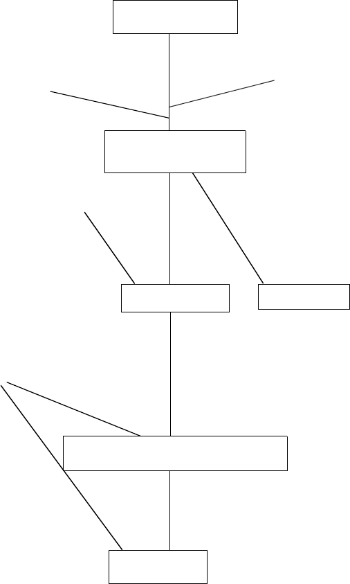
Large bowel carcinogenesis is a multistage process with at least three dis-
tinct histological stages (Hill and Morson, 1978; Hill, 1991; Hill et al., 2001).
These are (a) adenoma formation, (b) adenoma growth, and (c) increasingly
severe dysplasia to malignancy and potential or actual metastatic disease.
The evidence for the dysplasia–carcinoma sequence (formerly known as
the adenoma–carcinoma sequence) was reviewed by Morson (1974) and by
Morson et al. (1983). The steps in this pathway are distinct (Figure 9.2), and
have different controlling factors.
Adenoma formation is an extremely common event in Western popu-
lations, and postmortem studies show their prevalence to be approximately
50% in men and 30% in women by age 70. The vast majority of these are
tiny, and are presumably not seen at endoscopy. They remain tiny and asymp-
tomatic and therefore are normally detected only at postmortem. The risk of
finding a malignant component in a tiny adenoma is very low (less that
1 per 1000 for adenomas less than 3 mm diameter), whereas it is high
in adenomas greater than 20 mm diameter (Morson et al., 1983). Clearly,
therefore, adenoma growth is an important step on the adenoma–carcinoma
sequence.
P1: JZP
052182091Xc09.xml CB786/Lax 0 521 82091 X November 4, 2005 5:3
212
christine p j caygill and michael j hill
MUCOSAL CELL
Genetic Predisposition
Environmental factors
E1
SMALL ADENOMA
Environmental factors Environmental factors
E2
C (rarely)
(Adenoma Growth Factors)
Environmental Factors
C
ADENOMA WITH SEVERE DYSPLASIA
LARGE
ADENOMA
CARCINOMA
CARCINOMA
Figure 9.2. The hypothesised mechanism of colorectal carcinogenesis based on that
proposed by Hill and Morson (1978).
P1: JZP
052182091Xc09.xml CB786/Lax 0 521 82091 X November 4, 2005 5:3
213
bacteria and cancer
There are differences in epidemiology between small adenomas, large
adenomas, and colorectal cancers. One of the most crucial differences is the
differing subsite distribution. In a very large number of postmortem studies
(Hill, 1986), small adenomas have been shown to be evenly distributed around
the colorectum, suggesting that the causal agents are delivered via the vascular
system, or have a genetic cause. In contrast, large adenomas and cancers are
concentrated in the distal colon and rectum. This is consistent with, but does
not prove, the hypothesis that the factors causing adenomas to increase in
size and in severity of epithelial dysplasia are luminal products of bacterial
metabolism; this view is supported by the fact that adenomas regress after
diversion of the faecal stream.
The colon lumen is a rich source of potential carcinogens, produced in situ
by bacterial action on benign substrates (Table 9.1). We were not able to find
evidence associating the risk of colon cancer with the faecal concentrations
of the amino acid metabolites, and the best evidence suggests a role for
secondary bile acids (Table 9.5)inthe adenoma growth stage (Hill, 1991).
This evidence comes from comparisons of populations with colon cancer
risk, case control studies, and studies of high-risk patient groups.
There were two major groups of high-risk patients. The adenoma patients
were part of a follow-up study, and were followed for 10 years. There was
no relation between bile acids and the rate of formation of new adenomas,
showing that bile acids have no role in adenoma formation. However there
was a good correlation with the size of the largest adenoma, supporting the
hypothesis that bile acids are implicated in adenoma growth. The colitics had
all been patients for more than 10 years and had been offered a colectomy.
They opted instead to join a follow-up clinic, and were followed for a further
10 years. During that time dysplasia (analogous to adenoma formation) was
detected in 44 out of 112 patients. Although there was no relation between
bile acid concentration and formation of dysplasia per se, there was a good
correlation with the severity of dysplasia.
In addition, there is a mass of supporting evidence from animal model
studies (Reddy, 1992). The evidence that bile acids are tumour promoters or
mutagens or co-mutagens is summarised in Table 9.2.
Gallbladder Cancer
Cancer of the gallbladder is rare in Western countries, but is most frequent
in the Andean countries of South and Central America and in American
Indian groups (Devor, 1982) and in some parts of India (Shukla et al., 2000).
It has a poor prognosis. The aetiology is not well understood, but known risk

P1: JZP
052182091Xc09.xml CB786/Lax 0 521 82091 X November 4, 2005 5:3
214
christine p j caygill and michael j hill
Table 9.5 The evidence implicating faecal bile acid (FBA) concentration in
colorectal cancer (CRC) risk (Hill, 1991)
Study type Observation
1. Comparison of populations In ten studies of populations, the FBA
concentration correlated with CRC risk
2 Case-control studies In some, but not all, case control studies, the
FBA concentration was higher in CRC cases
than in controls.
3. Diet studies Diet factors that decrease risk of CRC (e.g.,
cereal fibre) also decrease FBA concentration
4. Animal diet studies Diet manipulations that increase CRC risk
increase FBA concentration and vice versa.
5. Patient groups Surgical treatments that increase CRC risk
(e.g., partial gastrectomy, cholecystectomy)
also increase FBA concentration.
6. Bile acid binding sites Bile acid binding sites were detected in 31% of
CRC patients and less than 2% of controls.
7. In vitro studies Bile acids are tumour promoters in animal
models and co-mutagenic in microbial
mutagenesis assays.
8. Mucosal toxicity Bile acids cause dysplastic changes in the
rodent colon.
factors are gallstones, Polya partial gastrectomy, and chronic infection with
Salmonella typhi/paratyphi for peptic ulcer. One feature common to all these
risk factors is the association with bacterial infection.
Devor (1982) reviewed 69 reports of series of gallbladder cancer cases. Of
these 59 reports (4184 cases) had details of gallstone status, and of these 77%
were associated with gallstone carriage. The nature of this association is not
clear, but it is known that gallstones are associated with bacterial infection of
the gallbladder (England and Rosenblatt, 1977).
The aim of Polya partial gastrectomy for peptic ulcer disease is to de-
crease acid secretion into the stomach. This is achieved by removal of most
of the lower part (including much of the acid-secreting section) of the stom-
ach and results in the stomach becoming hyperchlorhydric with a pH of
about 4.5. This is a perfect milieu for bacterial overgrowth and formation of
N-Nitroso compounds (see p. 202), which have been shown to be carcinogenic
P1: JZP
052182091Xc09.xml CB786/Lax 0 521 82091 X November 4, 2005 5:3
215
bacteria and cancer
in all species in which they have been studied. Polya partial gastrectomy is as-
sociated with a 10-fold excess risk of gallbladder cancer with a 20-year latency
period (Caygill et al., 1988).
There is a growing body of evidence that typhoid carriers are at an in-
creased risk of biliary tract cancer. The largest study reported to date is a
case-control study of 471 carriers registered by the New York City Health
Department and 942 age- and sex-matched controls. It showed that chronic
carriers were six times as liable to die of hepatobiliary cancer as controls
(Welton, 1979) and this has been confirmed by others (Mellemgaard and
Gaarslev, 1988; Caygill et al., 1994; Nath et al., 1997).
We investigated the long-term cancer risk in two cohorts – one a cohort
of 386 acute typhoid cases from a single outbreak and the other 83 typhoid
carriers from a number of different outbreaks (Caygill et al., 1994; Caygill
et al., 1995). Tables 9.6 and 9.7 show cancer risk in the two cohorts. In the
case of acute infection in the 1964 Aberdeen outbreak, there was no excess
risk from cancer of the gallbladder, nor indeed from any other cancer, but in
the cohort with chronic infection there was an almost 200-fold excess risk of
cancer of the gallbladder.
There is no doubt that gallbladder cancer has a multifactorial aetiology,
which may well differ according to the individual’s environmental exposure.
One important aspect of this appears to be exposure to chronic bacterial
infection of the gallbladder.
Oesophageal Cancer
Although H. pylori infection is accepted as a risk factor in gastric cancer,
its role in oesophageal cancer is less well established. Siman et al. (2001)
found that infection with H. pylori was associated with a decreased risk
of oesophageal malignancy, the protective effect being more pronounced
for oesophageal adenocarcinoma (odds ratio 0.16) than for squamous cell
carcinoma (odds ratio 0.41). In a case control study (22 resection patients
with adenocarcinoma compared to 22 age- and sex-matched resection patients
with squamous cell carcinoma), H. pylori was seen in one adenocarcinoma
case and five squamous cell cancer controls (Cameron et al., 2002).
Adenocarcinoma of the oesophagus results from persistent gastroe-
sophageal reflux disease, particularly Barrett’s oesophagus, and an increase
in this condition in the United States and Europe is concomitant with a de-
cline in the prevalence of infection with H. pylori in these populations. This
and the effect of therapy are discussed in two reviews by Sharma (2001) and
Koop (2002).