Lax Alistair J. Bacterial protein toxins: Role in the interference with cell growth regulation (Бактериальные токсины белков: роль в регуляции роста клеток)
Подождите немного. Документ загружается.

P1: IwX
052182091Xc07.xml CB786/Lax 0 521 82091 X November 4, 2005 2:53
156
m
c
gowan, harmey, coxon, stenbeck,rogers, and grigoriadis
Several signalling cascades are affected by PMT, both directly and indirectly
(reviewed by Ward et al., 1998) (see also Chapter 2). The toxin stimulates sig-
nalling cascades linked to phospholipase Cβ, leading to increased formation
of diacylglycerol and inositol phosphates, increased protein kinase C activity,
and ultimately mobilisation of intracellular calcium. Interestingly with re-
gard to osteoclast function, PMT also stimulates signalling pathways linked
to the cytoskeleton, via a mechanism that involves Rho GTPase. Inhibitors
of Rho and downstream effectors block the cytoskeletal rearrangements in-
duced by PMT (Lacerda et al., 1997), although Rho itself does not appear to
be a direct target for this toxin. Despite the molecular target for PMT being
unknown, recent studies in transgenic mice have provided strong evidence
to suggest that activation by PMT requires a G
q
specific event (Zywietz et al.,
2001). Furthermore, cAMP levels are not affected by PMT, suggesting that
unlike cholera toxin (from Vibrio cholerae) and pertussis toxin (from Bordetella
pertussis), PMT does not target the G proteins, G
i
and or G
s
(Rozengurt et al.,
1990).
Thus, with regard to the bone pathology observed in pigs, it is not yet
clear whether this toxin stimulates processes resulting in excessive bone re-
sorption, or alternatively, defective bone formation because either or both
could explain the observed phenotype. Previous studies have utilised in vitro
and in vivo bone cell and organ culture systems to address these questions,
although the findings are not clear and often contradictory, reflecting the
diversity and heterogeneity of the particular culture systems employed (see
below). The challenge therefore is to identify the precise cellular targets (i.e.,
osteoblasts vs osteoclasts) responding to PMT, and to delineate which of the
proposed signal transduction pathways activated by PMT control the differ-
entiation and activity of these bone cell populations.
PMT and Osteoblasts
As mentioned above, PMT is a potent mitogen for fibroblasts (Rozengurt
et al., 1990) and it is possible that the proliferation of other mesenchy-
mal cell derivatives may also be stimulated by PMT. Analysis of PMT ef-
fects of cell proliferation and DNA synthesis in different osteoblast pop-
ulations, such as primary rat and chick osteoblasts and osteoblast-like os-
teosarcoma cells, demonstrated that PMT is clearly mitogenic for osteoblasts
(Figure 7.4a; see also Harmey et al., 2004). Moreover, in vitro bone formation
assays, whereby primary osteoblast precursors are stimulated to undergo in
vitro differentiation into functional osteoblasts and lay down bone matrix in
three-dimensional nodules, demonstrated that PMT was a potent inhibitor
P1: IwX
052182091Xc07.xml CB786/Lax 0 521 82091 X November 4, 2005 2:53
157
bacterial toxins and bone
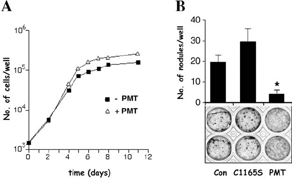
Figure 7.4. PMT regulates osteoblast proliferation and differentiation. (A) Effects of PMT
on the proliferation of primary rat calvarial osteoblasts. Cells were plated in the absence or
presence of recombinant PMT (20 ng/ml) and cell number was quantified on the indicated
days. (B) Effects of PMT on in vitro differentiation of rat calvarial osteoblasts into bone
nodules. Cells were cultured for 15 days in the absence (Con) or presence of recombinant
PMT (20 ng/ml) or inactive mutant PMT C1165S (20 ng/ml), and mineralised bone
nodules were quantified (upper panel) and visualised using the von Kossa technique
(black staining – lower panel). (
∗
p < 0.05 vs control) (see also Harmey et al., 2004).
of osteoblast differentiation and bone nodule formation (Figure 7.4B; see
also Harmey et al., 2004). The inhibitory effect of PMT was also confirmed
by the downregulation of the expression and activity of several osteoblast
markers (e.g., osteonectin, type I collagen, alkaline phosphatase, osteocalcin)
(Sterner-Kock et al., 1995; Mullan and Lax, 1996; Harmey et al., 2004). We
have also recently demonstrated that PMT downregulates the expression of
the runx2/cbfa-1 transcription factor, which, as shown in Figure 7.1,isanes-
sential gene for osteoblast differentiation and bone formation (Harmey et al.,
2004). The mechanism of PMT inhibition of osteoblast differentiation is not
well understood, although the signalling pathways known to be triggered by
PMT might offer some clue as to its mechanism of action in osteoblasts.
Indeed, PMT treatment of osteoblasts induced cytoskeletal rearrangements,
suggesting activation of the Rho GTPase, and our recent data have demon-
strated that the PMT-induced inhibition of osteoblast differentiation was re-
stored by treatment with the Rho inhibitor, C3 transferase (Table 7.1 and
Harmey et al., 2004).

P1: IwX
052182091Xc07.xml CB786/Lax 0 521 82091 X November 4, 2005 2:53
158
m
c
gowan, harmey, coxon, stenbeck,rogers, and grigoriadis
Table 7.1 The effects of PMT on in vitro bone
nodule formation of primary osteoblasts
Treatment No. of bone nodules
Control 44 ± 4
PMT 2 ± 0.5
PMT + C3 transferase 50 ± 12.5
Primary mouse osteoblasts were treated in the absence
(Control) or presence of recombinant PMT (20 ng/ml)
and the number of bone nodules were assessed. Treat-
ment with the Rho inhibitor C3 transferase abolishes
the inhibitory effects of PMT. (
∗
p < 0.05 vs control).
Taken together, these findings have demonstrated that activation of the
Rho GTPase negatively regulates osteoblast differentiation, and have also
demonstrated the usefulness of PMT in identifying a signalling pathway in
osteoblasts that is important for their function in synthesising bone.
PMT and Osteoclasts
PMT causes bone loss in vivo, and the results described in the previous section
suggest that one mechanism that may contribute to this phenotype is the
ability of PMT to inhibit bone formation in the bone remodelling cascade.
However, the progressive and rapid loss of nasal turbinate bones in the pig
is well established, and points to some effects of PMT on bone-resorbing
osteoclasts. Bone remains a principal target for PMT, an observation that
occurs apparently regardless of the route of administration. The effects of
PMT, however, are not restricted to pigs, or indeed to the nasal turbinate
bones, as pathological resorption has also been detected in rats and the long
bones of foetal mice (Kimman et al., 1987; Ackermann et al., 1991; Felix
et al., 1992; Martineau-Doize et al., 1993). The pro-resorptive effects of PMT
are therefore well established, but the effects of PMT on the osteoclast itself
remain unclear. Despite the slight increases in vivo in osteoclast size and
number observed by some (Martineau-Doize et al., 1993), findings to the
contrary have also been reported (Ackermann et al., 1993). Moreover, in vitro
studies have been unable to elaborate further on the direct effects of PMT
on osteoclast differentiation and activity, relative to the bone loss observed
in vivo.
P1: IwX
052182091Xc07.xml CB786/Lax 0 521 82091 X November 4, 2005 2:53
159
bacterial toxins and bone
As described earlier, osteoblasts are essential in osteoclastogenesis, and
any pathological alteration in bone mass could be attributed to effects on os-
teoblasts, osteoclasts, or both. Long-term porcine and murine bone marrow
cultures in the presence of PMT have shown increases in osteoclast-like cell
formation (TRAP positive multinucleated cells) (Jutras and Martineau-Doize,
1996; Gwaltney et al., 1997). However, in the studies by Gwaltney et al. (1997),
no increase in resorption was observed relative to the increase in osteoclast-
like cell number. Moreover, resorption data were not included in the findings
reported by Jutras and Martineau-Doize (1996). The only truly defining charac-
teristic of bona fide osteoclasts requires functional criteria, namely, the ability
to form resorption lacunae on a mineralised substrate (i.e., bone or dentine).
In this regard, it is imperative that osteoclast formation assays incorporate a
functional element. Furthermore, the heterogeneous nature of bone marrow
cultures and high probability of stromal cell contamination in these culture
systems could be contributing indirectly to the pro-resorptive effects.
It is interesting to note therefore that in the studies by Gwaltney et al.
(1997)itwas suggested that marrow stromal cells may be involved in the
observed increase in osteoclast formation. Subsequent studies by Mullan
and Lax, using partly purified populations of chick osteoblasts and osteo-
clasts, found no effect of PMT on bone resorption when either cell population
was cultured separately (Mullan and Lax, 1998). However, when co-cultured
with cell contact permitted, bone resorption was stimulated by PMT in a
concentration-dependent manner, indicating that the bone-resorbing effects
of PMT appear to be mediated, at least in part, via an interaction with osteo-
blasts (see below) (Mullan and Lax, 1998). This suggested that PMT induced
a pro-resorptive signal via osteoblasts expressed on the membrane of these
cells. By analogy with a variety of other mediators that act via the osteoblast
to induce bone resorption, this would suggest that PMT may increase the ex-
pression ratio of RANKL to OPG, although our studies in murine osteoblasts
suggest that the ratio of RANKL relative to OPG is decreased by PMT, indicat-
ing an overall negative effect on osteoclastogenesis (Harmey and Grigoriadis,
unpublished).
Following the discovery of the RANKL/OPG system of soluble regulators
of osteoclastogenesis, it has been possible to investigate osteoclast differen-
tiation much more efficiently and unambiguously in purified populations
of osteoclast precursors – for example, using macrophage precursors from
murine bone marrow preparations or human monocytes from peripheral
blood. Recently our group has investigated the role of PMT on osteoclast
differentiation and activity using this system, which has so far not been
P1: IwX
052182091Xc07.xml CB786/Lax 0 521 82091 X November 4, 2005 2:53
160
m
c
gowan, harmey, coxon, stenbeck,rogers, and grigoriadis
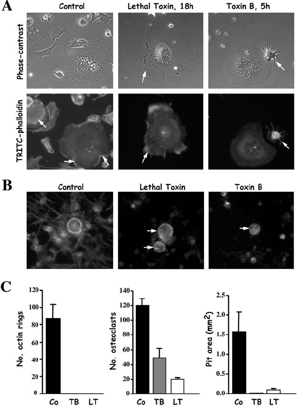
Figure 7.5. Toxin B and Lethal Toxin interfere with osteoclast morphology and function.
(A) Images of rabbit osteoclasts on plastic dishes were taken at the indicated time periods,
and cells were then fixed and stained with TRITC-phalloidin. Both lethal toxin and toxin B
disrupted the distribution of peripheral actin (arrows in Control) and caused retraction of
osteoclasts (retraction fibres indicated by arrows). Longer incubation with both toxins
resulted in complete retraction and loss of adherence (not shown). (B) Rabbit osteoclasts
were treated for 20 hours on dentine slices, then fixed and stained with TRITC-phalloidin.
Both lethal toxin and toxin B caused disruption of F-Actin rings and retraction of

P1: IwX
052182091Xc07.xml CB786/Lax 0 521 82091 X November 4, 2005 2:53
161
bacterial toxins and bone
addressed by others. Following the demonstration of the potent inhibitory
effect of PMT on osteoblast differentiation – an effect partially mediated by
activation of Rho – we found that PMT exerted a potent inhibitory effect on
murine osteoclastogenesis and resorption directly via acting on the osteo-
clast precursor population (Harmey and Grigoriadis, unpublished). Further-
more, PMT also inhibited human osteoclast formation from peripheral blood-
derived osteoclast precursors (McGowan and Grigoriadis, unpublished), and
in both precursor populations, these direct inhibitory actions of PMT were re-
stricted to the early stages of culture. Interestingly, preliminary data also sug-
gest that PMT may elicit an activating effect in mature human osteoclasts –
an effect also associated with morphological changes in the F-Actin rings
expressed by these cells (McGowan and Grigoriadis, unpublished). These
results suggest that PMT may have significantly different effects on the dif-
ferentiation of osteoclasts from haematopoietic precursors, versus its effects
on mature osteoclasts. It remains to be seen which signalling mechanisms
activated by PMT are involved in these divergent effects.
GTPases AND OSTEOCLASTS
With the important role of Rho GTPases in the physiological regulation of
the actin cytoskeleton, and the requirement for unique cytoskeletal arrange-
ments in normal osteoclast function (polarisation, ruffled border formation,
and subsequent resorption), it would be predicted that bacterial toxins might
influence osteoclast morphology and activity. Indeed, treatment of primary
mature osteoclasts with either Clostridium difficile toxin B or lethal toxin from
Clostridium sordellii, which inactivate all Rho family members (Rho, Rac, and
Cdc42), disrupted normal F-Actin ring formation and inhibited bone resorp-
tion (Figure 7.5; Coxon and Rogers, unpublished observations). These data
are in support of previous studies by Zhang and colleagues, who, using C3 ex-
oenzyme, were the first to suggest a dependence on Rho for normal F-Actin
ring (sealing zone) formation and resorption in vitro (Zhang et al., 1995).
RhoA is highly expressed in osteoclasts and appears to be actively involved in
the formation of podosomes – punctate membrane protrusions that are rich
in F-Actin and proteins such as vinculin and talin. Chellaiah and colleagues,
osteoclasts (arrows). (C) Rabbit osteoclasts on dentine were treated for 48 hours, and the
number of F-Actin rings, osteoclasts, and resorption area were determined. Both toxin B
(TB) and lethal toxin (LT) completely disrupted actin rings, reduced the number of
adherent osteoclasts on dentine, and dramatically inhibited bone resorption.
P1: IwX
052182091Xc07.xml CB786/Lax 0 521 82091 X November 4, 2005 2:53
162
m
c
gowan, harmey, coxon, stenbeck,rogers, and grigoriadis
using C3 exoenzyme in addition to constitutively active (Rho
Val-14
) and dom-
inant negative forms of Rho (Rho
Asn-19
), demonstrated that Rho activation
stimulated podosome assembly, osteoclast motility, and the resorptive activ-
ity of avian osteoclasts. Accordingly, opposite effects were observed in the
absence of functional Rho (Chellaiah et al., 2000).
Other small GTPase family members (Ras, Rab) also appear to have im-
portant roles in physiological osteoclast function (for a detailed review, see
Coxon and Rogers, 2003). Ras GTPases may be essential for osteoclast sur-
vival as dominant negative forms of Ras-induced apoptosis in murine osteo-
clasts (Miyazaki et al., 2000). Moreover, there is evidence to suggest that the
downstream mitogen activated protein kinase (MAPK) cascades (MEK/ERK)
are required for normal osteoclast differentiation (Miyazaki et al., 2000; Lee
et al., 2002), although reports to the contrary have also been published
(Matsumoto et al., 2000; Hotokezaka et al., 2002). Rab proteins are likely
to be essential for normal osteoclast function, given their role in vesicular
transport – a mechanism employed by the ruffled border in the acidification
of resorption lacunae. Rab 3b/c and Rab 7 are both expressed in osteoclasts,
with the latter being localised specifically to the ruffled border. Furthermore,
inhibition of Rab 7 by antisense was shown by Zhao and co-workers to inhibit
bone resorption in vitro (Zhao et al., 2001). Taken together, these data extend
the list of small GTPases that have specific effects on osteoclast function. De-
lineating how each of these signalling pathways interact under physiological
conditions to regulate bone resorption provides the basis for exciting future
experiments.
CONCLUDING REMARKS
Specific signalling pathways that regulate multiple aspects of cell differen-
tiation and cell physiology are being uncovered very rapidly by many labo-
ratories. The pronounced effects of several bacterial toxins on the differen-
tiation and activity of the two major bone cell populations, osteoblasts and
osteoclasts, have provided researchers with valuable insights for the elucida-
tion of signalling pathways utilised by small GTPases during the process of
bone remodelling. This has highlighted the usefulness of bacterial protein
toxins as tools to perturb specific signalling pathways in bone cells, result-
ing in altered cell behaviour. Elucidating further the mechanism of action
and specificity of such bacterial toxins in osteoblasts and osteoclasts will un-
doubtedly provide critical insights for understanding their roles in diseases
of the skeleton, and ultimately, for the development of novel therapeutic
strategies.
P1: IwX
052182091Xc07.xml CB786/Lax 0 521 82091 X November 4, 2005 2:53
163
bacterial toxins and bone
REFERENCES
Ackermann M R, Adams D A, Gerken L L, Beckman M J, and RimlerRB(1993).
Purified Pasteurella multocida protein toxin reduces acid phosphatase-positive
osteoclasts in the ventral nasal concha of gnotobiotic pigs. Calcif. Tissue Int.,
52, 455–459.
Ackermann M R, Rimler R B, and Thurston J R (1991). Experimental model of
atrophic rhinitis in gnotobiotic pigs. Infect. Immun., 59, 3626–3629.
Anderson D M, Maraskovsky E, Billingsley W L, Dougall W C, Tometsko M E,
Roux E R, Teepe M C, DuBose R F, Cosman D, and Galibert L (1997). A
homologue of the TNF receptor and its ligand enhance T-cell growth and
dendritic-cell function. Nature, 390, 175–179.
Athanasou N A (1996). Cellular biology of bone-resorbing cells. J. Bone. Joint.
Surg. Am., 78, 1096–1112.
Aubin J E (1998). Advances in the osteoblast lineage. Biochem. Cell Biol., 76, 899–
910.
BishopALand Hall A (2000). Rho GTPases and their effector proteins. Biochem.
J., 348, 241–255.
Blair H C, Teitelbaum S L, Ghiselli R, and Gluck S (1989). Osteoclastic bone
resorption by a polarized vacuolar proton pump. Science, 245, 855–857.
Boquet P (2000). The cytotoxic necrotizing factor 1 (CNF1). From uropathogenic
Escherichia Coli. Adv. Exp. Med. Biol., 485, 45–51.
Boyce B F, Yoneda T, Lowe C, Soriano P, and MundyGR(1992). Requirement
of pp 60c-src expression for osteoclasts to form ruffled borders and resorb
bone in mice. J. Clin. Invest, 90, 1622–1627.
Chellaiah M A, Soga N, Swanson S, McAllister S, Alvarez U, Wang D, Dowdy
SF,and Hruska K A (2000). Rho-A is critical for osteoclast podosome orga-
nization, motility, and bone resorption. J. Biol. Chem., 275, 11993–12002.
CoxonFPand Rogers M J (2003). The Role of Prenylated Small GTP-Binding
Proteins in the Regulation of Osteoclast Function. Calcif. Tissue Int., 72, 80–
84.
Darnay B G, Haridas V, Ni J, Moore P A, and Aggarwal B B (1998). Character-
ization of the intracellular domain of receptor activator of NF-κB (RANK).
Interaction with tumor necrosis factor receptor- associated factors and ac-
tivation of NF-κB and c-Jun N-terminal kinase. J. Biol. Chem., 273, 20551–
20555.
Darnay B G, Ni J, Moore P A, and Aggarwal B B (1999). Activation of NF-κB
by RANK requires tumor necrosis factor receptor-associated factor (TRAF)
6 and NF-κB-inducing kinase. Identification of a novel TRAF6 interaction
motif. J Biol. Chem., 274, 7724–7731.
P1: IwX
052182091Xc07.xml CB786/Lax 0 521 82091 X November 4, 2005 2:53
164
m
c
gowan, harmey, coxon, stenbeck,rogers, and grigoriadis
Ducy P, Schinke T, and Karsenty G (2000). The osteoblast: a sophisticated fibro-
blast under central surveillance. Science, 289, 1501–1504.
Duong L T, Lakkakorpi P, Nakamura I, and RodanGA(2000). Integrins and
signaling in osteoclast function. Matrix Biol., 19, 97–105.
Etienne-Manneville S and Hall A (2002). Rho GTPases in cell biology. Nature,
420, 629–635.
Felix R, Fleisch H, and Frandsen P L (1992). Effect of Pasteurella multocida toxin
on bone resorption in vitro. Infect. Immun., 60, 4984–4988.
Flatau G, Lemichez E, Gauthier M, Chardin P, Paris S, Fiorentini C, and Boquet
P (1997). Toxin-induced activation of the G protein p21 Rho by deamidation
of glutamine. Nature, 387, 729–733.
Fujikawa Y, Sabokbar A, Neale S D, Itonaga I, Torisu T, and Athanasou N A
(2001). The effect of macrophage-colony stimulating factor and other hu-
moral factors (interleukin-1, -3, -6, and -11, tumor necrosis factor-alpha, and
granulocyte macrophage-colony stimulating factor) on human osteoclast for-
mation from circulating cells. Bone, 28, 261–267.
Grigoriadis A E, Wang Z Q, Cecchini M G, Hofstetter W, Felix R, Fleisch H A, and
Wagner E F (1994). c-Fos: A key regulator of osteoclast-macrophage lineage
determination and bone remodeling. Science, 266, 443–448.
Gwaltney S M, Galvin R J, Register K B, Rimler R B, and AckermannMR(1997).
Effects of Pasteurella multocida toxin on porcine bone marrow cell differenti-
ation into osteoclasts and osteoblasts. Vet. Pathol., 34, 421–430.
Harmey D, Stenbeck G, Nobes C D, Lax A J, and Grigoriadis A E (2004). Regulation
of osteoblast differentiation by Pasteurella multocida toxin (PMT): A role for
Rho GTPase in bone formation. J. Bone Miner. Res., 19, 661–670.
Helfrich M H and HortonMA(1999). Integrins and adhesion molecules. In
Dynamics of Bone and Cartilage Metabolism, ed. M J Siebel,SPRobins, and
JPBileziken, pp. 111–125, Academic Press, San Diego.
Henderson B and Nair S P (2003). Hard labour: Bacterial infection of the skeleton.
Trends Microbiol., 11, 570–577.
Hofbauer L C and Heufelder A E (1998). Osteoprotegerin and its cognate ligand:
A new paradigm of osteoclastogenesis. Eur. J. Endocrinol., 139, 152–154.
Horiguchi Y, Inoue N, Masuda M, Kashimoto T, Katahira J, Sugimoto N, and
Matsuda M (1997). Bordetella bronchiseptica dermonecrotizing toxin induces
reorganization of actin stress fibers through deamidation of Gln-63 of the
GTP-binding protein Rho. Proc. Natl. Acad. Sci. USA, 94, 11623–11626.
Horiguchi Y, Okada T, Sugimoto N, Morikawa Y, Katahira J, and Matsuda M
(1995). Effects of Bordetella bronchiseptica dermonecrotizing toxin on bone
formation in calvaria of neonatal rats. FEMS Immunol. Med. Mic., 12, 29–32.
P1: IwX
052182091Xc07.xml CB786/Lax 0 521 82091 X November 4, 2005 2:53
165
bacterial toxins and bone
Hotokezaka H, Sakai E, Kanaoka K, Saito K, Matsuo K, Kitaura H, Yoshida N,
and Nakayama K (2002). U0126 and PD98059, specific inhibitors of MEK,
accelerate differentiation of RAW264.7 cells into osteoclast-like cells. J. Biol.
Chem., 277, 47366–47372.
Hsu H, Lacey D L, Dunstan C R, Solovyev I, Colombero A, Timms E, Tan H L,
Elliott G, Kelley M J, Sarosi I, Wang L, Xia X Z, Elliott R, Chiu L, Black
T, Scully S, Capparelli C, Morony S, Shimamoto G, Bass M B, and Boyle
WJ(1999). Tumor necrosis factor receptor family member RANK mediates
osteoclast differentiation and activation induced by osteoprotegerin ligand.
Proc. Natl. Acad. Sci. USA, 96, 3540–3545.
Iotsova V, Caamano J, Loy J, Yang Y, Lewin A, and Bravo R (1997). Osteopetrosis
in mice lacking NF-κB1 and NF-κB2. Nat. Med., 3,1285–1289.
Jutras I, and Martineau-Doize B (1996). Stimulation of osteoclast-like cell for-
mation by Pasteurella multocida toxin from hemopoietic progenitor cells in
mouse bone marrow cultures. Can. J. Vet. Res., 60, 34–39.
Karsenty G, and Wagner E F (2002). Reaching a genetic and molecular under-
standing of skeletal development. Dev. Cell, 2, 389–406.
Kimman T G, Lowik C W, van de Wee-Pals L J, Thesingh C W, Defize P, Kamp
EM,and Bijvoet O L (1987). Stimulation of bone resorption by inflamed
nasal mucosa, dermonecrotic toxin-containing conditioned medium from
Pasteurella multocida, and purified dermonecrotic toxin from P. multocida.
Infect. Immun., 55, 2110–2116.
Lacerda H M, Pullinger G D, Lax A J, and Rozengurt E (1997). Cytotoxic necro-
tizing factor 1 from Escherichia coli and dermonecrotic toxin from Bordetella
bronchiseptica induce p21(rho)-dependent tyrosine phosphorylation of focal
adhesion kinase and paxillin in Swiss 3T3 cells. J. Biol. Chem., 272, 9587–
9596.
Lacey D L, Timms E, Tan H L, Kelley M J, Dunstan C R, Burgess T, Elliott R,
Colombero A, Elliott G, Scully S, Hsu H, Sullivan J, Hawkins N, Davy E,
Capparelli C, Eli A, Qian Y X, Kaufman S, Sarosi I, Shalhoub V, Senaldi G,
Guo J, Delaney J, and Boyle W J (1998). Osteoprotegerin ligand is a cytokine
that regulates osteoclast differentiation and activation. Cell, 93,165–176.
Lax A J, and Chanter N (1990). Cloning of the toxin gene from Pasteurella multocida
and its role in atrophic rhinitis. J. Gen. Microbiol., 136, 81–87.
Lee S E, Woo K M, Kim S Y, Kim H M, Kwack K, Lee Z H, and Kim H H (2002). The
phosphatidylinositol 3-kinase, p38, and extracellular signal-regulated kinase
pathways are involved in osteoclast differentiation. Bone, 30, 71–77.
Lerm M, Schmidt G, and Aktories K (2000). Bacterial protein in toxins targetting
rho GTPases. FEMS Microbiol. Lett., 188, 1–6.