Lax Alistair J. Bacterial protein toxins: Role in the interference with cell growth regulation (Бактериальные токсины белков: роль в регуляции роста клеток)
Подождите немного. Документ загружается.

P1: IwX
052182091Xc07.xml CB786/Lax 0 521 82091 X November 4, 2005 2:53
166
m
c
gowan, harmey, coxon, stenbeck,rogers, and grigoriadis
Lerm M, Schmidt G, Goehring U M, Schirmer J, and Aktories K (1999). Iden-
tification of the region of rho involved in substrate recognition by Es-
cherichia coli cytotoxic necrotizing factor 1 (CNF1). J. Biol. Chem., 274, 28999–
29004.
Lomaga M A, Yeh W C, Sarosi I, Duncan G S, Furlonger C, Ho A, Morony S,
Capparelli C, Van G, Kaufman S, van der Heiden A, Itie A, Wakeham A,
Khoo W, Sasaki T, Cao Z D, Penninger J M, Paige C J, Lacey D L, Dunstan
CR,Boyle W J, Goeddel D V, and MakTW(1999). TRAF6 deficiency results
in osteopetrosis and defective interleukin-1, CD40, and LPS signaling. Gene
Dev., 13, 1015–1024.
Lowe C, Yoneda T, Boyce B F, Chen H, Mundy G R, and Soriano P (1993).
Osteopetrosis in Src-deficient mice is due to an autonomous defect of osteo-
clasts. Proc. Natl. Acad. Sci. USA, 90, 4485–4489.
Mackay D J and Hall A (1998). Rho GTPases. J. Biol. Chem., 273, 20685–
20688.
Manolagas S C (2000). Birth and death of bone cells: Basic regulatory mechanisms
and implications for the pathogenesis and treatment of osteoporosis. Endocr.
Rev., 21, 115–137.
Martineau-Doize B, Caya I, Gagne S, Jutras I, and Dumas G (1993). Effects of
Pasteurella multocida toxin on the osteoclast population of the rat. J. Comp.
Pathol., 108, 81–91.
Matsumoto M, Sudo T, Saito T, Osada H, and Tsujimoto M (2000). Involvement
of p38 mitogen-activated protein kinase signaling pathway in osteoclastogen-
esis mediated by receptor activator of NF-κB ligand (RANKL). J. Biol. Chem.,
275, 31155–31161.
Miyazaki T, Katagiri H, Kanegae Y, Takayanagi H, Sawada Y, Yamamoto A, Pando
MP,Asano T, Verma I M, Oda H, Nakamura K, and Tanaka S (2000).
Reciprocal role of ERK and NF-κB pathways in survival and activation of
osteoclasts. J. Cell Biol., 148, 333–342.
Mullan P B and Lax A J (1996). Pasteurella multocida toxin is a mitogen for bone
cells in primary culture. Infect. Immun., 64, 959–965.
Mullan P B and Lax A J (1998). Pasteurella multocida toxin stimulates bone re-
sorption by osteoclasts via interaction with osteoblasts. Calcified. Tissue Int.,
63, 340–345.
Mundy G R (1998). Bone remodelling. In Primer on the metabolic bone diseases and
disorders of mineral metabolism, 4th ed., ed.MJFavus, pp. 30–39, Lippincott
Williams & Wilkins, Philadelphia.
Nair S P, Meghji S, Wilson M, Reddi K, White P, and Henderson B (1996). Bac-
terially induced bone destruction: Mechanisms and misconceptions. Infect.
Immun., 64, 2371–2380.
P1: IwX
052182091Xc07.xml CB786/Lax 0 521 82091 X November 4, 2005 2:53
167
bacterial toxins and bone
Nesbitt S A and Horton M A (1997). Trafficking of matrix collagens through
bone-resorbing osteoclasts. Science, 276, 266–269.
Nobes C D and Hall A (1995). Rho, rac, and cdc42 GTPases regulate the assem-
bly of multimolecular focal complexes associated with actin stress fibers,
lamellipodia, and filopodia. Cell, 81, 53–62.
Raisz L G (1999). Physiology and pathophysiology of bone remodeling. Clin.
Chem., 45, 1353–1358.
Rodan G A and Martin T J (1981). Role of osteoblasts in hormonal control of bone
resorption–A hypothesis. Calcif. Tissue Int., 33, 349–351.
Rozengurt E, Higgins T, Chanter N, Lax A J, and Staddon J M (1990). Pasteurella
multocida toxin: Potent mitogen for cultured fibroblasts. Proc. Natl. Acad. Sci.
USA, 87, 123–127.
Salo J, Lehenkari P, Mulari M, Metsikko K, and Vaananen H K (1997). Removal
of osteoclast bone resorption products by transcytosis. Science, 276, 270–
273.
Sarma U and Flanagan A M (1996). Macrophage colony-stimulating factor in-
duces substantial osteoclast generation and bone resorption in human bone
marrow cultures. Blood, 88, 2531–2540.
Schmidt G, Goehring U M, Schirmer J, Lerm M, and Aktories K (1999). Identifi-
cation of the C-terminal part of Bordetella dermonecrotic toxin as a transglu-
taminase for rho GTPases. J. Biol. Chem., 274, 31875–31881.
Schmidt G, Sehr P, Wilm M, Selzer J, Mann M, and Aktories K (1997). Gln 63 of
Rho is deamidated by Escherichia coli cytotoxic necrotizing factor-1. Nature,
387, 725–729.
Schmidt G, Selzer J, Lerm M, and Aktories K (1998). The Rho-deamidating cy-
totoxic necrotizing factor 1 from Escherichia coli possesses transglutaminase
activity. Cysteine 866 and histidine 881 are essential for enzyme activity.
J. Biol. Chem., 273,13669–13674.
Simonet W S, Lacey D L, Dunstan C R, Kelley M, Chang M S, Luthy R, Nguyen
HQ,Wooden S, Bennett L, Boone T, Shimamoto G, DeRose M, Elliott R,
Colombero A, Tan H L, Trail G, Sullivan J, Davy E, Bucay N, Renshaw-Gegg
L, Hughes T M, Hill D, Pattison W, Campbell P, and Boyle W J (1997).
Osteoprotegerin: A novel secreted protein involved in the regulation of bone
density. Cell, 89, 309–319.
Soriano P, Montgomery C, Geske R, and Bradley A (1991). Targeted disrup-
tion of the c-src proto-oncogene leads to osteopetrosis in mice. Cell, 64,
693–702.
Sterner-Kock A, Lanske B, Uberschar S, and AtkinsonMJ(1995). Effects of
the Pasteurella multocida toxin on osteoblastic cells in vitro. Vet. Pathol., 32,
274–279.
P1: IwX
052182091Xc07.xml CB786/Lax 0 521 82091 X November 4, 2005 2:53
168
m
c
gowan, harmey, coxon, stenbeck,rogers, and grigoriadis
Suda T, Nakamura I, Jimi E, and Takahashi N (1997). Regulation of osteoclast
function. J. Bone Miner. Res., 12, 869–879.
Suda T, Takahashi N, and Martin T J (1992). Modulation of osteoclast differenti-
ation. Endocr. Rev., 13, 66–80.
Suda T, Takahashi N, Udagawa N, Jimi E, Gillespie M T, and Martin T J (1999).
Modulation of osteoclast differentiation and function by the new members
of the tumor necrosis factor receptor and ligand families. Endocr. Rev., 20,
345–357.
Takahashi N, Akatsu T, Udagawa N, Sasaki T, Yamaguchi A, Moseley J M, Martin
TJ,and Suda T (1988). Osteoblastic cells are involved in osteoclast formation.
Endocrinology, 123, 2600–2602.
Takai Y, Sasaki T, and Matozaki T (2001). Small GTP-binding proteins. Physiol.
Rev., 81, 153–208.
Teitelbaum S L (2000). Bone resorption by osteoclasts. Science, 289, 1504–1508.
Vaananen H K and Horton M (1995). The osteoclast clear zone is a specialized
cell-extracellular matrix adhesion structure. J. Cell Sci., 108, 2729–2732.
Ward P N, Miles A J, Sumner I G, Thomas L H, and Lax A J (1998). Activity
of the mitogenic Pasteurella multocida toxin requires an essential C-terminal
residue. Infect. Immun., 66, 5636–5642.
Wei S, Teitelbaum S L, Wang M W, and RossFP(2001). Receptor activator of
nuclear factor-kappa b ligand activates nuclear factor-κBinosteoclast pre-
cursors. Endocrinology, 142, 1290–1295.
Yasuda H, Shima N, Nakagawa N, Yamaguchi K, Kinosaki M, Mochizuki S,
Tomoyasu A, Yano K, Goto M, Murakami A, Tsuda E, Morinaga T, Higashio
K, Udagawa N, Takahashi N and Suda T (1998). Osteoclast differentiation
factor is a ligand for osteoprotegerin/osteoclastogenesis-inhibitory factor and
is identical to TRANCE/RANKL. Proc. Natl. Acad. Sci. USA, 95, 3597–3602.
Zhang D, Udagawa N, Nakamura I, Murakami H, Saito S, Yamasaki K, Shibasaki
Y, Morii N, Narumiya S, and Takahashi N (1995). The small GTP-binding
protein, rho p21, is involved in bone resorption by regulating cytoskeletal
organization in osteoclasts. J. Cell Sci., 108, 2285–2292.
Zhao H, Laitala-Leinonen T, Parikka V, and Vaananen H K (2001). Downregula-
tion of small GTPase Rab7 impairs osteoclast polarization and bone resorp-
tion. J. Biol. Chem., 276, 39295–39302.
Zywietz A, Gohla A, Schmelz M, Schultz G, and Offermanns S (2001). Pleiotropic
effects of Pasteurella multocida toxin are mediated by Gq-dependent and
-independent mechanisms: Involvement of Gq but not G11. J. Biol. Chem.,
276, 3840–3845.
P1: IwX
052182091Xc08.xml CB786/Lax 0 521 82091 X November 4, 2005 3:13
169
CHAPTER 8
Helicobacter pylori mechanisms for inducing
epithelial cell proliferation
Michael Naumann and Jean E Crabtree
Helicobacter pylori, the first bacterium to be designated a Class I carcino-
gen, has a major aetiological role in human gastric carcinogenesis. H. pylori
infection is acquired primarily in childhood and, in the majority of in-
stances, infection and associated chronic gastritis are lifelong. A key feature of
H. pylori infection of relevance to the associated increased risk of developing
gastric cancer is the hyperproliferation of gastric epithelial cells induced by
the bacterium. Infection is associated with increased gastric epithelial cell
proliferation in both humans and in experimental animal models.
Clinically, there is a marked diversity in the outcome of H. pylori infec-
tion and only a few infected subjects will develop gastric cancer (reviewed
Peek and Blaser, 2002). Recent studies in Japan show that the risk of cancer
with H. pylori infection is greatest in infected subjects with nonulcer dys-
pesia or gastric ulceration who develop severe gastric atrophy and intestinal
metaplasia (Uemura et al., 2001). Bacterial virulence factors such as the cag
pathogenicity island (PAI) (Blaser et al.,1995; Kuipers et al., 1995; Webb et al.,
1999) and genetic polymorphisms in the interleukin-1β and IL-1 receptor an-
tagonist genes associated with overexpression of IL-1 and hypochlorhydria
(El-Omar et al., 2000; Machado et al., 2001; Furuta et al., 2002) have each been
linked to an increased risk of developing gastric atrophy and/or intestinal type
gastric cancer.
H. pylori is one of several chronic infections that have recently been as-
sociated with the development of neoplasia (see Chapter 9). The cellular and
molecular pathways by which H. pylori infection promotes epithelial hyper-
proliferative responses and transformation are the subject of active investiga-
tion. H. pylori represents an excellent model system to investigate pathogen-
induced epithelial cell signalling pathways of relevance to neoplasia. Recent
studies have focused on identifying bacterial and host factors involved in the
P1: IwX
052182091Xc08.xml CB786/Lax 0 521 82091 X November 4, 2005 3:13
170
michael naumann and jean e crabtree
epithelial hyperproliferative responses. Both in vitro bacterial–epithelial co-
culture systems, and in vivo studies in humans and animal models, have been
used to examine bacterial factors involved in the hyperproliferative response
and the mechanisms by which H. pylori alters cell cycle control in the gastric
epithelium.
CLINICAL STUDIES
Many clinical studies have demonstrated that infection with H. pylori is as-
sociated with increased epithelial cell proliferation in humans (Lynch et al.,
1995a; Lynch et al., 1995b; Cahill et al., 1996; Jones et al., 1997; Moss et al.,
2001). Increased epithelial proliferation is observed early in the natural his-
tory of infection, being present in H. pylori–infected children (Jones et al.,
1997). Lifelong increased cell turnover is likely to be an important risk factor
for the development of gastric cancer. Patients with H. pylori negative gas-
tritis do not have increased gastric epithelial cell proliferation compared to
uninfected controls (Lynch et al., 1995a; Lynch et al., 1995b; Cahill et al., 1996;
Panella et al., 1996). Epithelial proliferation indices have been positively cor-
related with the degree of histological inflammation in both the antrum and
corpus mucosa in H. pylori–infected patients (Peek et al., 1997; Lynch et al.,
1999; Moss et al., 2001). However, stepwise multiple regression analysis has
indicated that the only independent predictor of epithelial cell proliferation
is the density of H. pylori colonisation (Lynch et al., 1999). This suggests that
in humans the enhanced epithelial proliferation observed with infection is
promoted both as a consequence of the inflammatory response, and by a
route independent of inflammation.
The effect of eradication of H. pylori infection on gastric epithelial cell
proliferation has also been investigated. A significant decrease in gastric ep-
ithelial cell proliferation has been observed following successful eradication
of H. pylori (Brenes et al., 1993; Fraser et al., 1994; Lynch et al., 1995b; Cahill
et al., 1995; Leung et al., 2000). In addition, attenuated levels of epithelial cell
proliferation have also been observed in patients where H. pylori eradication
was unsuccessful (Fraser et al., 1994; Lynch et al., 1995b). This may be due
to a decrease in the intensity of inflammation and/or bacterial density after
therapy in those patients in whom treatment failed. Only one study to date
has failed to demonstrate a reduction in gastric epithelial cell proliferation
after eradication of H. pylori infection (El-Zimaity et al., 2000). Divergent
results may be attributed to varying patient populations, labelling tech-
niques, and/or treatment with pharmacological agents such as proton pump
inhibitors.
P1: IwX
052182091Xc08.xml CB786/Lax 0 521 82091 X November 4, 2005 3:13
171
helicobacter pylori
AB
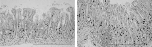
Figure 8.1. Gastric epithelial cell proliferation in the antrum of Mongolian gerbils
infected with H. pylori SS1 strain. Epithelial cell proliferation in the antral gastric mucosa
of A) uninfected control Mongolian gerbil, B) Mongolian gerbil, 4 weeks post-infection
with H. pylori. Bar = 500 µm. Mongolian gerbils were injected 1 hour prior to sacrifice
with bromodeoxyuridine (BrdU), and the presence of proliferating epithelial cells was
detected by immunohistochemistry using a monoclonal antibody to BrdU. Adapted from
Court et al., 2002.
ANIMAL MODELS
Experimental infection with H. pylori,orthe related gastric Helicobacter sp.,
H. mustelae,orH. felis, increases gastric epithelial cell proliferation in a variety
of animal species, including Japanese monkeys (Fujiyama et al., 1995), ferrets
(Yu et al., 1995), Mongolian gerbils (Peek et al., 2000; Israel et al., 2001; Court
et al., 2002) (Figure 8.1), and mice (Fox et al., 1996; Wang et al., 1998; Fox et al.,
1999). Dietary factors such as salt intake have also been shown to increase
gastric epithelial cell proliferation in the H. pylori murine model (Fox et al.,
1999).
The most detailed studies to date on Helicobacter-induced gastric epithe-
lial cell proliferation have been undertaken in the murine H. felis model.
H. felis infection in mice causes severe inflammation and marked epithelial
hyperplasia (Wang et al., 1998; Ferrero et al., 2000). In C57BL/6 mice, H. felis
induces greater gastric epithelial cell proliferative responses than H. pylori
(Court et al., 2002). Both the severity of the gastritis induced by H. felis in-
fection (Sakagami et al., 1996), and the extent of epithelial proliferation and
apoptosis (Wang et al., 1998), are dependent on the strain of mouse infected.
In C57BL/6 mice, H. felis induces extensive epithelial hyperproliferation in
the corpus mucosa, with parietal and chief cells being replaced with mucous
secreting cells (Sakagami et al., 1996). Wang et al. (1998) demonstrated that
both epithelial proliferative responses and apoptosis regulation are increased
in H. felis infected C57BL/6 mice, which they considered could relate to the
absence of phospholipase A2 in these mice (Wang et al., 1998). Interestingly,
P1: IwX
052182091Xc08.xml CB786/Lax 0 521 82091 X November 4, 2005 3:13
172
michael naumann and jean e crabtree
detailed analysis of H. felis stimulated gastric pathology, epithelial cell prolif-
eration, and apoptosis regulation over a one-year period in C57BL/6 mice has
revealed marked gender differences in responses (Court et al., 2003). Signifi-
cant increases in epithelial proliferation and apoptosis in response to H. felis
were observed only in female mice, possibly reflecting sex differences in the
immune responses and cytokine production. Gender differences in ethanol-
induced ulceration and gastritis have been previously observed in rats, with
females having increased levels of gastric epithelial cell proliferation com-
pared to males (Liu et al., 2001). The functional importance of gender should
be considered in future murine studies on H. felis– and H. pylori–induced
chronic gastritis.
H. pylori infection is associated with elevated plasma gastrin (El-Omar
et al., 1997), a protein known to promote gastric epithelial hyperproliferation
(Wang et al., 1996; Tsutsui et al., 1997; Miyazaki et al., 1999). In gerbils,
H. pylori–induced epithelial cell proliferation has been correlated with ele-
vated serum gastrin levels (Peek et al., 2000). In a transgenic mouse model
that over-expresses gastrin, H. felis infection accelerates the development of
gastric adenocarcinoma (Wang et al., 2000b). However, the effects of H. felis
infection on gastric epithelial cell proliferation in wild-type and hypergastri-
naemic mice have not been examined. The contributions of gastric Helicobac-
ter infection, hypergastrinaemia, and the chronic inflammatory response to
epithelial hyperproliferation are areas of active research.
H. PYLORI VIRULENCE FACTORS
H. pylori is a genomically diverse pathogen (Suerbaum et al., 1998), and sev-
eral bacterial virulence factors are now considered to have a key role on the
epithelial response to infection. It has become apparent in recent years that
the epithelial cellular response to H. pylori is variable. Only strains contain-
ing the 40 kb cag PAI (Censini et al., 1996; Akopypants et al., 1998) trigger
signalling cascades in gastric epithelial cells resulting in AP-1 and NF-κB
activation (Naumann et al., 1999; Foryst-Ludwig and Naumann, 2000) and
multiple associated changes in gene expression. Of particular interest has
been the observation that chemokines such as IL-8 are upregulated in gastric
epithelial cells by cag PAI positive H. pylori strains (Crabtree et al., 1994a;
Sharma et al., 1995). The upregulation of IL-8 and other neutrophil chemo-
tactic C-X-C chemokines, such as GRO-α (Crabtree et al., 1994b; Eck et al.
2000), in human gastric epithelial cells is probably critical to the association
among infection with cag PAI positive strains, neutrophilic responses, and
more severe gastroduodenal disease (Crabtree et al., 1991; Weel et al., 1996).
P1: IwX
052182091Xc08.xml CB786/Lax 0 521 82091 X November 4, 2005 3:13
173
helicobacter pylori
The gene products of the cag PAI encode a type IV secretory system that
translocates CagA (Segal et al., 1999; Asahi et al., 2000; Backert et al., 2000;
Odenbreit et al., 2000; Stein et al., 2000) and presumably unknown factors
into the gastric epithelial cells. The profile of gene expression, as determined
by cDNA array analysis, in gastric epithelial cells differs markedly after cul-
ture with wild-type cag PAI positive and wild-type cag PAI negative strains,
with many genes involved with cell cycle control and apoptosis being dif-
ferentially expressed (Cox et al., 2001). There has been considerable interest
in whether in vivo specific H. pylori strains are associated with enhanced
epithelial proliferation and apoptosis.
Evidence for H. pylori Strain Related Differences in Gastric
Epithelial Cell Proliferation in vivo
The contribution of bacterial factors to the induction of gastric epithelial cell
proliferation and the regulation of apoptosis is under active investigation.
One approach has been to examine the effects of H. pylori strains of spe-
cific genotype and isogenic mutants on proliferation and apoptosis in gastric
epithelial cell lines in vitro (Peek et al., 1999). However, there is marked vari-
ability in the expression of cell cycle regulatory proteins such as p27
KIP1
and
cyclin E/cdk2 activity induced by H. pylori in different gastric epithelial cell
lines (Sommi et al., 2002), making extrapolation of in vitro studies to events
in vivo difficult. In addition, the majority of studies have documented inhibi-
tion of proliferation by H. pylori in vitro (Shirin et al., 1999; Peek et al., 1999),
which will not necessarily reflect events in vivo.
It is currently unclear from the clinical studies carried out whether gastric
epithelial cell proliferation varies according to the cag PAI status of the infect-
ing strain. Two studies have reported that gastric epithelial cell proliferation
is greater in patients infected with cagA positive strains than cagA negative
strains (Peek et al., 1997; Rokkas et al., 1999), although another study in pa-
tients with non-ulcer dyspepsia failed to confirm these observations (Moss
et al., 2001). A recent study in a Chinese population where 98% of patients
were infected with cagA+ H. pylori strains identified increased epithelial cell
proliferation in those infected with strains expressing the blood group anti-
gen binding adhesin babA2 (Yu et al., 2002). The presence or absence of
adhesins may account for earlier discrepancies.
There have also been similarly divergent results with respect to the ef-
fects of the cag PAI on apoptosis in human gastric epithelial cells. Two studies
reported that apoptosis was greater in patients infected with cagA negative
strains than cagA+ strains (Peek et al., 1997; Rokkas et al., 1999), whilst one
P1: IwX
052182091Xc08.xml CB786/Lax 0 521 82091 X November 4, 2005 3:13
174
michael naumann and jean e crabtree
reported the converse (Moss et al., 2001). Lipopolysaccharide (LPS) of H. pylori
cag PAI positive strains induced apoptosis in isolated guinea pig–derived gas-
tric pit cells in vitro, whereas LPS from cag PAI negative strains did not induce
apoptosis (Kawahara et al., 2001). Other recent in vitro studies indicate that
H. pylori induces apoptosis via NF-κB dependant cascades (Gupta et al.,
2001)–observations not in accordance with the observed reduced apoptosis in
the gastric mucosa of patients with cag PAI positive strains compared to those
with cag PAI negative strains (Peek et al., 1997; Rokkas et al., 1999). Activa-
tion of the peroxisome proliferator-activated receptor γ (PPARγ ) suppressed
NF-κB mediated apoptosis in vitro (Gupta et al., 2001), but this inhibition was
independent of cag PAI status. From the limited clinical data, the effects of
bacterial virulence factors, such as the cag PAI, on gastric epithelial prolif-
eration and apoptosis in vivo are unresolved. There are several confounding
factors, including probably variable usage of non-steroidal anti-inflammatory
drugs, which will effect apoptosis (Leung et al., 2000).
Infection of animal models with genetically defined H. pylori strains is an
alternative approach to investigate the importance of the cag PAI on gastric
epithelial proliferation and apoptosis. Studies in mice have mainly used the
SS1 H. pylori strain (Lee et al., 1997). Despite being cagA+, this strain appears
to lack a functional cag PAI (Crabtree et al., 2002), and fails to induce IL-8
secretion in human gastric epithelial cells in vitro (van Doorn et al., 1999). In
the mouse, host-induced changes in cag PAI genotype and related functions
have also been reported (Sozzi et al., 2001). In addition, recent studies indicate
that the mouse is preferentially colonised by cagA negative strains which
induce reduced inflammatory responses (Philpott et al., 2002), thus making
it an unsuitable model for investigating cag PAI effects on host responses.
Mongolian gerbils can be successfully colonised with H. pylori strains,
with or without a functional cag pathogenicity island (Wirth et al., 1998;
Peek et al., 2000; Israel et al., 2001; Akanuma et al., 2002; Wang et al., 2003)
(Figure 8.2). Long-term infection is associated with the development of in-
testinal metaplasia (Ikeno et al., 1999) and gastric adenocarcinoma (Watanabe
et al., 1998). In the gerbil, cag PAI strains of H. pylori have been shown to
induce more severe gastritis (Ogura et al., 2000; Israel et al., 2001; Akanuma
et al., 2002) and increased epithelial proliferation and apoptosis regulation
(Israel et al., 2001), compared to strains lacking a functional cag PAI. Gastric
epithelial cell proliferation in the gerbil in response to H. pylori SS1 strain is
significantly greater than in the mouse (Court et al., 2002). During the early
stages of infection in the gerbil, the epithelial hyperproliferative responses
are confined to the antrum, but with time, progress to the corpus (Israel
et al., 2001; Court et al., 2002), mirroring the pathology and proliferative
P1: IwX
052182091Xc08.xml CB786/Lax 0 521 82091 X November 4, 2005 3:13
175
helicobacter pylori
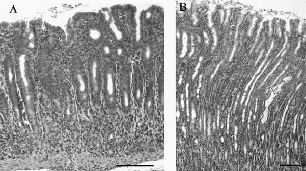
Figure 8.2. Gastric histology in Mongolian gerbil infected with H. pylori strain SS1 for
36 weeks. Haematoxylin and Eosin stained sections of A) antral mucosa and B) corpus
mucosa. Bar = 100 µm. Adapted from Naumann and Crabtree (2004). (See color section.)
responses observed with human infection (Moss et al., 2001). The gerbil is
thus a useful model to analyse the role of H. pylori virulence factors on gastric
epithelial proliferation and apoptosis. Further studies with isogenic mutants
of other virulence factors will be important to delineate their importance in
the epithelial hyperproliferative response.
H. PYLORI INTERFERENCE WITH HOST CELL SIGNALLING
Activation of Proliferation-Associated Kinases ERK/MEK in
H. pylori Infection and the Role of COX-2 Induction
While clinical and animal model studies have investigated several aspects
of the bacterial-induced hyperproliferative responses, recent in vitro studies
with gastric epithelial cells have begun to delineate the importance of specific
signalling pathways. Furthermore, the contribution of these pathways to over-
expression of key genes potentially involved in gastric neoplasia has been
examined.
Pathways of great current interest are the induction of nitric oxide syn-
thase and cyclooxygenase 2 (COX-2) in H. pylori infection (Fu et al., 1999).
As in many human tumour cells, gastric cancer cells over-express COX-2
(Sung et al., 2000), which represents an enzyme responsible for the release
of prostaglandin E
2
(PGE
2
). PGE
2
is implicated in maintaining the function
and structure of the gastric mucosa by modulating diverse cellular functions,