Koster G., Rijnders G. (Eds.) In situ Characterization of Thin Film Growth
Подождите немного. Документ загружается.

184 In situ characterization of thin film growth
© Woodhead Publishing Limited, 2011
available for download from the web, allows customized calculation of
the BSE factors for user specic geometries and materials. The calculated
electron trajectories show how the BSE ux is the result of multiple small
angle scattering events rather than one single large angle scattering event,
as is the case for Bragg diffraction. There is the formation of a shower-like
excitation volume under the surface. PE kinetic energy is progressively
dissipated inside the excitation volume until the electron becomes part of
the valence or conduction band.
7.2.2 Primary electron range and penetration depth
The range of primary electrons of kinetic energy E
p
is a measure for the
maximum penetration depth of the beam. It can be described, at normal
incidence, using a simple power formula (Reimer,1985, p96)
R = a E
p
n
[7.1]
where a and n are material specic constants. The range R is expressed in
mg cm
–2
. This relation is valid in an energy range 5–25 kV (Everhart and
Hoff, 1971; Sogard, 1980). For Al or Si, the range is given by R = 4.0 E
p
1.5
with E in kV. The range for Cu is given by R = 9.0 E
p
1.5
showing that the
energy variation of R is quite independent of the atomic number Z when
measured as mass-thickness in units of mg cm
–2
. This range corresponds to
the maximum penetration of the PE beam at normal incidence. The range in
units of cm is obtained by the density r in g cm
–3
. More accurate calculations
performed by Werner (2003) are available from the database (Werner, 2010).
The range values, as shown in Fig. 7.2 for Si, are much larger than the EMFP
or inelastic mean free path (IMFP).
7.2.3 Inelastic scattering processes and characteristic
energy losses (CEL)
The PE can lose energy in several ways. The largest energy losses are the
ionization of core energy levels and bremsstrahlung emission processes.
Smaller loss values are related to plasmon and band excitation which are
characteristic energy losses (CEL). Finally, small energy losses are due to
phonon interactions and the creation of electron–hole pairs.
Inner shell ionization processes
The PE of energy E
p
ejects an inner shell electron from an energy level E
i
,
leaving a vacancy in an inner shell energy level. The cross-section for this
core level ionization process can be calculated using the formula given by
Gryzinski (1965):

185Spectroscopies combined with RHEED
© Woodhead Publishing Limited, 2011
s
i
= 6.51 ¥ 10
–14
N
i
G(U
i
)/E
i
2
[7.2]
with G(U
i
) a universal function depending only on the overvoltage parameter
U
i
= E
p
/E
i
. N
i
is the number of electrons in the ionized shell. K shells
contribute with two electrons and M
45
shells with six electrons. Examples
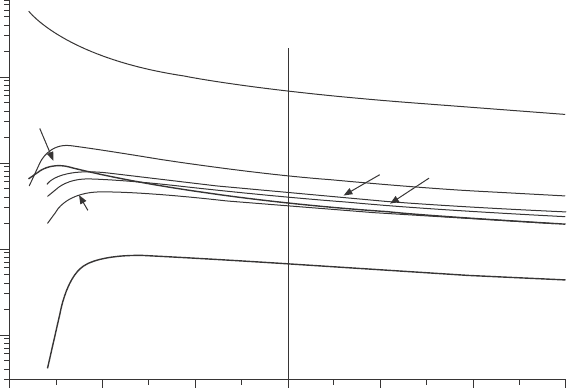
of calculated cross-sections as a function of the PE energy are shown in Fig.
7.3. The two major characteristics of the distributions are:
∑ The maximum cross-section for ionization is reached for primary electron
energies 3 to 6 times larger than the ionization energy. For solids, the
position of the maximum ionization yield is shifted toward higher PE
energies because of the backscattered electrons as discussed below.
∑ The value of the maximum cross-section is larger for lower ionization
energies E
i
, as shown in Fig. 7.3 in the case of silicon. The cross-section
at E
p
= 15 keV for the low-energy L
23
level (E
i
= 104 eV, six electrons)
is 100 times larger than for the K level (two electrons, E
i
=1844 eV).
The difference in the values of the Si cross-sections is the dominant
factor explaining the large variations of the signal intensities for different
transitions in AES and X-ray emission spectroscopy (XES). On the
other hand, the variation of s for L
23
shell ionization shows a weaker
dependence on Z for neighboring elements like Fe to As for PE energies
above 10 keV.
Cross-section (¥10
–19
c m
2
)
10
1
0.1
0.01
0 5 10 15 20 25 30
Energy (kV)
O K
As L
23
Fe L
23
Si L
23
Zn L
23
Ga L
23
Si K
7.3 Ionization cross-sections for the K and L
23
shell of selected
elements as a function of the energy of the RHEED electron beam.
186 In situ characterization of thin film growth
© Woodhead Publishing Limited, 2011
CEL: plasmons and band transitions
Electrons located in outer energy bands can be excited either as collective
density oscillations known as plasmon excitation, or as single electron inter-
and intra-band transitions. The result is the creation of CEL having a well-
dened energy DE generally in the range from a few eV up to about 40 eV.
The PE loses this amount of kinetic energy and is scattered by a small angle
as required by the conservation of energy and momentum.
Plasmon losses correspond to the eigenmodes of resonance of the electron
gas forming the outer electron shell, valence, and conduction bands. The
plasmon frequency for a metal with an ideal free electron gas, like aluminum,
is given by
w
p
= (4p e
2
/m)
1/2
(n)
1/2
[7.3]
with e the electron charge, m the effective mass of the electrons, and n
the electron density of the conduction electrons (Raether, 1980; Ferguson,
1989). The excitation of plasmons is not restricted to free electron gas, but
also exists for insulators and semiconductors because the plasmon energy,
commonly in the range 5–30 eV, is larger than the electron binding or gap
energy (Raether, 1980, p15). The plasmon loss energy is DE =
h
/
w
p
and
is directly related to the density of state n and, therefore, to the chemical
environment of the surface atoms. For instance, CEL of al and al
2
O
3
are
very different with plasmon loss energies of 15 and 23 eV respectively.
Plasmon excitations come in two forms, surface and volume plasmons.
The surface plasmon w
p,s
corresponds to electron density uctuations in the
boundary surface and the loss energy is
DE
p,s
= DE
p,v
/√2 [7.4]
for a free electron gas model. Plasmon losses are generally observed as
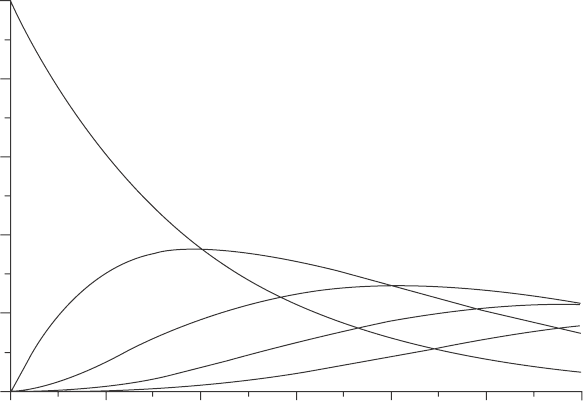
multiple losses. The probability for an electron to suffer n plasmon losses
depends on the ratio between the path length d traveled and the IMFP l and
is given by the Poisson distribution
Pn = 1/n! d/l [7.5]
as represented in Fig. 7.4. The no-loss probability P
0
decreases very rapidly
even for small values of d/l. For d = l, P
0
is reduced by about 37% and
equals P
1
, the rst loss peak. Even a layer thickness of 0.5 nm will cause
the no-loss peak to decrease to 60% of its value. This is a sensitive method
for measuring the thickness of deposits like a gas adsorbed on the surface.
The ratio between intensities of multiple plasmon losses can be compared
to give an estimate of d/l. A plasmon excitation can also occur during the
ionization-recombination process (intrinsic excitation). An atomic layer
adsorbed on the surface will suffer characteristic energy losses even if the
escape distance traveled is negligible compared with the IMFP. This process

187Spectroscopies combined with RHEED
© Woodhead Publishing Limited, 2011
adds to the excitation of extrinsic losses occurring during travel through
surface layers.
Band transitions are excitations of outer shell electrons. Ionization occurs
when a valence or conduction electron is ejected, resulting in the creation of
a vacancy. In contrast to optical absorption, the available momentum of the
PE allows for direct and indirect band transitions. Band transition losses often
occur in the same energy range as plasmons and overlap. The energy loss
distribution is closely related to the optical properties of the surface. The energy
loss function is given by the imaginary part of the complex dielectric constant
(Raether, 1980, p35) and CEL distributions can be deduced from optical data.
The strength of the energy losses is given by their IMFP which is the mean
distance traveled by the electron between two CEL. The mean free path for a
plasmon excitation is proportional to the electron kinetic energy, except in the
low kinetic energy range (<50 eV) where it increases. The IMFP shown for
Si in Fig. 7.2 has a value of about 4 nm at 1800 eV and only 0.6 nm at about
100 eV. This is a large difference in escape depth for Auger or photoelectrons
and is a major factor in the calculation of atom densities.
Continuous X-ray emission (bremsstrahlung)
Fast PE elastically scattered by nuclei in the crystal lattice are subject to
strong acceleration and can emit X-ray photons of energy ranging up to the
Probability
1.0
0.8
0.6
0.4
0.2
0.0
0.0 0.5 1.0 1.5 2.0 2.5 3.0
d/l
P0
P1
P2
P3
P4
7.4 Probability for multiple energy loss as a function of the ratio
between the path length d and the mean free path l.
188 In situ characterization of thin film growth
© Woodhead Publishing Limited, 2011
PE energy (Reimer, 1985, p158). The probability for this process is small
and the process does not contribute signicantly to the stopping power, but
it generates a signicant background continuum of X-rays. This background
distribution adds to the characteristic X-ray lines and lowers the signal to
noise ratio and the detection limit. The production rate In is given by Small
et al. (1987)
In = 10
5
¥ e
B
[Z (U–1)]
M
[7.6]
with e the natural log base, Z the atomic number, and U = E
p
/E
i
the overvoltage
parameter. The constants are M = 1.05 and B = 5.80 in the PE range 5 to
40 kV. The contribution increases almost linearly with Z and the detection
of a low-Z element deposited on a high-Z substrate is more difcult than
the other way around.
7.3 Recombination and emission processes
Core level vacancies created by fast PE impact are lled by electrons from
higher shells and the energy is released either as X-ray photons or Auger
electrons. The probability for the X-ray emission rather than an Auger
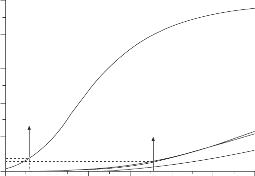
electron emission is given by the uorescence yield factor w. Figure 7.5
shows the uorescence yield for different shells plotted against the atomic
number Z using data from Krause (1979) and Segre (MUCAL). The X-ray
photon energy ranges up to several 10 keV, but the kinetic energy range of
Auger electrons is much narrower. A range up to 2500 eV is sufcient to
include the main Auger lines from all elements. The K shell Auger lines
extend up to Z = 16 (S), the L shells up to Z = 46 (Rh), the M and N shells
up to Z = 85 (At). These ranges are indicated in Fig. 7.5 and show that the
uorescence yields for the K, L, M shells are correspondingly low. For
instance, the uorescence yield for oxygen K is 0.83% and for silicon K is
5%. The uorescence yields are small for low-energy transitions and Auger
electron emission is the dominating process. Theoretical treatment of the
Auger process is complex because of the existence of relaxation effects
due to coulomb screening of the double ionized atom and possible inter-
atomic cross-transitions (Coster–Kronig transitions). General descriptions of
these emission processes are given in Reimer (1985) and Ferguson (1989)
and are more detailed for XPS and AES applications in Briggs and Grant
(2003).
Auger and characteristic X-ray lines generally have a multiplet structure.
This structure can be resolved with a suitable energy resolution of the
detector system. This is normally the case for AES energy analyzers and
X-ray dispersive (XDS) spectrometers, with resolution in the range 1–5 eV.
Energy dispersive X-ray detectors (EDS) have limited energy resolution,
about 100–150 eV, and many ne structures will appear as a single peak.

189Spectroscopies combined with RHEED
© Woodhead Publishing Limited, 2011
7.3.1 Quantification of the signal intensities
The emission yield is primarily given by the cross-section for ionization, the
escape depth, and the backscattering factor. The major experimental difference
between XRF, AES, and REELS is the large difference in escape depth of
photons or electrons. X-ray photons can escape from several micrometers
below the surface whereas AE will be limited to a few nanometers. The
absorption of X-rays is dominated by photo-ionization (mostly self-absorption)
and Compton scattering. The resulting MFP value is in the range of several
micrometers. In practice, the X-ray escape depth can be larger than the
penetration of the PE beam for an energy of 10 keV. Most of the emitted
X-ray photons generated near the surface are able to escape into vacuum
and have a nearly isotropic angular distribution as shown in Fig. 7.1. The
surface sensitivity of XES can be improved using grazing takeoff angles as
discussed below for TRAXS. In contrast, the IMFP for Auger electrons is
in the nanometer range (Ferguson, 1989, p25; Kanter, 1970) and the angular
distribution more closely follows Lambert’s cosine law; see Fig. 7.1.
A convenient way to quantify the data is to use the ratio method based on
reference spectra. Figure 7.6 shows the basic experimental conditions found
in growth experiments. In the rst case, Fig. 7.6(b), two elements A and B
with atomic numbers Z
a
and Z
b
are forming an alloy A + B. The reference
spectra from each pure element, corresponding to an atomic concentration
Fluorescence yield
1.0
0.8
0.6
0.4
0.2
0.0
10 20 30 40 50 60 70
Atomic number Z
K
S
Rh
L2, L3
L1
7.5 Fluorescence yields for the K and L shells as a function of the
atomic number Z. The ranges corresponding to the KLL and Lmm
Auger transitions are marked and correspond to low fluorescence
yield. After m.O. Krause and C. Segre.

190 In situ characterization of thin film growth
© Woodhead Publishing Limited, 2011
of 100%, is measured and gives the signal intensities I
a
(100) and I
b
(100).
An alloy of concentration x of A and 1 – x of B gives the signal intensities
I
a
(x) and I
b
(1 – x). The signal intensity for the pure element A of volume
density N
a
is given by
I
a
x
= N
a
x
I
p
s
A
M
a
D
a
[7.7]
with s
a
the ionization cross-section for A for a PE beam of energy E
p
and intensity I
p
. M
a
is a correction for matrix effects that include multiple
effects such as the backscattering coefcient, the uorescence yield, and
the escape probability of the particle to be emitted from the surface. The
escape probability depends on the IMFP for Auger and REELS electrons or
on the absorption for X-rays. It also depends on the angle of detection q
d
.
the instrumental factor D
a
is the detection efciency of the spectrometer
for element A and includes the acceptance angle and transmission of the
spectrometer. The detailed calculation of each correcting factor is complex
and it can be simplied using reference data from pure elements. The ratio
between the signals of an alloy of concentration x and the pure element is
I
a
(x)/I
a
(100) = N
a
(x)/N
a
(100) · M
a
/M
a
(x),
b
(1–x) [7.8]
The correcting factor is now the ratio between the matrix factor for the alloy
M
a
(x),
b
(1 – x) to the pure element M
a
. the major variations are those of
the attenuation length and backscattering factors, and the correcting factor
can be expressed for a signal from A as
d
a
(x), B
a
(x)/[d
a
(100) · B
a
(100)] [7.9]
where d
a
is the escape depth of a signal from A at concentration x and 100%
and B
a
is the backscattering factor for the same.
The second example is shown in Fig. 7.6(d) where element B is deposited
on top of substrate A. Signal I
b
from element B will grow linearly at the
beginning of the growth until the surface coverage reaches 1 ML (monolayer).
For a thicker layer, I
b
will reach a steady value for deposits thicker than
(a) (b) (c) (d) (e)
PE beam PE beam
q
i
q
i
q
d
q
d
1
3
4
2
A + BA B
B B
A A + B
7.6 models for calibration of atomic concentrations for a compound
A + B (a, b, c), for a deposited layer A on B (d) and for a layer of B
on A + B (e).
191Spectroscopies combined with RHEED
© Woodhead Publishing Limited, 2011
the escape depth of signal B in layer B. Concurrently, the signal A will
decrease as the signal I
a
has to cross the layer B and nally will vanish.
The absorption is characterized by the IMPF of I
a
through layer B and by
the detector take-off angle q
d
. Signal I
a
will decrease as
I
a
(d) = I
a
(0) exp – d/[cos(q
d
) l] [7.10]
with d the thickness (coverage) of B and l the IMFP at the energy of line
a in element b.
A frequent case is when growing a compound using a larger ux of one
element, like As in GaAs or like O and O
2
in oxide growth, where one
element may accumulate on the surface. Figure 7.6(e) shows the case of the
growth of element B on A + B where element B builds up on the surface.
The signal I
b
comes from two contributions, one from the bulk and the other
from the surface layer. I
b
will grow as long as the surface coverage remains
low, below 1 ML, because the attenuation in layer B is small and both
contributions can add. The signal from A will not change much. When the
layer thickness is larger than the attenuation length l, signal I
b
will saturate
to its value for pure B material and signal A will vanish. Interestingly, in
the case that the thickness is near l and that the atomic number Z of a
is larger than that of B (as for instance an oxygen layer on a metal oxide
substrate), the I
b
signal will show a peak because the ux of BSE from A
+ B into the surface layer B is larger than the BSE ux from pure B. This
example shows that the deduction of atomic densities from signal strengths
requires the proper knowledge and modeling of the growth conditions on
the surface.
7.4 Descriptions and results of in situ
spectroscopies combined with reflection high-
energy electron diffraction (RHEED)
This review focuses on recent combined techniques implemented in situ with
RHEED and able to deliver results during the growth process, excluding
analytical instruments built in situ but requiring sample transfer or interruption
of the deposition process for acquisition of data. The environment of a
growth chamber puts specic constraints on the instrument design in order
to operate in situ and during growth.
∑ A large working distance between the sample and the detector system
is required in order not to impair the atomic uxes from the deposition
sources.
∑ A good detection sensitivity allowing real-time data acquisition, fast
enough to follow the growth process with acquisition times in the range
1–30 s is necessary for most processes.
192 In situ characterization of thin film growth
© Woodhead Publishing Limited, 2011
∑ An outstanding resistance against material deposition is needed to ensure
long-term stable operation over several months.
∑ The capability to operate in a wide range of pressures, from ultra-high
vacuum (UHV) up to millitorrs, in order to be compatible with the
operation of gaseous sources, is important.
Standard instruments are not directly suitable and must be modied to
t these applications. New instruments, specially designed for operation in
growth chambers, are now being tested and will add new capabilities. The
techniques presented here are CL, XRF, TRAXS, REELS, and AES.
7.4.1 In situ CL spectroscopy
CL is similar to photoluminescence (PL) spectroscopy because the
recombination process is the same for both. There is, however, a major
difference in the excitation process because, in the case of PL excited by a
laser, the photon energy is fully absorbed, whereas electron excitation leads
to a wide distribution of energy transferred. Further, the cross-sections for
photon absorption have a peak at the transition threshold, but for electrons
the increase is smooth near the threshold. PL can selectively excite specic
transitions and CL will excite a wide range of transitions. The recombination
process involves band transitions between the valence and conduction bands
(Reimer, 1985, p289). Electrons from the valence band are excited into
unoccupied states of the conduction band. A cascade of non-radiative phonon
and electron excitations reduces their energy until they reach the bottom of
the conduction band. The luminescence decay processes involve the creation
of electron–hole pairs and excitons (Lightowlers, 1990). The transition can
be direct or an indirect transition involving phonons in order to insure the
conservation of momentum. The spatial extent of the region producing the
CL signal is very large because all of the PE, BSE, and even most of the
secondary electrons have sufcient energy to excite interband transitions
and generate CL emission. The spatial area covers the full range of the
cascade shown in Fig. 7.1. An additional broadening, due to the diffusion of
the carriers during their lifetime before decay, further extends the emission
volume (Reimer, 1985, p292). Therefore, the CL method basically delivers
bulk information, except when observed transitions involve surface states.
Experimental set-up for CL spectroscopy
The CL set-up was developed for use in electron microscopes and involves
the imaging of the beam spot through a large aperture optical collector
(Reimer, 1985, p210). CL designs must be modied for in situ applications
to accept larger sample size and to increase the clearance required between
193Spectroscopies combined with RHEED
© Woodhead Publishing Limited, 2011
the sample and the optical system. In practice, only a limited beam aperture
is focused onto the detector. In contrast with electron microscope chambers
that are extremely dark, growth chambers contain various sources of stray
light and in addition the sample may radiate when heated during the growth.
Optical lters, adapted to the emitted CL spectral range can lter out the
parasite light outside the CL emission range. A more efcient suppression
method is the modulation of the PE beam intensity, using beam blanking,
and measuring the signal using a lock-in amplier detection technique (Lee
and Myers, 2007).
Application of CL to the measurement of the substrate temperature
GaN having a direct band gap is a good CL emitter and the spectral distribution
shows a peak energy corresponding to the band gap energy (Lee and Myers,
2007). The position of the maximum is very temperature sensitive. The
peak shifts toward lower energy as the sample temperature increases. The
temperature measured by CL represents the actual temperature of the very
surface layer and represents a way to calibrate sample temperature between
different chambers. Once calibrated, CL is able to detect variations of the
surface temperature more precisely than the usual thermocouple measurement.
CL provides a way to cross-calibrate temperature measured in different GaN
growth chambers.
7.4.2 In situ XES combined with RHEED
XRF, also XES, is performed in situ using different detectors. One of the
rst experiments combining RHEED and XRF used a wavelength dispersive
spectrometer (Sewell and Cohen, 1967). The wavelength dispersive
spectrometer (WDS) is a windowless device and can detect low-energy
X-ray lines, such as oxygen Ka. The energy resolution is sufcient to
resolve the multiplet structure of the lines, but the major disadvantage is
that the detected signal is low and the acquisition requires a long measuring
time. More recent experiments use the EDS detector with the advantage of
detecting all incoming X-ray photons and delivering a signal pulse with an
amplitude proportional to the photon energy. Integration times are much
shorter and compatible with the speed of growth of the surface. The energy
resolution of the detector is, however, not as good as for the WDS system
and lines of neighboring Z elements often strongly overlap.
Experimental set-up for RHEED-XRF in situ monitoring
The angular distribution of characteristic X-rays is basically isotropic (see
Fig. 7.1), except in the range of very grazing angle of emission where the
