Koren B., Vuik K. (Editors) Advanced Computational Methods in Science and Engineering
Подождите немного. Документ загружается.

S. van der Hoeven, B.J. Boersma, and D.J.E.M. Roekaerts
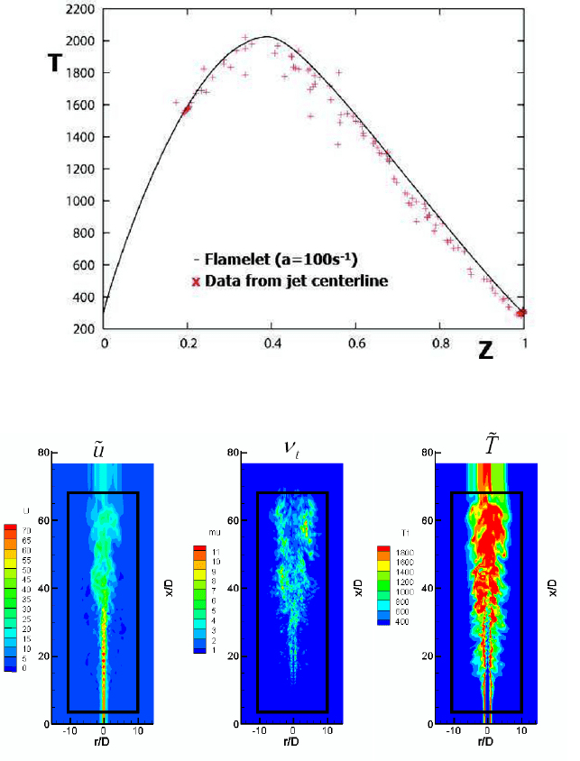
Fig. 9 The flamelet model temperature profile as a function of the mixture fraction Z (solid line),
and the local centerline temperature obtained from the LES model (dots).
Fig. 10 Instantaneous plots of resolved axial velocity (left), eddy viscosity (middle) and resolved
mixture fraction (right).
Mach number LES [7] in combination with steady flamelet modeling. Better results
are expected when the present model is combined with a more precise representation
of the turbulent inflow.
284
Large Eddy Simulation of Jet Flames
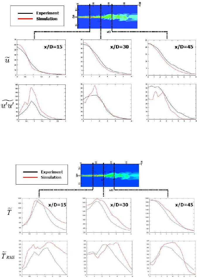
Fig. 11 Radial profiles of the time averaged axial velocity and the variance of axial velocity at
three axial positions.
Fig. 12 Radial profiles of the time averaged temperature and the standard deviation of temperature
at three axial positions.
285
S. van der Hoeven, B.J. Boersma, and D.J.E.M. Roekaerts
7 Summary and conclusions
A new compressible combustion code for DNS/LES of turbulent non-premixed jet
flames has been developed, based on a high-order compact finite difference formu-
lation. The Favre-filtered transport equations were solved and the SGS fluxes were
closed with a constant Smagorinksy model. The steady flamelet model was used for
the chemistry. In order to couple the flamelet solution with the compressible code a
relaxation term was added to the total non-chemical energy equation. Special atten-
tion was given to the treatment of the computational boundaries. We propose adding
a convective term with supersonic velocity in a small region near the in- and out-
flow boundaries together with explicit filtering of the velocity. Using this approach
it was observed that the simulation kept on running stable and reflections where too
low to have an effect on the interior domain. Furthermore, the computational costs
(measured in time) compared to the NSCBC method were reduced with a factor 1.5-
2.0 for this simulation configuration. Nevertheless, it must be mentioned that such a
comparison depends strongly on the simulation parameters (number of grid points,
number of processors, number of open boundaries) and the implementation (numer-
ical efficiency and used algorithms). To obtain better agreement with experimental
results a more accurate representation of the turbulent inflow conditions is needed.
8 Acknowledgements
The Dutch national computing facilities foundation NCF is acknowledged for the
allotted computing time on the TERAS/ASTER system, under project nr. SG-07-
279.
References
1. B.J. Boersma, A staggered compact finite difference approach for the compressible Navier-
Stokes equations, J. Comp. Phys., 208, 675, (2005)
2. S.K. Lele, Compact finite differences with spectral-like resolution, J. Comp. Phys., 103, 16,
(1992)
3. P. Chu, C. Fan, A three point combined compact difference scheme, J. Comp. Phys., 140, 370,
(1998)
4. S. Nagarajan, S.K. Lele, J.H. Ferziger, A robust high-order compact method for large eddy
simulation, J. Comp. Phys., 191, 392, (2003)
5. R.W.C.P. Verstappen, A.E.P. Veldman, Symmetry-preserving discretization of turbulent flow,
J. Comp. Phys., 187, 343, (2003)
6. International Workshop on Measurement and Computation of Turbulent Nonpremixed Flames,
http://www.ca.sandia.gov/TNF/DataArch/FlameD.html
7. H. Pitsch, H. Steiner, Large-eddy simulation of a turbulent piloted methane/air diffusion flame
(Sandia flame D), Phys. Fluids., 12, 2541, (2000)
8. N. Branley, W.P. Jones, Large Eddy Simulation of a turbulent non-premixed flame, Comb.
Flame, 127, 1914-1934, (2001)
286
Large Eddy Simulation of Jet Flames
9. T. Poinsot, S.K. Lele, Boundary conditions for direct simulations of compressible viscous
flows, J. Comp. Phys., 101, 1042 (1992)
10. C.S. Yoo, Y. Wang, A. Trouve, H.G. Im, Characteristic boundary conditions for direct simula-
tions of turbulent counterflow flames, Comb. Th. Mod., 9, 617-646, (2005)
11. T. Colonius, S.K. Lele, P. Moin, Boundary conditions for direct computation of aerodynamic
sound computation, AIAA Journal, 31, 1574-1582, (1993)
12. S. Ta’asan, D.M. Nark, An absorbing buffer zone technique for acoustic wave propagation,
AIAA Paper 1995-0146, (1995)
13. J.P. Berenger, A perfectly matched layer for the absorption of electromagnetic waves, J. Comp.
Phys., 114, 185-200, (1994)
14. J.B. Freund, Proposed inflow/outflow boundary condition for direct computation of aerody-
namic sound, AIAA Journal tech. notes, 34, 740-742, (1997)
15. D.J. Bodony, S.K. Lele, Jet noise prediction of cold and hot subsonic jets using Large-Eddy
Simulation, AIAA Paper 2004-3022 (2004)
16. N. Peters, Turbulent Combustion, Cambridge University Press (2000)
17. K.K. Kuo, Principles of Combustion, John Wiley and sons (2005)
18. T. Poinsot and D. Veynante, Theoretical and Numerical Combustion, R.T. Edwards Inc. (2005)
19. R.A. Vreman, An eddy-viscosity subgrid-scale model for turbulent shear flow: Algebraic the-
ory and applications, Phys. Fluids, 16, 3670 (2004)
20. P.J. Coelho, O.J. Teerling and D. Roekaerts, Spectral radiative effects and turbulence/radiation
interaction in a non-luminous turbulent jet diffusion flame, Combust. Flame, 133, 75-91,
(2003)
21. A. Habibi, B. Merci and D. Roekaerts, Turbulence/radiation interaction in RANS simulations
of non-premixed piloted turbulent laboratory scale flames, Combust. Flame, 151, 303-320,
(2007)
287

A Suite of Mathematical Models for Bone
Ingrowth, Bone Fracture Healing and
Intra-Osseous Wound Healing
F.J. Vermolen, A. Andreykiv, E.M. van Aken, J.C. van der Linden, E. Javierre, and
A. van Keulen
Abstract In this paper, some modeling aspects with respect to bone ingrowth, frac-
ture healing and intra-osseous wound healing are described. We consider a finite el-
ement method for a model of bone ingrowth into a prosthesis. Such a model can be
used as a tool for a surgeon to investigate the bone ingrowth kinetics when position-
ing a prosthesis. The overall model consists of two coupled models: the biological
part that consists of non-linear diffusion-reaction equations for the various cell den-
sities and the mechanical part that contains the equations for poro-elasticity. The two
models are coupled and in this paper the model is presented with some preliminary
academic results. The model is used to carry out a parameter sensitivity analysis
of ingrowth kinetics with respect to the parameters involved. Further, we consider
a Finite Element model due to Bailon-Plaza and Van der Meulen for fracture heal-
ing in bone. This model is based on a set of coupled convection-diffusion-reaction
equations and mechanical issues have not been incorporated. A parameter sensitiv-
ity analysis has been carried out. Finally, we consider a simplified model due to
Adam to simulate intra-osseous wound healing. This model treats the wound edge
as a moving boundary. To solve the moving boundary problem, the level set method
is used. For the mesh points in the vicinity of the wound edge, a local adaptive mesh
refinement is applied.
F.J. Vermolen, E.M. van Aken, E. Javierre
Delft Institute of Applied Mathematics, Faculty of Electrical Engineering, Mathematics and Com-
puter Science, Delft University of Technology, Mekelweg 4, 2628 CD Delft, The Netherlands
e-mail: F.J.Vermolen@tudelft.nl,
A. Andreykiv, J.C. van der Linden, F. van Keulen
Mechanical, Maritime and Materials Engineering, Delft University of Technology, Mekelweg 2,
2628 CD Delft, The Netherlands e-mail: A.Andreykiv@tudelft.nl
289
Lecture Notes in Computational Science and Engineering 71,
DOI 10.1007/978-3-642-03344-5_10,
B. Koren and C. Vuik (eds.), Advanced Computational Methods in Science and Engineering,
© Springer-Verlag Berlin Heidelberg 2010
F.J. Vermolen et al.
1 Introduction
In osteoporosis, fracture risk is high, after a hip fracture a prosthesis that replaces
the joint is often the only remedy. In the case of osteoarthritis and rheumatoid arthri-
tis, the cartilage degrades and moving the joints becomes painful. Ultimately, most
patients will receive a prosthesis to restore the function of a diseased joint. Pros-
theses are usually attached to the host bone by means of surgical screws to obtain
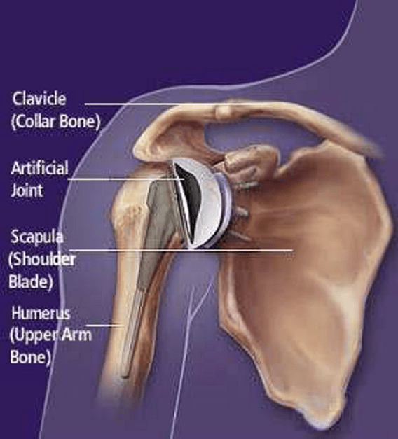
sufficient initial stability. A schematic of a prosthesis of the shoulder cavity, embed-
ded within an artificial joint is shown in Figure 1. In the course of time, bone will
grow into a porous tantalum layer and hence more stability of the prosthesis is ob-
tained. To investigate the quality and life time of such an artificial joint, one needs
to study the effects of the placement of the prosthesis and of the materials that are
involved in the joint. At present, these effects are often studied using large amounts
of data derived from patients. To predict the life span and performance of artificial
joints, numerical simulations are of great value since these simulations give many
qualitative insights by means of parameter sensitivity analysis. These insights are
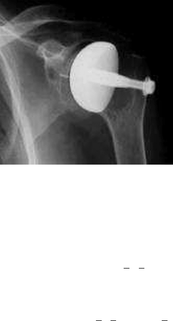
hard to obtain by experiments. In Figure 2, an X-ray picture of the prosthesis of the
shoulder cavity is shown.
In the case of a shoulder prosthesis, the angle at which the prosthesis is posi-
tioned by the surgeon is crucially important. The angle is important for the ability
of moving the arm by the patient, but also to have the right strain pattern for (op-
timal) bone ingrowth. The latter fact is due to the fact that the mammalian bone is
only generated if a certain strain is exceeded, but also smaller than a certain upper
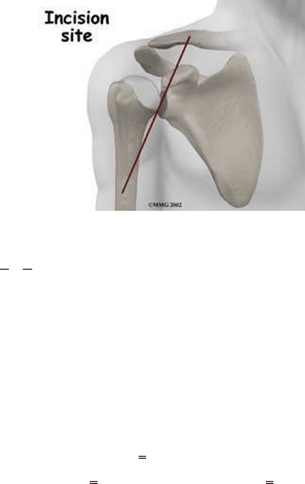
bound. For the surgery on the shoulder, the incision on the shoulder is made at the
front in order to save crucial organs and muscles of the patient. The location of the
incision is shown in Figure 3.
As a result of a limited visibility of the orthopaedic surgeon, the angle of place-
ment of the prosthesis is a crucial issue. Currently the stress and strain behavior
of the shoulder blade is studied at the Delft University of Technology, using three-
dimensional Finite Element simulations as a function of the angle of placement of
the prosthesis. An example of a computational domain is shown in Figure 4.
Several studies have been done to simulate bone-ingrowth or fracture healing of
bones. To list a few of them, we mention the model due to Adam [1], Ament and
Hofer [3], Bailon-Plaza et al. [7], Prendergast et al. [10] and recently by Andreykiv
[4]. The model due to Prendergast et al. and LaCroix et al. [10, 13] will be treated
in more detail, since we expect that this model contains most of the biologically
relevant processes, such as cell division and differentiation, tissue regeneration, and
cell mobility. Many ideas from modeling fracture healing of bones are used in these
models, since bone-ingrowth into a prosthesis resembles the fracture healing pro-
cess. In the model due to Prendergast, the influence of the mechanical properties on
the biological processes are incorporated. Further, we note that Prendergast’s model
has been compared to animal experiments.
Next to bone-ingrowth into a prosthesis, we present the model due to Bailon-
Plaza and Van der Meulen [7] for fracture healing in bone. This model is not cou-
pled with the equations from (poro-)elasticity. Andreykiv [6] applies the model for
290
Models for Bone Ingrowth and Healing
Fig. 1 An example of a schematic of an artificial shoulder joint.
bone-ingrowth to fracture healing in which coupling with mechanics has been ac-
complished.
Finally, the issue of intra-osseous wound healing is modeled using the simplified
formalism due to Adam [1]. An intra-osseous wound may result from surgery (think
of a wound on a skull due to brain surgery) or an injury caused by an accident.
The equations have a rather simple nature, though obtaining a numerical solution is
challenging since the wound edge is treated as a moving boundary.
In this paper, we will see a calibrated existing bone ingrowth model in terms
of a system of nonlinearly coupled diffusion-reaction equations, for which the me-
chanical strain and fluid flow are important input parameters. In order to compute
the aforementioned parameters, the poro-elasticity equations are solved. These two
classes of models will be treated separately. This paper concerns a compilation of
preliminary results, with some data for a shoulder prosthesis. Further, the paper
considers a model for fracture healing in bone and finally a model for healing of an
intra-osseous wound is described.
291
F.J. Vermolen et al.
Fig. 2 An example of an X-ray picture of the prosthesis of the shoulder cavity.
2 The bone-ingrowth model
We consider a prosthesis for the shoulder cavity. A sketch of the prosthesis is given
in Figure 5. The top part of the prosthesis consists of polyethylene. This part is in
actual contact with the upper arm, which exerts a loading on it. The second part con-
sists of a tantalum mesh and polyethylene. In these two parts, we solve an equation
for mechanical equilibrium combined with Hooke’s Law only. The third part con-
tains the porous tantalum. Into this part, bone ingrowth takes place from the glenoid,
which is the part of the scapula, in which the humeral head rotates. We denote the
entire computational domain by
Ω
, which consists of
Ω
E
(the elastic domain) and
Ω
P
(the poro-elastic domain), hence
Ω
=
Ω
E
∪
Ω
P
∪(
Ω
E
∩
Ω
P
). The overlines
indicate the closure of the (sub) domain. Further, we assume
Ω
E
and
Ω
P
to be dis-
joint, that is
Ω
E
∩
Ω
P
= /0. The domain
Ω
E
represents the part of the computational
domain on which only the elastic equations are solved. On
Ω
P
, one solves the poro-
elastic equations. Further, in
Ω
P
, we solve the equations for bone-ingrowth, which
is referred to as the biological part of the model.
Further, we note that
Ω
E
=
Ω
P
E
∪
Ω
B
E
, where
Ω
P
E
and
Ω
B
E
are the elastic part of
the prosthesis and bone respectively. Further,
Ω
P
E
∩
Ω
B
E
are separated by
Ω
P
, hence
292
Models for Bone Ingrowth and Healing
Fig. 3 The location at which the incision to put the shoulder prosthesis is carried out by the sur-
geon.
Ω
P
E
∩
Ω
B
E
= /0. The boundary of the computational domain,
Ω
, is denoted by
Γ
, and
of the subdomains
Ω
E
and
Ω
P
, their respective boundaries are represented by
Γ
E
and
Γ
P
.
The biological part of the model involving cell growth, division, differentiation
and formation of bone and cartilage, applies for the porous tantalum. The coef-
ficients in the biological part of the model depend on the local strains and fluid
flow velocity, which establishes a nonlinear coupled problem. First, the mechanical
model is presented and subsequently we give the biological model.
2.1 The mechanical model
Assuming mechanical equilibrium in both the poro-elastic part and the elastic part
of the domain, we solve
−div
σ
= f, x ∈
Ω
. (1)
In the above equation,
σ
and f respectively denote the stress tensor
σ
=
σ
xx
σ
xy
σ
yx
σ
yy
in two dimensions and f represents the internal body force. In our application, we
disregard the internal body force, hence we take f = 0. At a part of the top boundary
of the prosthesis, which is a part of the computational domain, a quadratic loading
is exerted, that is
293
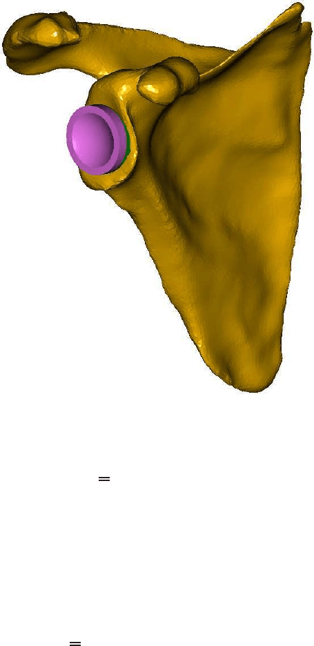
F.J. Vermolen et al.
Fig. 4 A three-dimensional computational domain of the shoulder blade.
σ
·n = t, x ∈
Γ
l
. (2)
Here, t denotes the exerted loading on
Γ
l
, which is the part of
Γ
on which the external
loading is applied. On the bottom boundary of the computational domain, which is
on the glenoid, we assume that the displacement is zero, that is
u = 0, x ∈
Γ
c
. (3)
Here
Γ
c
denotes the part of the outer boundary that is fixed to the host bone. On all
the other parts of the boundary, it is assumed that there is no loading, that is
σ
·n = 0, x ∈
Γ
\(
Γ
l
∪
Γ
c
). (4)
The material properties vary strongly over the various parts of the computational
domain. The displacement and traction are assumed to be continuous.
294
