Kim Y.J. (Ed.) Advanced Environmental Monitoring
Подождите немного. Документ загружается.

22 Monitoring of Dissolved Organic Carbon 293
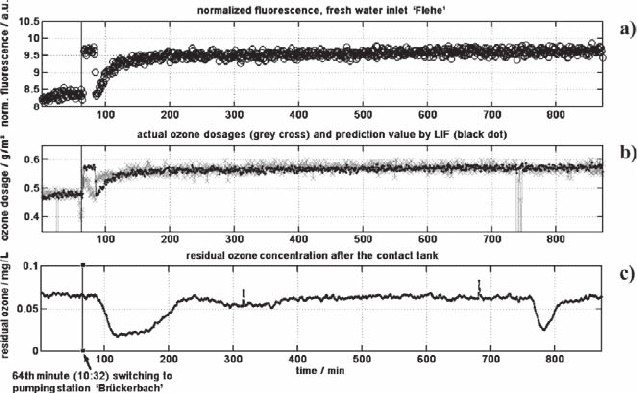
LIF-signal recorded at the freshwater inlet at the cleaning facility; the grey crosses
in Fig. 22.12 (b) represent the actually applied ozone dosage and the black dots show
the recommended ozone dosage determined by LIF-spectroscopy. After switching
one pumping line to the pumping station ‘Brückerbach,’ there is an enhanced need
for ozone in the first place. The ozone dosage is adjusted, but the demand is too high
resulting in a very low residual ozone concentration. Low residual ozone concentra-
tions are also unfavourable because the disinfection capacity is too low.
At minute 720, there seems to be a short failure at the ozone gas generator,
resulting in a difference between the recommended and the actual ozone dosage
again. In both cases where the actual ozone dosage differs from the recommended
value deduced by LIF spectroscopy, 20 min later a very low residual ozone concen-
tration in the water is recorded. The time delay of 20 min corresponds well to the
retention time for the water in the contact tank (see Fig. 22.6).
These examples document well that the LIF-signal can be used for predicting the
ozone consumption during the oxidation process using different raw waters from
the incoming pumping stations.
22.5 Summary
The investigations have shown that LIF is suitable for the determination of
DOC-values of raw waters. Prerequisite is a calibration to the local conditions of
the raw water. The demonstrated LIF set-up was successfully used for on-line
measurements of DOC at different pumping stations.
Fig. 22.12 (a) Normalized fluorescence, (b) actual ozone dosage (grey cross) and prediction
value (black dot) determined by LIF and (c) residual ozone concentration
294 U. Wachsmuth et al.
Furthermore LIF is a sensitive probe for revealing differences in water quality.
It was possible to distinguish between stagnating water and freshwater in the case
of waterworks in Düsseldorf-Flehe. The investigations at the ‘Grind’ show changes
of LIF from starting water withdrawal to equilibrium.
The investigations demonstrate also a direct correlation between LIF-signal and
ozone consumption. The normalized LIF-signal can be used to determine a
prediction value of the ozone demand for oxidation processes. These measure-
ments have been performed directly at the raw water inlet of the cleaning facility
at the water works ‘Flehe’ of the Stadtwerke Düsseldorf AG. Following this predic-
tion value the ozone dose can be adjusted in real time.
In the future, LIF could be applied to control the input and output of drinking
water plants to get further information about the water cleaning process which is an
important goal in the water safety plans of the suppliers.
References
Baker A. (2002), Spectrophotometric discrimination of river dissolved organic matter. Hydrol.
Process., 3203–3213.
Bünting U., Lewitzka F., and Karlitschek P. (1999), Mathematical model of laser-induced
fluorescence fiber-optic-sensor head for trace detection of pollutants in soil. Appl. Spectrosc.,
53, 49–56.
Esteves V.I., and Duarte A.C. (2001), Differences between Humic substances from riverine,
estuarine, and marine environments observed by fluorescence spectroscopy. Acta Hydrochem.
Et Hydrobiol., 28, 359–363.
Lewitzka F., Niederkrüger M., and Marowsky G. (2004), Application of two-dimensional LIF for
the analysis of aromatic molecules in water. In P. Hering, J.P. Lay and S. Stry (Eds), Laser in
Environmental and Life Sciences, Springer-Verlag: Berlin, pp.141–161.
Marowsky G., Lewitzka F., Bünting U., and Niederkrüger M. (2000), Quantitative analysis of
aromatic compounds by laser induced fluorescence spectroscopy. Proc. SPIE, 42, 218–223.
Niederkrüger M., Wachsmuth U., Konradt N., Rohns H.-P., Nelles T., and Irmscher R. (2004),
Mobiles Laserfluoreszenzspektrometer zum Monitoring auf gelöste organische Verbindungen
bei der Wassergewinnung aus Uferfiltrat. VDI-Berichte, 1863, 17–24.
Peuravuori J., Koivikko R., and Pihlaja K. (2002), Characterization, differentiation and classi-
fication of aquatic humic matter separated with different sorbents: synchronous scanning
fluorescence spectroscopy. Water Res., 36, 4552–4562.
Section 4
Biosensors, Bioanalytical
and Biomonitoring Systems

Chapter 23
Biosensors for Environmental
and Human Health
Peter-D. Hansen
Abstract Sensors and biosensors as well as biochemical responses (biomarkers) in
ecosystems owing to environmental stress provide us with signals (environmental
signalling) of a potential damage in the environment. These responses are per-
ceived in this early stage, but in ecosystems, the eventual damage can be prevented.
Once ecosystem damage has occurred, the remedial action processes for recovery
could be expensive and pose certain logistical problems. Prevention of ecosystem
deterioration is always better than curing. Ideally, “early warning signals” in eco-
systems using sensing systems and biochemical responses (biomarkers) would not
only tell us the initial levels of damage, but these signals will provide us as well
with answers to develop control strategies and precautionary measures with respect
to the water framework directive. To understand the complexity of the structure
of populations and processes behind the health of populations, communities and
ecosystems, we have to direct our efforts to promote rapid and cost-effective new
emerging parameters of ecological health. New emerging parameters are bio-
chemical effect (biomarker) related parameters in the field of immunotoxicity and
endocrine disruption. Environmental effects such as genotoxicity and clastogenic-
ity were detected in organisms from various “hot spots”. Vital fluorescence tests
are one means that allow us to unmask adverse events (i.e., genetic alterations in
field-collected animals or in situ-exposed organisms) by a caging technique. New
emerging ecosystem health parameters are closely linked to biomarkers of organ-
isms measured in monitored areas. One problem is always to find the relevant
interpretation and risk assessment tools for the environment.
Keywords: Biosensor, effect assessment, ecotoxicological classification in sedi-
ment, endocrine effects, assessment of “good ecological status”, drug exposure.
Technische Universität Berlin, Faculty VI, Department of Ecotoxicology,
Franklin Strasse 29 (OE4), D-10587 Berlin, Germany
(Tel: +493031421463, Fax: +49308318113)
297
Y.J. Kim and U. Platt (eds.), Advanced Environmental Monitoring,
297–311.
© Springer 2008
298 P.-D. Hansen
23.1 Introduction
For risk assessment and risk assessment tools new recommendations are described
in the Technical Guidance Document of EU – Edition 2, in the new EU Chemicals
Legislation REACH and in the status report for toxicological methods of the
European Centre for the Validation of Alternative Methods. In the EU water frame-
work directive (2000) a general requirement for ecological protection, and a gen-
eral minimum chemical standard was introduced to cover all surface waters. For the
description of “good ecological status” and “good chemical status”, effects moni-
toring tools are needed for the description of the ecological status of river basin
systems. In some cases, biomarkers were very helpful to promote an environmen-
tally sensitive and sustainable use in studies with coastal zone samples (Baumard
et al. 1999, Hansen et al. 1985, 1990, 1995, 2006). A very promising tool is the
scale classification based on toxicity studies and environmental monitoring to clas-
sify sediments: i.e., “Ecotoxicological Classification of Sediments by Bioassays”
(Krebs 2005a, 2005b). The currently available biomarkers or biochemical responses
are used as environmental monitoring tools to assess information on early responses
of living organisms to environmental stressors, and to deliver signals of ecosystem
damage and pathology owing to both man-made and natural pollutions. Biomarkers
are already being applied to the water matrix and to benthic organisms that include
new emerging biomarkers such as endocrine effects and immunotoxicity. Usefulness
for new emerging effect parameters such as immunotoxicity (phagocytosis) in the
context of biotoxins is demonstrated by mussels exposed to sediments in coastal
areas and exposure of mussels under controlled conditions to the water matrix.
Here, results of immunotoxic effects in mussels and the endocrine load of the sedi-
ments are demonstrated. Immunotoxic response of the blue mussel was quantified
by phagocytosis activity (Phagocytosis Index) of its hemocytes. It appears that both
immunosuppressive and immunostimulative effects are likely to occur at specific
sites and that responses will be influenced by the type and intensity of contaminants
present. Immune system function in bivalves can be adversely affected by long-
term exposure to environmental contaminants. Investigating alterations in immu-
nity can therefore yield relevant information about the relationship between
exposure to environmental contaminants and susceptibility to infectious diseases.
23.2 Biochemical Responses and Biosensors for Effects
Monitoring in the Environment
Use of biomarkers (biochemical responses) in multi-arrays for environmental
monitoring is complementary to chemical analysis since they can alert for presence
of toxic compounds that require further instrumental analysis or “bioresponse-
linked instrumental analysis”. Biosensors are by definition analytical devices
incorporating a biological component like micro-organisms, organelles, cell receptors,

23 Biosensors for Environmental and Human Health 299
enzymes, antibodies, nucleic acids and a physicochemical transducer system:
optical, electrochemical, thermometric, piezoelectric or magnetic. An enzyme
linked recombinant receptor assay like the ELRA is a biosensor according to this
definition: the biological component is the enzyme and the transducer is the optical
component. Biosensor and biochemical responses for the assessment of environ-
mental health are listed in Table 23.1. It is rather difficult to transfer the monitored
biochemical responses or the sensor responses into an operational effect related
standard (EQN = environmental quality norm) for environmental monitoring.
Sensing systems based on the induction and inhibition of a functional system relat-
ing to function, interference and effect related endpoints are listed in Table 23.1 and
depicted in Fig. 23.1.
Table 23.1 Environmental monitoring of effect related biochemical responses (biomarkers) and
potential sensing systems with their endpoints (“effects at the level of” neurotoxicity etc.)
(Modified after Bilitewski et al. 2000.)
Targets Example Interference by compounds Endpoints
Proteins Acetylcholine esterase Organophosphorus and Neurotoxicity
Enzymes carbamic compounds
Protein phosphatase Microcystins Hepatotoxicity
1 and 2A
Ion channels Na+ channel (voltage- Saxitoxin, tetrodotoxin, Neurotoxicity
gated channel) procaine
Transport SHBG, CBG, TBG Endocrine disruptors Growth,
protein reproduction
Receptors Estrogen receptor Endocrine disruptors (e.g. Growth,
o,p-DDT, nonylphenol) reproduction
Nicotinic acetylcholine Anatoxins neurotoxicity
(ACh) receptor
(ligand-gated
channel)
Electron QB protein Photosynthesis II herbicides Photosynthesis
carriers (e.g. s-triazines,
phenylureas), phytotoxins
Nucleic acids DNA double strands PAHs, Pesticides, PCBs, Genotoxicity
DNA EDCs, intercalating
polycyclic aromates
(ethidium, acridine, caffeine);
DNA adducts (metabolites
of chloracetamide
herbicides)
Cytoskeleton Tubulin Colchicin, taxol; anti-tubulin Cytotoxicity
herbicides (e.g. trifluralin,
oryzalin)
Ribosomes rRNA (ricin) Ribotoxins (ricin, abrin, cytotoxicity
Shga toxin)
300 P.-D. Hansen
Biosensors together with effect related parameters or biochemical responses for
environmental monitoring are very complex but they will give a clear picture of the
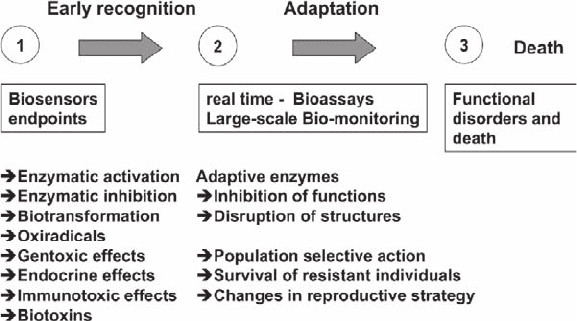
health status of the investigated system. In Fig. 23.1 the biochemical responses are
demonstrated by three phases of reaction by the organisms. The enzymes and
oxyradicals are helpful tools for a final biosensor. Enzyme biosensors are sensors
based on enzymatic bio-recognition elements. Therefore the definition of “biosensors”
can be widened: “Biosensors are analytical devices incorporating a biologically
derived and/or bio-mimicking material (i.e., cell receptors, enzymes etc.), associated
with or integrated within a physicochemical transducer or transducing microsystem,
which may be optical, electrochemical, thermometric, piezoelectric or magnetic.
Biosensors are distinct from bioassays in that the transducer is not an integral part of
the analytical system. Figure 23.1 shows an example of biochemical responses for
diagnosis of the health status of environmental systems (Hansen 2003).
The effect related parameters or biochemical responses are complex but they will
give a clear picture of the health status of the investigated system. “Ecosystem
Health” is synonymous with “environmental integrity”, from which it follows that
the scope of ecosystem health (EH) research encompasses all the tools and
approaches which are efficacious in increasing the cognitive, curative and preventive
knowledge as the goal to the preservation of environmental integrity. Ecosystem
health research thus directs its attention to the prediction of reversible and irreversible
insults which human or other activities could potentially inflict on the environment.
For the assessment of ecosystem health, very promising biomarker approaches are
centred on quantifying biochemical effects in organisms and populations. One
important tool for the acceptance of biomarkers in science, technology and govern-
mental legislature are the so-called inter-laboratory comparison studies of measurements
Fig. 23.1 Environmental signalling and diagnosis by biosensors and biochemical responses
(biosensors endpoints) In figure: Genotoxic effects; Oxyradicals
23 Biosensors for Environmental and Human Health 301
of biomarkers. Laboratory studies have established a strong causal link between
exposure of fish to PAHs and co-planar PCBs and the expression of cytochrome
P
450
1A1 and its associated 7-ethoxyresorufin-O-deethylase (EROD) activity. The
induction of EROD activity in fish liver has particularly been used as a biomarker
for the effects of these organic contaminants in several inter-calibration exercises
(see Stagg and Addison 1995). EROD induction is a classical biomarker and well
established for MFOs (mixed function oxigenases) and biotransformation in
ecotoxicology. So far, however, only phase I of the MFOs is investigated and not
phase II with conjugation and the real detoxification process. Additionally, another
often used biomarker is cholinesterase inhibition. The basic concept is that organo-
phosphorus pesticides and carbamates inhibit cholinesterase at different levels
(Sturm and Hansen 1999; Sturm et al. 1999). For the quantification of neurotoxicity
there are two well known cholinesterases (acetyl cholinesterase and butryl
cholinesterase) and the methodology in principle is standardised after DIN (German
Institute for Norming: DIN 38415-T1 1995). There is an application of validated
biomarkers available with endpoints including new emerging biomarkers for endocrine
effects and immunotoxicity in addition to genotoxicity. There is a high potential of
biochemical responses and the development of fast and reliable biochemical tools
(biosensors) for on-site screening in environmental and human health analysis. The
principles associated with the different scales of biochemical processes relating to
ecosystem and human health are shown in Table 23.2.
23.3 The River System of Berlin and the “Good Ecological
Status” Classified by a Biosensor System
In considering the impact of either natural stress or man-made stress we always
encounter detoxification, disease defence, regulation and adaptation processes. This
situation makes the assessment approach by biomarkers rather complicated. However,
in symptoms analysis including functional (behaviour, activity and metabolism) and
structural changes in organisms (cells, tissues and organs), biomarkers do have a
significant ecological assessment potential. For landscape planning and environmental
management it is necessary to get significant data from biochemical responses for
relevant and sustainable actions. As an example of transfer of new substances from
sediments to groundwater – the River Havel case study in Germany has demonstrated
close aspects between environmental monitoring and ecosystem health aspects.
23.3.1 Description of the River System of Berlin
and the Biosensor (Receptor Assay–Sensor) Application
The River Havel is an intensively monitored waterway because of its multiple use
for beach filtration and finally as a source to produce drinking water. The River

302 P.-D. Hansen
Havel is a water stretch of 30 km in length with a surface of more than 20 km
2
. It is
a slow flowing river with several conflicting uses such as (1) receiving waterway
for secondary effluent and rain water, (2) active beach filtration and drinking
water production, (3) sport- and professional fisheries, (4) leisure and recreation
concerning water sports and the EU water directive for swimming areas with
beaches along the river and (5) waterway for leisure boats and commercial cargo
vessels. With an extended surface, a low water depth (mean water depth 7 m) and
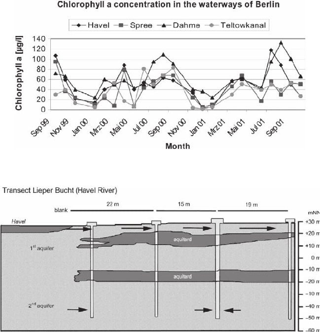
a steady input of nutrients, the river Havel has a potential for algal blooms.
Blooms begin in February (Fig. 23.2) and continue until May. During May and
June the algae growing are reduced and a second bloom of blue greens with
microcystins start at the end of June. There is a combination of increasing
temperature (up to 30°C) and decreasing flow of water (approx. 30 m
3
/s), which
trigger increases in biomass and in the amount of suspended solids. Finally, the
latter are eventually deposited as sediments.
Table 23.2 Selected biochemical responses for assessment of environmental health and the
biomarker methods used for their examination (Modified after Bresler et al. 1999)
Method Characteristic of health
Measurement of blue and green fluorescence
of NADH and FAD in living tissues
Metabolic state of mitochondria, cells or
tissues respiration and glycolysis
Quantitative fluorescent cytochemistry DNA, RNA, proteins and lipids content
Using permeable fluorogenic substrates of
enzymes, specific inhibitors, and kinetic
analysis
Enzyme activity in living cells in situ:
a. Non-specific esterases b. Detoxifying
enzymes c. Marker enzymes
Using special fluorescent anionic markers Alterations of permeability of plasma
membranes, epithelial layers and histo-
hematic barriers
Using specific fluorescent transport substrates,
inhibitors and kinetic analysis
State of carrier-mediated transport system
for xenobiotics elimination
Using fluorescent xenobiotics or fluorescent
analogue of xenobiotics
Xenobiotics distribution, extra- and intrac-
ellular accumulation and storage
Using special fluorescent xenobiotics or
fluorescent analogues of xenobiotics
State and function of xenobiotic-binding
proteins
Vital tests with Acridine Orange or
Neutral Red
State of lysosomes and cell viability
Metachromatic fluorescence of intercalated
or bound Acridine Orange, 590/530 nm
Microfluorometry
Functional rate of nuclear chromatin, DNA
denaturation
Complete cyto- and histopathological
examination
Early pathological alterations and signs of
environmental pathology
Electron microscopy Cell structures and organoids
Cytogenetic examinations Detection of environmental genotoxicity
and clastogenicity
Mass Spectrometry (MALDI/TOF/MS;
ESI-TOF-MS/MS)
Identification and detection of membrane
proteins, epitope-binding areas of
proteins
23 Biosensors for Environmental and Human Health 303
Besides algal blooms, there is a second source for suspended solids in the river
because of effluent loading by sewage treatment plant emissions. The river consists of
10%, 20%, 30% and 40% treated effluents (tertiary treatment and sometimes membrane
filtration). In winter (high water flow = approx. 100 m
3
/s) loading corresponds to
10% effluent to 20%–30% in spring and autumn (medium water flow = approx.
30 m
3
/s) and to 40% of treated effluent in summer (low water flow = approx. 5 m
3
/s).
The second source of suspended solids in the receiving river Havel on top of
extreme eutrophication by algae (see Fig. 23.2) is the outlet of the Berlin-Ruhleben
sewage treatment plant. The effluent contains 10 mg/l suspended solids in treated
sewage sludge. The sewage plant close to the river Havel is one of the main sewage
plants in Berlin with an output of 240,000 m
3
of treated effluent per day. Together
Fig. 23.2 Algal blooms in the Berlin waterways River Havel, Spree, Dahme and the Teltowkanal
Fig. 23.3 Schematic hydro geologic section of the transect at the River Havel (Lieper Bucht),
location of monitoring wells (shallow and deep) and of the raw-water supply well for drinking
water (Modified after Heberer and Mechlindki 2003)