Jan Lindhe. Clinical Periodontology
Подождите немного. Документ загружается.

MODIFYING FACTORS: DIABETES, PUBERTY, PREGNANCY AND THE MENOPAUSE AND TOBACCO SMOKING • 183
to stimuli, resulting in excessive release of cytokines.
Altered macrophage phenotype due to cell surface
binding with AGE, prevents the development of
macrophages associated with repair. This could con-
tribute to delayed wound healing seen in diabetic
patients (Iacopino 1995).
Connective tissue
A hyperglycemic environment, due to decreased pro-
duction or utilization of insulin, can reduce growth,
proliferation and matrix synthesis by gingival and
periodontal ligament fibroblasts and osteoblasts. The
formation of AGE results in reactive oxygen species,
which are damaging to cellular function in gingival
tissues, due to oxidative stress (Schmidt et al. 1996).
The accumulation of AGE in tissues alters the function
of
several intercellular matrix components, including
vascular wall collagen, resulting in deleterious com-
plications (Ulrich & Cerami 2001). This has adverse
effects on cell-matrix interactions and vascular integ-
rity, potentially affecting periodontal disease presen-
tation and treatment responses in uncontrolled diabet-
ics. Vascular changes such as thickening of the capil-
lary basement membrane in a hyperglycemic environ-
ment can impair oxygen diffusion, metabolic waste
elimination, PMN migration and diffusion of antibod-
ies. Binding of AGE to vascular endothelial cells can
trigger responses that induce coagulation, leading to
vasoconstriction and microthrombus formation (Es-
posito et al. 1992), resulting in impaired perfusion of
tissues.
Effects on healing and treatment response
Wound healing is impaired due to the cumulative
effects on cellular functions as described above. In
summary, these factors include:
1. Decreased synthesis of collagen by fibroblasts
2. Increased degradation by collagenase
3. Glycosylation of existing collagen at wound mar-
gins
4. Defective remodeling and rapid degradation of
newly synthesized, poorly cross-linked collagen.
Periodontal treatment
The treatment of well-controlled DM patients would
be similar to that of non-diabetic patients for most
routine dental procedures. The short-term non-surgi-
cal treatment response of stable diabetics has been
found to be similar to that of non-diabetic controls,
with similar trends in improved probing depths, at-
tachment gain and altered sub-gingival microbiota (
Christgau et al. 1998). Well-controlled diabetics with
regular supportive therapy have been shown to main-
tain treatment results 5 years after a combination of
non-surgical and surgical treatment (Westfelt et al.
1996). However, a less favorable treatment outcome
may occur in long-term maintenance therapy of
poorly-controlled diabetics, who may succumb to
more rapid recurrence of initially deep pockets (Ter-
vonen & Karjalainen 1997).
PUBERTY, PREGNANCY AND THE
MENOPAUSE
The hormonal variations experienced by women dur-
ing physiological and non-physiological conditions (
such as hormone replacement therapy and use of
hormonal contraceptives) result in significant changes
in the periodontium, particularly in the presence of
pre-existing, plaque-induced gingival inflammation.
Periods of hormonal flux are known to occur during
puberty, menstruation, pregnancy and the meno-
pause. Changes in hormone levels occur when the
anterior pituitary secretes follicle stimulating hor-
mone (FSH) and luteinizing hormone (LH), resulting
in the maturation of the ovary and cyclical production
of estrogen and progesterone.
The gingiva is a target tissue for the actions of
steroid hormones. Clinical changes in the tissues of the
periodontium have been identified during periods of
hormonal fluctuation. The effects of estrogen and pro-
gesterone on the periodontium have received signifi-
cant research attention. The main potential effects of
these hormones on the periodontal tissues can be
summarized as:
• Estrogen affects salivary peroxidases, which are ac-
tive against a variety of microorganisms (Kimura et
al. 1983), by changing the redox potential.
• Estrogen has stimulatory effects on the metabolism
of collagen and angiogenesis (Sultan et al. 1986).
• Estrogen can trigger autocrine or paracrine poly-
peptide growth factor signaling pathways, whose
effects may be partially mediated by the estrogen
receptor itself (Chau et al. 1998).
• Estrogen and progesterone can modulate vascular
responses and connective tissue turnover in the
periodontium, associated with interaction with in-
flammatory mediators (Soory 2000b).
The interaction of estrogen and progesterone with
inflammatory mediators may help to explain the in-
creased levels of inflammation seen during periods of
hormonal fluctuation. For example, when cultured
human gingival fibroblasts were incubated with pro-
gesterone concentrations common in late pregnancy,
there was a 50% reduction in the formation of the
inflammatory mediator IL-6, compared with control
values (Lapp et al. 1995). IL-6 induces the synthesis of
tissue inhibitor of metallo-proteinases (TIMP) in fi-
broblasts (Lotz & Guerne 1991), reduces the levels of
TNF and enhances the formation of acute phase pro-
teins (Le & Vilcek 1989). A progesterone-induced re-
duction in IL-6 levels could result in less TIMP, more
proteolytic enzyme activity and higher levels of TNF

184 • CHAPTER 6
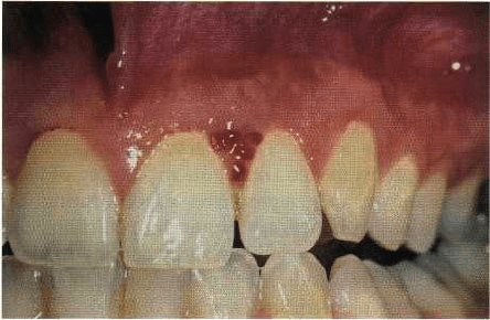
Fig. 6-6. Gingivitis associated with pregnancy. (a) A patient in the last trimester of pregnancy with very
inflamed edematous gingival tissue which tended to bleed with the slightest provocation. (b) The improvement
in gingival health 6 months after birth of the baby and an intensive course of non-surgical periodontal
treatment.
at the affected sites, due to less inhibition, resulting in
inflammation and obvious clinical manifestations.
Puberty and menstruation
During puberty, there are raised levels of testosterone
in males and estradiol in females. Several studies have
demonstrated an increase in gingival inflammation in
children of circum-pubertal age, with no change in
plaque levels (Sutcliffe 1972). In a longitudinal study,
Mombelli et al. (1989) reported that the mean papillary
bleeding scores and percentage of interdental bleed-
ing sites correlated with the development of secon-
dary sexual characteristics at puberty, while other
studies did not find a significant correlation between
the onset of puberty and gingival changes in parapu-
bescent women (Tiainen et al. 1992). These discrepan-
cies may be attributed to factors such as the oral
hygiene status of the population and study design.
The prevalence of certain periodontal pathogens
reported during puberty may have a direct association
with the hormones present and their utilization by
selected pathogens. For example
Prevotella intermedia
is able to substitute progesterone and estrogen for
menadione (vitamin K) as an essential nutrient (Korn-
man & Loesche 1979). An association between puber-
tal gingivitis,
Prevotella intermedia
and serum levels of
testosterone, estrogen and progesterone has been re-
ported in a longitudinal study (Nakagawa et al. 1994).
Pre-existing plaque-induced gingivitis may be an
important factor in detecting hormone-induced
changes during the menstrual cycle. Holm-Pedersen
& Loe (1967) demonstrated that women with gingivi-
tis experienced increased inflammation with an asso-
ciated increase in crevicular fluid exudate during
menstruation compared with healthy controls. Most
female patients are not aware of any changes in their
gingivae during the menstrual cycle (Amar & Chung
1994), while a few experience enlarged hemorrhagic
gingivae in the days preceding menstrual flow. This
has been associated with more gingivitis, increased
crevicular fluid flow and tooth mobility (Grant et al.
1988). Early studies demonstrated similar findings
during the menstrual cycle in a population with pre-
existing gingivitis, in response to fluctuations in the
levels of estrogen and progesterone (Lindhe &
Attstrom 1967).
Pregnancy
During pregnancy, the increased levels of sex steroid
hormones are maintained from the luteal phase which
results in implantation of the embryo, until parturi-
tion. Pregnant women, near or at term, produce large
quantities of estradiol (20 mg/day), estriol (80
mg/day) and progesterone (300 mg/day). Gingival
inflammation initiated by plaque, and exacerbated by
these hormonal changes in the second and third tri-
mester of pregnancy, is referred to as pregnancy gin-
givitis. Parameters such as gingival probing depths (
Hugoson 1970, Miyazaki et al. 1991), bleeding on
probing (Miyazaki et al. 1991) and crevicular fluid
flow (Hugoson 1970) were found to be increased.
These inflammatory features can be minimized by
maintaining good plaque control.
According to early reports, the prevalence of preg-
nancy gingivitis ranges from 35% (Hasson 1966) to
100% (Lundgren et al. 1973). In a study of 130 pregnant
women, Machuca et al. (1999) demonstrated gingivitis
in 68% of the population, ranging from 46% in techni-
cal executives to 88% in manual workers. Cross-sec-
tional studies examining pregnant and postpartum
women have shown that pregnancy is associated with
significantly more gingivitis than at postpartum, de-
spite similar plaque scores (Silness & Loe 1963). Fur-
ther observations were made by Hugoson (1970) in a
longitudinal study of 26 women during and following
pregnancy, which also demonstrated that the severity
of gingival inflammation correlated with the gesta-
tional hormone levels during pregnancy (Fig. 6-6). A
more recent study of a rural population of Sri Lankan
women (Tilakaratne et al. 2000a) showed increased
gingivitis of varying degrees of significance amongst
all the pregnant women investigated, compared with

MODIFYING FACTORS: DIABETES, PUBERTY, PREGNANCY AND THE MENOPAUSE AND TOBACCO SMOKING • 185
Fig. 6-7. Multilobulated appearance of an early preg-
nancy epulis, demonstrating vascular elements and tis
sue edema.
matched non-pregnant controls. There was a progres-
sive increase in inflammation with advancing preg-
nancy which was more significant in the second and
third trimester of pregnancy, despite the plaque levels
remaining unchanged. At the third month after partu-
rition, the level of gingival inflammation was similar
to that observed in the first trimester of pregnancy.
This suggests a direct correlation between gingivitis
and sustained, raised levels of gestational hormones
during pregnancy, with regression during the post-
partum period. In investigations by Cohen et al. (1969)
and Tilakaratne et al. (2000a), the values for loss of
attachment remained unchanged during pregnancy
and three months postpartum.
Effects on the microbiota
There is an increase in the selective growth of peri-
odontal pathogens such as
Prevotella
in termedia in sub-
gingival plaque during the onset of pregnancy gingi-
vitis at the third to fourth month of pregnancy. The
gestational hormones act as growth factors, by satis-
fying the naphthoquinone requirement for bacteria (
Di Placido et al. 1998). These findings were also con-
firmed by Muramatsu & Takaesu (1994) who showed
that from the third to fifth month of pregnancy, the
number of gingival sites which bled on probing corre-
sponded with the percentage increase in
Prevotella
intermedia.
During pregnancy, progesterone is less ac
tively catabolized to its inactive products, resulting
in higher levels of the active hormone (Ojanotko-
Harri et al. 1991). A 55-fold increase in the
proportion of P. in termedia has been demonstrated in
pregnant women compared with non-pregnant
controls (Jensen et al. 1981), implying a role for
gestational hormones in causing a change in
microbial ecology in the gingival pocket. Although an
overall association has been demonstrated, a cause
and effect relationship may be less clear.
Effects on the tissues and host response
The increase in severity of gingivitis during preg-
nancy has been partly attributed to the increased cir-
culatory levels of progesterone and its effects on the
capillary vessels (Lundgren et al. 1973). Elevated pro
gesterone levels in pregnancy enhance capillary per-
meability and dilatation, resulting in increased gingi-
val exudate. The effects of progesterone in stimulating
prostaglandin synthesis can account for some of the
vascular changes (Miyagi et al. 1993).
The elevated levels of estrogen and progesterone in
pregnancy affect the degree of keratinization of the
gingival epithelium and alter the connective tissue
ground substance. The decreased keratinization of the
gingivae, together with an increase in epithelial gly-
cogen, are thought to result in decreased effectiveness
of the epithelial barrier in pregnant women (Abra-
ham-Inpijn et al. 1996). Hormonal factors that affect
the epithelium and increase vascular permeability can
contribute to an exaggerated response to bacterial
plaque during pregnancy.
The influence of gestational hormones on the im-
mune system can contribute further to the initiation
and progression of pregnancy gingivitis. High levels
of progesterone and estrogen associated with preg-
nancy (and the use of some oral contraceptives) have
been shown to suppress the immune response to
plaque (Sooriyamoorthy & Gower 1989). Neutrophil
chemotaxis and phagocytosis, along with antibody
and T-cell responses have been reported to be de-
pressed in response to high levels of gestational hor-
mones (Raber-Durlacher et al. 1993).
Pregnancy granuloma or epulis
A pedunculated, fibro-granulomatous lesion can
sometimes develop during pregnancy and is referred
to as a pregnancy granuloma or epulis. A combination
of the vascular response induced by progesterone and
the matrix stimulatory effects of estradiol, contribute
to the development of pregnancy granulomas, usually
at sites with pre-existing gingivitis (Fig. 6-7). The vas-
cular effects result in a bright red, hyperemic and
edematous presentation. The lesions often occur in the
anterior papillae of the maxillary teeth and usually do
not exceed 2 cm in diameter. They can bleed when
traumatized and their removal is best deferred until
after parturition, when there is often considerable
regression in their size (Wang et al. 1997). Surgical
removal of the granuloma during pregnancy can re-
sult in recurrence due to a combination of poor plaque
control and hormone mediated growth of the lesion.
Careful oral hygiene and debridement during preg-

186 • CHAPTER 6
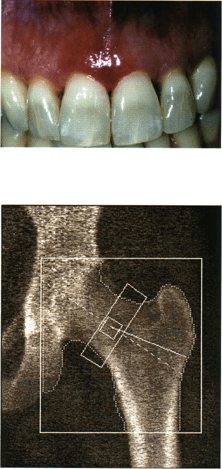
Fig. 6-8. Clinical appearance of anterior maxillary
gingiva with pronounced desquamation in a woman
during menopause.
Fig. 6-9. A DEXA scan used to measure mineral bone
density in the hip. This is not routinely applied to the
jaws.
nancy are important in preventing its occurrence (
Wang et al. 1997).
Periodontal treatment during pregnancy
Pregnant women need to be educated on the conse-
quences of pregnancy on gingival tissues and thor-
oughly motivated in plaque control measures, with
professional treatment as required. They are likely to
be more comfortable to receive dental treatment dur-
ing the second trimester than in the first or third
trimester of pregnancy, although emergency treat-
ment is permissible at any stage during pregnancy (
Amar & Chung 1994). Since most medications cross
the placental barrier and organogenesis occurs mainly
in the first trimester, pregnant women are best treated
in the second trimester, to avoid the occurrence of
developmental defects. Any form of medication dur-
ing pregnancy must only be used if the gravity of the
condition being treated outweighs the consequences.
Amongst the antibiotics, tetracycline, vancomycin
and streptomycin can contribute to staining of teeth
and ototoxic and nephrotoxic effects during 4-9
months of pregnancy; erythromycin, penicillins and
cephalosporins are relatively safer, but any medica-
tion must only be administered in consultation with
the patient's obstetrician (Lynch et al. 1991).
Menopause and osteoporosis
During menopause there is a decline in hormonal
levels due to decreased ovarian function. This is char-
acterized by tissue changes such as desquamation of
gingival epithelium (Fig. 6-8) and osteoporosis (Fig.
6-9) which may be attributed to hormone deficiency.
It has been demonstrated that women with early onset
of menopause have a higher incidence of osteoporosis
and significantly lower bone mineral density (Kritz-
Silverstein & Barrett-Connor 1993).
A third of women over age 60 are affected by post-
menopausal osteoporosis (Baxter 1987). The changes
involved are a reduction in bone density, affecting its
mass and strength without significantly affecting its
chemical composition. An alteration in the calcium-
phosphate equilibrium due to deficient absorption of
dietary calcium and increased excretion due to dimin-
ished estrogen levels can account for some of the bone
changes seen in postmenopausal women (Shapiro et
al. 1985), usually involving the mandible more than
the maxilla.
Estrogen replacement therapy has been shown to
prevent osteoporosis and maintain bone mineral con-
tent at several sites throughout the skeleton (Moore et
al. 1990), with a 5% increase in bone mineral content
in the region of the head compared to those taking
placebo (Gotfredsen at al. 1986). The influence of es-
trogen on bone mineral density has been demon-
strated in these studies, but a cause and effect relation-
ship with periodontal disease is less clear.
A 2-year follow-up study of 42171 postmenopausal
women (Grodstein et al. 1996) showed that the risk of
tooth loss was significantly lower amongst hormone
users. These findings reinforce those of Paganini-Hill
(1995), who showed a 36% decrease in tooth loss in
estrogen users compared with non-users. There is
evidence to suggest that use of estrogen is necessary
to protect against bone loss (Grady et al. 1992). Al-
though osteoporosis in postmenopausal women may
not be the cause of periodontal disease, it may affect
the severity of pre-existing disease. The circulating
levels of estrogen have been shown to have an influ-
ence on alveolar bone density in postmenopausal
women (Payne et al. 1997).
MODIFYING FACTORS: DIABETES, PUBERTY, PREGNANCY AND THE MENOPAUSE AND TOBACCO SMOKING • 187
Effect of smoking on osteoporosis
A negative association between smoking and bone
density has been demonstrated by Krall & Dawson-
Hughes (1991). Smokers can differ from non-smokers
in weight, caffeine intake, age at menopause and alco-
hol consumption (Rigotti 1989, Lindquist & Bengtsson
1979); all these factors can potentially confound an
association between smoking and bone density. A
study on female twins by Hopper & Seeman (1994)
showed that in the 20 pairs who varied most, by 20 or
more pack years, the differences in bone density
within pairs were 9.3% at the lumbar spine, 5.8% at
the femoral neck and 6.5% at the femoral shaft. This
study also demonstrated increased serum levels of
follicle stimulating hormone and luteinizing hormone
in smokers, implying reduced circulating levels of
estrogen, leading to increased bone resorption. Other
investigators have demonstrated the effects of smok-
ing on the synthesis and degradation of estrogen (Jen-
sen et al. 1985). The study by Jensen et al. (1985)
investigated 136 postmenopausal women who were
treated with three different doses of estrogen-proges-
terone or placebo. They showed reduced levels of
estrogen in smokers (range of 1-30 cigarettes/day in
the previous 6 months, mean 12.4), compared with
non-smokers (not smoked in the previous 3 months).
There was also a significant inverse correlation be-
tween the number of cigarettes smoked per day and
the serum levels of estrogen, suggestive of increased
hepatic metabolism of estrogen in postmenopausal
smokers, resulting in lower serum levels of these hor-
mones.
Treatment of osteoporosis
In osteoporotic patients, the rate of bone loss during
the early postmenopausal period increases to 3-4% per
year. Estrogen replacement therapy, which slows bone
turnover, results in increased bone density in the tra-
becular spaces during remodeling (Frost 1989). The
increased skeletal bone mass which occurs in response
to estrogen replacement therapy is apparent in the
first 2 years of treatment and maintained with con-
tinuation of treatment (Kimmel et al. 1994). The effects
of estrogen in regaining bone mass to premenopausal
levels and in preventing/reversing postmenopausal
osteoporotic changes in the long bones and spine have
been demonstrated in several studies (Takahashi et al.
1994, Armamento-Villareal et al. 1992).
There is some controversy with regard to the bene-
fits of hormone replacement due to the risk factors
involved. Fractures due to osteoporosis and heart dis-
ease in postmenopausal women can be reduced by
50% with estrogen replacement therapy. However,
hormone replacement with estrogen alone exposes
such patients to the risk of endometrial cancer. Long-
term hormone replacement therapy has been shown
to correlate with an increased risk of breast cancer.
Modern formulations utilize combined therapy with a
suitable dose of progesterone in combination with
estrogen in order to minimize some of these risk fac-
tors (Whitehead & Lobo 1988).
Hormonal contraceptives
Contraceptives utilize synthetic gestational hormones (
estrogen and progesterone), to reduce the likelihood
of ovulation/implantation (Guyton 1987). Less dra-
matic but similar effects to pregnancy are sometimes
observed in the gingivae of hormonal contraceptive
users. The most common oral manifestation of ele-
vated levels of ovarian hormones is an increase in
gingival inflammation with an accompanying in-
crease in gingival exudate (Mariotti 1994).
There are reported systemic risk factors associated
with long-term use of hormonal contraceptives. The
correlation between hormonal contraceptive use and
significant cardiovascular disease associated with ar-
terial and venous thromboembolic episodes has been
reviewed by Westhoff (1996). Estrogen is responsible
for both arterial and venous effects, while progester-
one affects arterial changes. Women using oral contra-
ceptives show elevated plasma levels of several clot-
ting factors, related to the dose of estrogen. Raised
levels of factors Vllc and XIlc are significant, since
they increase the likelihood of coagulation and in men
these factors have a strong positive correlation with
ischemic heart disease. However, the relative risk is
dependent on the contraceptive formulation used and
there may not be a consistent biological plausibility to
explain this association (Davis 2000).
There are several different formulations of hormo-
nal contraceptives (Davis 2000) including:
1. Combined oral contraceptives containing artificial
analogues of estrogen and progesterone
2. Progesterone based mini-pill
3. Slow release progesterone implants placed sub-
dermally that last up to 5 years (e.g Norplant)
4. Depo Provera, a very effective progestin injection
given by a doctor every 3 months.
Current combined oral contraceptives consist of low
doses of estrogens of 50 ltg/day and/or progestins of
1.5 mg/day (Mariotti 1994). The formulations used in
the early periodontal studies contained higher con-
centrations of gestational hormones, e.g. 50 µg estro-
gen with 4 mg progestin (El-Ashiry et al.1971),100 µg
estrogen with 5 mg progestin (Lindhe & Bjorn 1967).
The results obtained in these studies would partly
reflect the contraceptive preparation used. In one
early study (Knight & Wade 1974) women who were
on hormonal contraceptives for more than 1.5 years
exhibited greater periodontal destruction compared to
the control group of comparable age and oral hy-
giene. This could partly reflect higher dose of
gestagens used in older contraceptive preparations.
However, a recent study on a population of rural Sri
Lankan women confirmed these findings (Tilakaratne
188 • CHAPTER 6
et al. 2000b), showing significantly higher levels of
gingivitis in contraceptive users (0.03 mg estradiol
and 0.15 mg of a progestin), than non-users, despite
similar plaque scores. There was also significant peri-
odontal breakdown in those who used the progester-
one injection (a depot preparation of 150 mg proges-
terone) 3 monthly for 2-4 years, compared with those
who used it for less than 2 years. These findings may
be attributed to the duration of use, and the effects of
progesterone in promoting tissue catabolism, result-
ing in increased periodontal attachment loss. How-
ever, if low plaque levels are established and main-
tained for the duration of use, these effects could be
minimized.
Effect on tissue response
Both estrogen and progesterone are known to cause
increased gingival exudate, associated with inflam-
matory edema (Lindhe & Bjorn 1967). A 53% increase
in crevicular fluid volume has been demonstrated in
hormonal contraceptive users compared with con-
trols. El-Ashiry et al. (1971) observed that the most
pronounced effects on the gingiva occurred in the first
3 months of contraceptive treatment, but the dose of
gestational hormones was higher in the older formu-
lations compared with those used currently (Davis
2000), accounting for a more florid response in the
tissues.
It has been suggested that the interaction of estro-
gen with progesterone results in the mediation of the
effects characteristic of progesterone. Human gingiva
has receptors for progesterone and estrogen (Vittek et
al. 1982, Staffolani et al. 1989), providing evidence that
gingiva is a target tissue for both gestational hor-
mones. In in vitro studies of cultured gingival fi-
broblasts, estrogen enhanced the formation of ana-
bolic androgen metabolites, while progesterone
caused a diminished response. The combined effect of
both gestational hormones on the yield of androgens
was less pronounced than with estrogen alone, imply-
ing a more catabolic role for progesterone (Tilakaratne
& Soory 1999).
Progesterone causes increased vascular permeabil-
ity, resulting in the infiltration of polymorphonuclear
leukocytes and raised levels of prostaglandin E
7
in the
sulcular fluid (Miyagi et al. 1993). Increased capillary
permeability maybe induced by estrogen by stimulat-
ing the release of mediators such as bradykinin, Pro-
staglandins and histamine. However, the main effects
of estrogen are in controlling blood flow. Hence the
combination of estrogen and progesterone in the con-
traceptive pill can contribute to vascular changes in
the gingivae. The resultant gingivitis can be mini-
mized by establishing low plaque levels at the begin-
ning of oral contraceptive therapy (Zachariasen 1993).
TOBACCO SMOKING
Tobacco smoking is very common, with cigarettes
being the main product smoked. In the European
Union, an average of 29% of the adult population
smoke. The figure is higher for men (34%) than for
women (24%). Most smokers start the habit as teenag
ers, with the highest prevalence in the 20-24 year old
age group. Socio-economic differences also exist with
higher smoking in the lower socio-economic groups.
These data are similar for the US population (Gar-
finkel 1997), but reported smoking rates for third
world countries are even higher. Smoking is associ-
ated with a wide spectrum of disease including stroke,
coronary artery disease, peripheral artery disease,
gastric ulcer and cancers of the mouth, larynx, esopha
gus, pancreas, bladder and uterine cervix. It is also a
major cause of chronic obstructive pulmonary disease
and a risk factor for low birth weight babies. Approxi-
mately 50% of regular smokers are killed by their habit
and smoking causes 30% of cancer deaths.
Cigarette smoke is a very complex mixture of sub-
stances with over 4000 known constituents. These
include carbon monoxide, hydrogen cyanide, reactive
oxidizing radicals, a high number of carcinogens and
the main psychoactive and addictive molecule – nico-
tine (Benowitz 1996). Many of these components
could modify the host response in periodontitis. In
most of the in vitro studies considered in the latter
parts of this chapter the experimenters utilized simple
models with nicotine alone. Tobacco smoke has a
gaseous phase and solid phase which contains tar
droplets. The tar and nicotine yields of cigarettes have
been reduced due to physical characteristics of the
filters. However, there has been little change in the tar
and nicotine content of the actual tobacco and the dose
an individual receives is largely dependent upon the
way in which they smoke (Benowitz 1989). Inter sub-
ject smoking variation includes: frequency of inhala-
tion, depth of inhalation, length of the cigarette stub
left, presence or absence of a filter and the brand of
cigarette (Benowitz 1988). The patient's exposure to
tobacco smoke can be measured in a number of ways
including interviewing the subject using simple ques-
tions or more sophisticated questionnaires and bio-
chemical analyses (Scott et al. 2001). The latter tests
include exhaled carbon monoxide in the breath, which
is commonly measured in smoking cessation clinics,
and cotinine (a metabolite of nicotine) in saliva,
plasma/serum or urine (Wall et al. 1988). Cotinine
measurements are more reliable in determining a sub-
ject's exposure to tobacco smoke because the half-life
is 14-20 hours compared with the shorter half-life of
nicotine which is 2-3 hours (Jarvis et al. 1988). The
mean plasma and salivary cotinine concentrations of
regular smokers are approximately 300 ng/ml and

MODIFYING FACTORS: DIABETES, PUBERTY, PREGNANCY AND THE MENOPAUSE AND TOBACCO SMOKING • 189
Fig. 6-10. The typical appearance of necrotizing ulcera
tive gingivitis in a heavy smoker with poor oral hy-
giene.
Fig. 6-11. The lingual aspects of the lower incisors
showing gross supragingival calculus formation and
relatively little gingival inflammation in a female pa-
tient who has smoked 20 cigarettes per day for over 20
years.
urine concentrations are about 1500 ng/ml. Non-
smokers typically have plasma/ saliva concentrations
under 2 ng/ml, but this may be raised slightly due to
environmental exposure (passive smoking).
Inhalation of tobacco smoke allows very rapid ab-
sorption of nicotine into the blood and transport to the
brain, which is faster than an intravenous infusion.
Nicotine in tobacco smoke from most cigarettes is not
well absorbed through the oral mucosa because the
nicotine is in an ionized form as a result of the pH (5.5).
In contrast cigar and pipe smoke is more alkaline (pH
8.5), which allows good absorption of un-ionized
nicotine through the buccal mucosa (Benowitz 1988).
Nicotine is absorbed rapidly in the lung where the
smoke is well buffered. The administration of nicotine
causes a rise in the blood pressure, an increase in heart
rate, an increase in respiratory rate and decreased skin
temperature due to peripheral vasoconstriction. How-
ever, at other body sites, such as skeletal muscle, nico-
tine produces vasodilatation. These differing actions
of nicotine have led to some controversy over its
action in the periodontal tissues. Clarke and co-work-
ers (1981) showed that the infusion of nicotine resulted
in a transient decrease in gingival blood flow in a
rabbit model. However, Baab and Oberg (1987) using
laser Doppler flowmetry to monitor relative gingival
flow in 12 young smokers, observed an immediate but
transient increase in relative gingival blood flow dur-
ing smoking, compared to the presmoking or resting
measurements. The authors hypothesized that the
steep rise in heart rate and blood pressure due to
smoking could lead to an increase in the gingival
circulation during smoking. These results were con-
firmed by Meekin et al. (2000) who showed that sub-
jects who smoked only very occasionally experienced
an increase in blood flow to the head, whereas regular
smokers showed no change in blood flow, demon-
strating tolerance in the regular smoker.
Periodontal disease in smokers
Pindborg (1947) was one of the first investigators to
study the relationship between smoking and peri-
odontal disease. He discovered a higher prevalence of
acute necrotizing ulcerative gingivitis, a finding that
was confirmed in many subsequent studies of this
condition (Pindborg 1949, Kowolik & Nisbet 1983,
Johnson & Engel 1984) (Fig. 6-10). Early studies
showed that smokers had higher levels of periodonti-
tis but they also had poorer levels of oral hygiene (
Brandzaeg & Jamison 1984) and higher levels of cal-
culus (Alexander 1970, Sheiham 1971) (Fig. 6-11).
Later studies which took account of oral hygiene
status and employed more sophisticated statistical
analyses showed that smokers had more disease re-
gardless of oral hygiene (Ismail et al. 1983, Bergstrom
1989, Bergstrom & Preber 1994).
A large number of studies have established that in
comparing smokers and non-smokers with periodon-
titis, smokers have:
1. Deeper probing depths and a larger number of deep
pockets (Feldman et al. 1983, Bergstrom &
Eliassson 1987a, Bergstrom et al. 2000a)
2. More attachment loss including more gingival re-
cession (Grossi et al. 1994, Linden & Mullally 1994,
Haffajee & Socransky 2001a)
3. More alveolar bone loss (Bergstrom & Floderus
Myhred 1983, Bergstrom & Eliasson 1987b, Feld-
man et al. 1987, Bergstrom et al. 1991, 2000b, Grossi
et al. 1995)
4. More tooth loss (Osterberg & Mellstrom 1986,
Krall et al. 1997)
5. Less gingivitis and less bleeding on probing (Feld-
man et al. 1983, Preber & Bergstrom 1985,
Bergstrom & Preber 1986, Haffajee & Socransky
2001a)
6. More teeth with furcation involvement (Mullally &
Linden 1996)

190 • CHAPTER 6
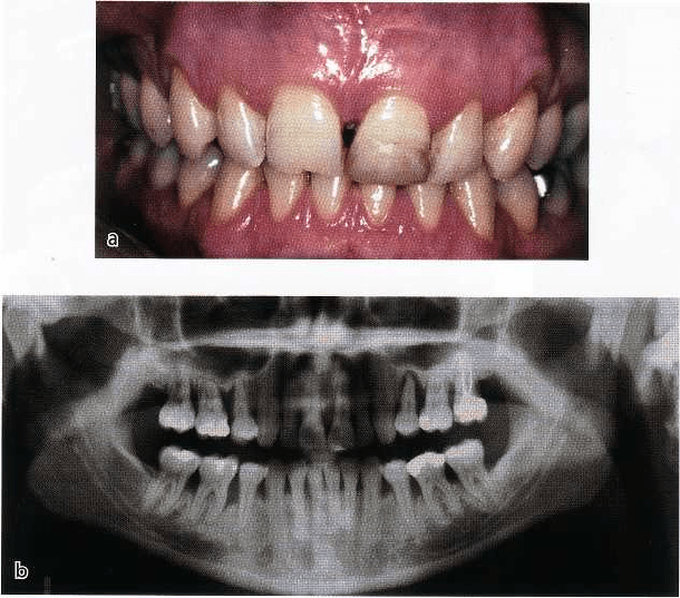
Fig. 6-12. A 30-year-old female
smoker with advanced periodonti
tis. (a) The clinical appearance
shows marginal gingiva with little
signs of inflammation. Probing
depths greater than 6 mm were
present at most interproximal
sites, but with little bleeding on
probing. (b) The generalized ad-
vanced bone loss in this patient.
The finding of less gingival bleeding on probing is
associated with less inflamed marginal tissue and
lower bleeding scores when probing the depth of the
pockets. The typical clinical appearance of the
smoker's gingival tissue is shown in Fig. 6-12, which
demonstrates relatively low levels of marginal inflam-
mation and a tendency to a more fibrotic appearance
with little edema. Despite the clinical appearance of
the gingival tissue, the patient has deep pockets, ad-
vanced attachment loss and bone loss as shown in Fig.
6-12b.
Modification of the host/bacteria
relationship in smoking
There are a number of theories as to why smokers have
more periodontal disease than non-smokers, involv-
ing both bacterial aspects and the host response.
Effects on plaque bacteria
Smokers may have higher levels of plaque than non-
smokers, which may be accounted for by poorer levels
of oral hygiene rather than higher rates of supragingi-
val plaque growth (Bergstrom 1981, Bergstrom & Pre
ber 1986). Several studies have shown that smokers
harbor more bacterial species which are associated
with periodontitis including
Porphyromonas gingivalis,
Actinobacillus actinomycetemcomitans, Bacteroides for-
sythus
(Zambon et al. 1996),
Prevotella intermedia, Pep-
tostreptococcus micros, Fusobacterium nucleatum, Cam-
pylobacter rectus
(van Winkelhoff et al. 2002),
coccus
aureus, Eschericia coli
and
Candida albicans
(Kamma et al. 1999) than non-smokers. Smokers may
have a higher proportion of sites harboring these pu-
tative periodontal pathogens, in particular the palatal
aspects of the maxillary teeth and the upper and lower
incisor regions (Haffajee & Socransky 2001a,b).
Effects on the host response
The relationship between plaque accumulation and
development of inflammation in smokers has been
studied in classical experimental gingivitis studies (
Bergstrom & Preber 1986). They demonstrated that
there is no difference in plaque accumulation when
comparing smokers and non-smokers. However, the
development of inflammation was very much re-
tarded in the smoking group with less sites exhibiting
redness or bleeding on probing. They also showed
lower amounts of gingival crevicular fluid during the
development of gingivitis. The reduced bleeding has
been proposed to be caused by nicotine induced vaso-
constriction, but as previously described in this chap-
ter, more recent evidence has failed to show a reduc-
tion in blood flow to the gingiva following smoking a
cigarette in regular smokers (Meekin et al. 2000). The
reduced bleeding on the other hand may be due to
long-term effects on the inflammatory lesion. Histo-
logical comparisons of the lesions from smokers and
non-smokers has shown fewer blood vessels in the
inflammatory lesions of smokers (Rezavandi et al.
2001).
Smoking has a profound effect on the immune and
inflammatory system (reviewed by Barbour et al.
1997). Smokers have an increased number of leuko-
cytes in the systemic circulation, but fewer cells may
migrate into the gingival crevice/pocket. Smoking is
associated with chronic obstructive pulmonary dis-
ease (Barnes 2000) and many of the mechanisms indi-
cated are paralleled in findings related to periodontal
disease. It is thought that the main cell type responsi-
ble for destruction of lung parenchyma is the neutro-
MODIFYING FACTORS: DIABETES, PUBERTY, PREGNANCY AND THE MENOPAUSE AND TOBACCO SMOKING • 191
phil, which is delayed in its transit through the pul-
monary vasculature (McNee et al. 1989) where it is
stimulated to release proteases including elastase,
cathepsins and matrix metalloproteases (Barnes 2000).
These destructive molecules are balanced by inhibi-
tors such as c -l-antitrypsin and tissue inhibitors of
matrix metalloproteases.
Studies in vitro have shown a direct inhibition of
neutrophil and monocyte-macrophage defensive
functions by high concentrations of nicotine that may
be achieved in patients using smokeless tobacco (
Pabst et al. 1995). MacFarlane and co-workers (1992)
examined patients with refractory periodontitis and
found a high proportion of smokers in this diagnostic
group. These investigators demonstrated abnormal
PMN phagocytosis associated with a high level of
cigarette smoking.
The PMN is a fundamental defense cell in the peri-
odontal tissue. There is a constant traffic of PMNs
from the gingival vasculature through the connective
tissue and junctional epithelium into the gingival sul-
cus/pocket. This is described in some detail in Chap-
ter 5. The PMN is the first line of defense and is
chemotactically attracted to bacterial challenge at the
dento-gingival junction. The PMN contains a power-
ful battery of enzymes including elastase and other
collagenases that have been implicated in tissue de-
struction in periodontitis and pulmonary disease.
Eichel and Shahrik (1969) suggested decreased PMN
migration into the oral cavity of smokers. Sub-
sequently, PMNs harvested from the gingival sulcus
of smokers were shown to have reduced phagocytic
capacity compared to PMNs from non-smokers (Ken-
ney et al. 1977). Neutrophil defects have been associ-
ated with an increased susceptibility to periodontitis,
including cyclic neutropenia where there is a reduc-
tion in the number of neutrophils, and conditions such
as leukocyte adhesion deficiency (LAD 1 and LAD 2),
which may be responsible for cases of generalized
prepubertal periodontitis as described by Page et al. (
1983). It is proposed that smoking causes alterations to
PMN function which could be considered to be minor
variations of these more profound defects.
The normal passage of the PMN from the microvas-
culature to the periodontal tissues involves a classic
series of events including capture, rolling on the en-
dothelium, firm adhesion to the endothelium and
transmigration through the vessel wall into the con-
nective tissue (Ley 1996). This involves a complex
interaction between receptors and ligands on the leu-
kocyte surface and endothelium including selectins,
ICAM-1 and LFA1 (CD18, CD11b) (Crawford &
Watanabe 1994, Gemmel et al. 1994). Defects in the
functional ligands for the selectins have been impli-
cated in LAD 2 and mutations in the gene encoding
CD18 resulting in absence of the (3 2 integrins with
LAD 1. Subjects with LAD are susceptible to serious
and life-threatening infections and have tremendous
destruction of the periodontal tissues, often leading to
total tooth loss in the deciduous dentition. These seri
ous and rare conditions illustrate the overwhelming
importance of the adhesion molecules and suggest
that minor defects in them may also give rise to more
subtle conditions that could lead to increased suscep-
tibility to periodontal destruction. In this respect, it
has been shown that smokers are affected by upregu-
lation of molecules such as ICAM-1 on the endothe-
lium and they have higher levels of circulating soluble
ICAM-1 which could interfere with the normal recep-
tor ligand binding and function of the leukocyte in the
defense of the periodontal tissue (Koundouros et al.
1996, Palmer et al. 1999, Scott et al. 2000a). A potential
destructive mechanism is the release of elastase from
neutrophils following binding of ICAM with CD18 (
Mac 1 and LFA 1) (Barnett et al. 1996). Lower levels
of elastase detected in the gingival fluid of smokers
compared to non-smokers, may indicate more elastase
release within the tissues (Alavi et al. 1995), and this
is especially important considering the effects of
smoking on protease inhibitors.
Tobacco smoking has a chronic effect on the ele-
vated levels of sICAM and there is evidence that the
subject may return to more normal levels after quitting
smoking (Scott et al. 2000b). These molecules can be
detected in the serum and in the gingival crevicular
fluid. It has also been shown that cotinine is present
in the gingival crevicular fluid in about the same
concentration as it appears in serum, but the levels of
sICAM are much lower in smokers despite very much
higher serum levels than non-smokers (Fraser et al.
2001).
The effects of smoking on lymphocyte function and
antibody production are very complex, with the vari-
ous components having the potential to cause immu-
nosuppression or stimulation. The leukocytosis ob-
served in smokers results in increased numbers of
circulating T and B lymphocytes (reviewed in Sopori
& Kozak 1998). Smoking appears to affect both B and
T cell function, inducing functional unresponsiveness
in T cells. Smoking has been shown to reduce most of
the immunoglobulin classes except for IgE and to
inhibit pro-inflammatory cytokines (Barbour et al.
1997, Quinn et al. 1998).
The clinical change in the tissues of smokers was
described above. It is not surprising that histological
evaluation of smokers' tissues has shown that there is a
decrease in the vascularity of the tissues (Rezavandi et
al. 2001). This is a chronic effect due to smoking and
may also be associated with alterations in the expres-
sion of adhesion molecules within the endothelium.
The effect of tobacco smoking on the expression of
adhesion molecules on leukocytes, within the inflam-
matory lesion, in the junctional epithelium and cells
of the pocket epithelium could have important impli-
cations on the progession of periodontitis in smokers.
The effect of smoking on macrovasular disease is well
documented (Powell 1998) and its effects on microvas-
cular disease could also be of importance in the peri-
odontal disease and in healing.
192 • CHAPTER 6
Effects on healing and treatment response
The healing potential of tissues has important impli-
cations in any chronic inflammatory lesion and in
repair following treatment. Smoking has been identi-
fied as an important cause of impaired healing in
orthopedic surgery, plastic surgery, dental implant
surgery (Bain & Moy 1993) and in all aspects of peri-
odontal treatment including non-surgical treatment,
basic periodontal surgery, regenerative periodontal
surgery and mucogingival plastic periodontal surgery
(Preber & Bergstrom 1986, Miller 1987, Grossi et al.
1996, 1997, Kaldahl et al. 1996, Tonetti et al. 1995,
Bostrom et al. 1998).
In non-surgical treatment, smoking is associated
with poorer reductions in probing depth and gains in
clinical attachment. In most studies the smokers at
baseline have a lower level of bleeding, and following
treatment bleeding scores are reduced in smokers in a
similar manner to non-smokers. The poorer reduc-
tions in probing depths and gains in attachment level
amount to a mean of approximately 0.5 mm. Much of
this may be due to less recession of the marginal
tissues in smokers as there is less edema and more
fibrosis in the gingiva. The same may be true for the
deeper tissues of the periodontium where there is less
of an inflammatory infiltrate and vascularity at the
depth of the pocket. These differences in the tissues
between smokers and non-smokers in the untreated
state may largely account for the differences in treat-
ment response in non-surgical treatment. It has been
proposed that these differences may be manifest by
differences in probe penetration in smokers and non-
smokers, particularly in deep pockets (Biddle et al.
2001).
The poor response to treatment in smokers in non-
surgical treatment may also apply to those treated
with adjunctive antibiotics (Kinane & Radvar 1997,
Palmer et al. 1999). Response to non-surgical treat-
ment may be seen merely as resolution of inflamma-
tion, improvement of the epithelial attachment to-
gether with some formation of collagen. However, the
response following periodontal surgery is more com-
plex and involves an initial inflammatory reaction
followed by organization of the clot, formation of
granulation tissue consisting of capillary buds and
fibroblasts laying down collagen. The surgical flaps
have to revascularize and the epithelial attachment
has to reform on the surface. In regenerative surgery
there also has to be formation of a connective tissue
attachment and cementogenesis. Tobacco smoke and
nicotine undoubtedly affect the microvasculature, the
fibroblasts and connective tissue matrix, the bone and
also the root surface itself. It has been shown in in vitro
studies that fibroblasts are affected by nicotine in that
they demonstrate reduced proliferation, reduced mi-
gration and matrix production and poor attachment to
surfaces (Raulin et al. 1988, Tipton & Dabbous 1995,
James et al. 1999). The root surfaces in smokers are
additionally contaminated by products of smoking
such as nicotine and cotinine and these molecules may
affect the attachment of cells (Raulin et al. 1988). Smok
ing has a direct effect on bone and is an established
risk factor in osteoporosis. It has also been proposed
that it may have a direct affect on bone loss in perio-
dontitis (Bergstrom et al. 1991) and it undoubtedly
delays healing of bone in fracture wound repair. It is
not surprising therefore that tobacco smoking has
been implicated in poorer responses to periodontal
surgical treatment.
Smoking cessation
All patients should be assessed for smoking status and
given advice to quit the habit. About 70% of people
who smoke would like to quit and should be assisted.
They should be referred to specialist cessation services
if the treating practitioner does not feel confident in
this area. They can be advised about nicotine replace-
ment therapy. People's success with quitting is consid-
erably improved using nicotine replacement therapy
and drugs such as buproprion hydrochloride. Former
smokers more closely resemble non-smokers in their
periodontal health status and response to treatment,
but the time required to revert to this status has not
been defined.
REFERENCES
Abraham-Inpijn, L., Polsacheva, D.V. & Raber-Durlacher, J.E. (
1996). The significance of endocrine factors and microorgan-
isms in the development of gingivitis in pregnant women.
Stomatolgiia 75, 15-18.
Alavi, Al., Palmer, R.M., Odell, E.W., Coward, P.Y. & Wilson,
R.F. (1995). Elastase in gingival crevicular fluid from smokers
and non-smokers with chronic inflammatory periodontal dis-
ease.
Oral Diseases 1,
103-105.
Aldridge, J.P, Lester, V, Watts, T.L., Collins, A., Viberti, G. &
Wilson, R.F. (1995). Single blind studies on the effects of
improved periodontal health on metabolic control in type 1
diabetes mellitus.
Journal of Clinical Periodontology 22,
271-275.
Alexander, A.G. (1970). The relationship between tobacco smok-
ing, calculus and plaque accumulation and gingivitis. Dental
Health 9, 6-9.
Amar, S. & Chung, K.M. (1994). Influence of hormonal variation
on the periodontium in women. Periodontology 2000 6, 79-87.
Armamento-Villareal, R., Villareal, D.T., Avioli, L.V. & Civitelli,
R. (1992). Estrogen status and heredity are major determi-
nants of premenopausal bone mass.
Journal of Clinical Investi-
gation 90, 2464-2471.
Atkinson, M.A. & Maclaren, N.K. (1990). What causes diabetes?
Scientific American 263, 62-63, 66-71.
Baab, D.A. & ()berg, P.A. (1987). The effect of cigarette smoking
on gingival blood flow in humans.
Journal of Clinical Periodon-
tology
14, 418-424.
Bain, C.A. & Moy, P.K. (1993). The association between the failure
