Jan Lindhe. Clinical Periodontology
Подождите немного. Документ загружается.


MICROBIOLOGY OF PERIODONTAL DISEASE • 113
Table 4-1. Summary of some of the types of data that suggest that Actinobacillus actinornycetenicomitans may
be an etiologic agent of destructive periodontal diseases (for literature citations see text and Haffajee &
Socransky 1994)
Association
Elevated in lesions of localized juvenile periodontitis, pre-pubertal or adolescent periodontal disease
Lower in
health, gingivitis and edentulous subjects or sites
Elevated in some adult periodontitis lesions
Elevated in active lesions of juvenile periodontitis
Detected in prospective studies
Detected in apical areas of pocket or in tissues from LJP lesions
Elimination
Elimination or suppression resulted in successful therapy
Recurrent lesions harbored the species
Host response
Elevated antibody in serum or saliva of LIP patients
Elevated antibody in serum or saliva of chronic periodontitis patients
Elevated local antibody in UP sites
Virulence factors
Leukotoxin; collagenase; endotoxin; epitheliotoxin; fibroblast inhibitory factor; bone resorption inducing factor; induction of cytokine
production from macrophages; modification of neutrophil function; degradation of immunoglobulins; cytolethal
distending toxin (Cdt);
induces apoptotic cell death
Invades epithelial and vascular endothelial cells
in vitro
and buccal epithelial cells
in vivo
Animal studies
Induced disease in gnotobiotic rats
Subcutaneous abscesses in mice
Table 4-2. Summary of some of the types of data that suggest that Porph yromonas gingivalis may be an etio-
logic agent of destructive periodontal diseases (for literature citations see text and Haffajee & Socransky 1994)
Association
Elevated in lesions of periodontitis
Lower in sites of health, gingivitis and edentulous subjects
Elevated in actively progressing lesions
Elevated in subjects exhibiting periodontal disease progression
Detected in cells or tissues of periodontal lesions
Presence indicates inc
r
eased risk for alveolar bone loss and attachment level loss
Elimination
Elimination resulted in successful therapy
Recurrent lesions harbored the species
Successful treatment lowered level and/or avidity of antibody
Host response
Elevated antibody in serum or saliva in subjects with various forms of periodontitis Altered
local antibody in periodontitis
Virulence factors
Collagenase; endotoxin; proteolytic trypsin-like activity; fibrinolysin; hemolysin; other proteases including gingipain; Phospholipase A;
degrades immunoglobulin; fibroblast inhibitory factor; H2S; NH3, fatty acids; factors that adversely affect PMNs; capsular
polysaccharide; bone resorption inducing factor; induction of cytokine production from various host cells; generates chemotactic
activities; inhibits migration of PMNs across epithelial barriers; invades epithelial cells
in vitro
Animal studies
Important in experimental pure or mixed subcutaneous infections
Induced disease in gnotobiotic rats
Studies in sheep, monkeys and dogs
Immunization diminished disease in experimental animals
possessed, by certain species may be suggestive that
that species could play a role in the disease process.
Animal model systems provide suggestive evi-
dence that a microbial species may play a role in
human disease. Particularly noteworthy are studies of
experimentally induced disease in dogs or monkeys,
which can be manipulated to favor selection of single
or subsets of species that may or may not induce
pathology. These models usually suggest a possible
etiologic role of a species indigenous to the test animal
that may have analogues in the human subgingival
microbiota. Finally, technological developments, such
as checkerboard DNA—DNA hybridization (Fig. 4-1)
and PCR, now permit assessment of specific microor-
ganisms in large numbers of subgingival plaque sam-
ples. This allows prospective studies to be performed

114 • CHAPTER 4
Table 4-3. Summary of some of the types of data that suggest that Bacteroides forsythus may be an etiologic
agent of destructive periodontal diseases (for literature citations see text and Haffajee & Socransky 1994)
Association
Elevated in lesions of periodontitis
Lower in sites of health or gingivitis
Elevated in actively progressing lesions
Elevated in periodontal abscesses
Increased in subjects with refractory periodontitis
Detected in epithelial cells of periodontal pockets
Presence indicates increased risk for alveolar bone loss, tooth and attachment level loss
Elimination
Elimination resulted in successful therapy
Recurrent
lesions harbored the species
Reduced in
successfully treated peri-implantitis
Host response
Elevated antibody in serum of periodontitis subjects and very high in a subset of subjects with refractory periodontitis
Virulence factors
Endotoxin; fatty acid and methylglyoxal production; induces apoptotic cell death; cytokine production from various host cells; invades
epithelial cells
in vitro
and
in vivo
Animal studies
Increased levels in ligature-induced periodontitis and peri-implantitis in dogs
Induced
disease in gnotobiotic rats
in which the risk of periodontal disease progression
conferred by the presence of an organism at given
levels may be assessed.
Periodontal pathogens
The World Workshop in Periodontology (Consensus
report 1996) designated A. actinomycetemcomitans, P.
gingivalis and B. forsythus as periodontal pathogens.
Tables 4-1 to 4-3 summarize some of the data that
indicate an etiologic role of these species in periodon-
tal diseases, categorized according to the criteria de-
fined above. The summary is by no means exhaustive
but does indicate that a growing literature suggests
some reasonable candidates as etiologic agents of de-
structive periodontal diseases.
Actinobacillus actinomycetemcomitans
One of the strongest associations between a
suspected pathogen and destructive periodontal
disease (at least in terms of number of publications) is
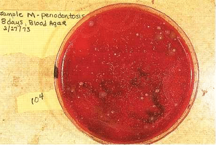
provided by A. actinomycetemcomitans. This is a small,
non-motile, Gram-negative, saccharolytic,
capnophilic, round-ended rod that forms small,
convex colonies with a "star-shaped" center when
grown on blood agar plates (Fig. 4-2). This species
was first recognized as a possible periodontal
pathogen by its increased frequency of detection and
higher numbers in lesions of localized juvenile
periodontitis (Newman et al. 1976, Slots 1976,
Newman & Socransky 1977, Slots et al. 1980,
Mandell & Socransky 1981, Zambon et al. 1983a,
Chung et al. 1989) when compared with numbers in
plaque samples from other clinical conditions includ-
ing periodontitis, gingivitis, and health. Soon after, it
was demonstrated that the majority of subjects with
LIP had an enormously elevated serum antibody re-
sponse to this species (Genco et al. 1980, Listgarten et
al. 1981, Tsai et al. 1981, Altman et al. 1982, Ebersole et
al. 1982, 1987) and that there was local synthesis of
antibody to this species (Schonfeld & Kagan 1982,
Ebersole et al. 1985, Smith et al. 1985, Tew et al. 1985a).
When subjects with this form of disease were treated
successfully, the species was eliminated or lowered in
level, while treatment failures were associated with
failure to lower the numbers of the species in treated
sites (Slots & Rosling 1983, Haffajee et al. 1984, Chris
tersson et al. 1985, Kornman & Robertson 1985, Man-
dell et al. 1986, Preus 1988, Shiloah et al. 1998, Tinoco
et al. 1998). The species produced a number of poten-
tially damaging metabolites including a leukotoxin (
Baehni et al. 1979), a cytolethal distending toxin (Saiki
et al. 2001, Shenker et al. 2001) and induced disease in
experimental animals (Irving et al. 1978). A. actinomy-
cetemcomitans has been shown, in vitro, to have the
ability to invade cultured human gingival epithelial
cells (Blix et al. 1992, Sreenivasan et al. 1993), human
vascular endothelial cells (Schenkein et al. 2000) and
buccal epithelial cells in vivo (Rudney et al. 2001).
Further, studies have shown that A. actinomycetem-
comitans induced apoptotic cell death (Arakawa et al.
2000, Kato et al. 2000).
Perhaps the strongest association data came from
studies of "active lesions" in which the species was
elevated in actively progressing periodontal lesions
when compared with non-progressing sites (Haffajee
et al. 1984, Mandell 1984, Mandell et al. 1987) and in
prospective studies of as yet undiseased siblings of
LJP subjects (DiRienzo et al. 1994). A. actinomycetem-
comitans was also elevated in studies of disease pro-
gression in young Indonesian subjects (Timmerman et
al. 2001). Collectively, the data suggest that A. acti-
nomycetemcomitans is a probable pathogen of LIP.
However, this should not be interpreted as meaning

MICROBIOLOGY OF PERIODONTAL DISEASE • 115
that it is the sole cause of this clinical condition since
a subset of subjects with LJP did not exhibit this spe-
cies in samples of their subgingival plaque and had no
elevated antibody response to the species (Loesche et
al. 1985, Moore 1987).
The possibility that only a subset of A. actinomy-
cetemcomitans clonal types is responsible for localized
juvenile periodontitis was raised by recent studies.
Strains of A. actinomycetemcomitans were isolated from
members of 18 families with at least one member with
active LJP as well as from 32 control subjects. Restric
tion fragment length polymorphisms (RFLP) indi-
cated 13 distinct RFLP groups of A.
tans (DiRienzo & McKay 1994). Isolates from LJP sub-
jects fell into predominantly RFLP pattern II, while
RFLP patterns XIII and XIV were seen exclusively in
isolates from periodontally healthy subjects. Further,
disease progression was related strongly to the pres-
ence of RFLP group II (DiRienzo et al. 1994).
Haubek et al. (1996) demonstrated that strains of A.
actinomycetemcomitans isolated from families of Afri-
can origin living in geographically different areas
were characterized by a 530 base pair deletion in the
leukotoxin gene operon leading to a significantly in-
creased production of leukotoxin. They speculated
that this virulent clonal type found in individuals of
African origin may account for an increased preva-
lence of LJP in African-Americans and other individu
als of African descent. The same investigators found a
strong association between the presence of A. acti-
nomycetemcomitans with the 530 bp deletion and early
onset periodontitis in Moroccan school children, but
no association between the presence of A. actinomy-
cetemcomitans without the deletion and early onset
periodontitis (Haubek et al. 2001). This deletion was
not detected in any strains of A. actinomycetemcomitans
isolated from adult Chinese subjects (Mombelli et al.
1999, Tan et al. 2001) or Asian subjects in the US (
Contreras et al. 2000). Subjects harboring A. actinomy-
cetemcomitans with the 530 bp deletion were 22.5 times
more likely to convert to LJP than subjects who had A.
actinomycetemcomitans variants containing the full
length leukotoxin promoter region (Bueno et al. 1998).
A. actinomycetemcomitans has also been implicated
in adult forms of destructive periodontal disease, but
its role is less clear. The species has been isolated from
Fig. 4-2. Photograph of a primary isolation plate of a
subgingival plaque sample from a diseased site in a
subject with LJP. A dilution of the plaque sample was
grown for 7 days at 35°C on an enriched blood agar
plate in an atmosphere of 80% N
2
, 10% H
2
and 10%
CO
2
. The majority of the small, round, convex colonies
on this plate are isolates of Actinobacillus
actinomycetemcomitans.
adult periodontitis lesions, but less frequently and in
lower numbers than from lesions in LJP subjects (
Rodenburg et al. 1990, Slots et al. 1990a). In addition,
its numbers in plaque samples from adult lesions were
often not as high as those observed for other suspected
pathogens in the same plaque samples. The most
frequently isolated serotype of A. actinomycetemcomi-
tans from lesions of LJP in American subjects was
serotype b (Zambon et al. 1983b), whereas serotype a
was more commonly detected in samples from adult
subjects (Zambon et al. 1983a). This finding was cor-
roborated indirectly by examination of serum anti-
body levels to the two serotypes. Most elevated re-
sponses to A. actinomycetemcomitans in LJP subjects
were to serotype b while elevated responses to sero-
type a were more common in subjects with adult
periodontal disease (Listgarten et al. 1981). Some sub-
jects in each group exhibited elevated serum antibody
responses to both serotypes. In Finnish subjects, sero-
types a and b were more frequently isolated from
subjects with periodontal disease and serotype c from
periodontally healthy subjects (Asikainen et al. 1991).
However, this pattern of serotype distribution was not
observed in Korea (Chung et al. 1989) or Japan (Saito
et al. 1993) where A. actinomycetemcomitans serotype c
was frequently observed in plaque samples from sites
of periodontal pathology. Recently, two other sero-
types, d and e, have been recognized (Dogan et al.
1999, Mombelli et al. 1999).
Antibody data and data from the treatment of A.
actinomycetemcomitans infected patients with adult or
refractory periodontitis provide the most convincing
evidence of a possible etiological role of A. actinomy-
cetemcomitans in adult forms of periodontal disease. 36
of 56 adults with destructive periodontal disease ex-
amined at multiple time periods at The Forsyth Insti-
tute exhibited an elevated serum antibody response to
A. actinomycetemcomitans serotypes a and/or b. Ele-
vated responses to other suspected periodontal patho-
gens were far less common. van Winkelhoff et al. (
1992) treated 50 adult subjects with "severe general-
ized periodontitis" and 40 subjects with refractory
periodontitis who were culture-positive for A. acti-
nomycetemcomitans using mechanical debridement and
systemically administered amoxicillin and
metronidazole. Only 1 of 90 subjects was culture posi-

116 • CHAPTER 4
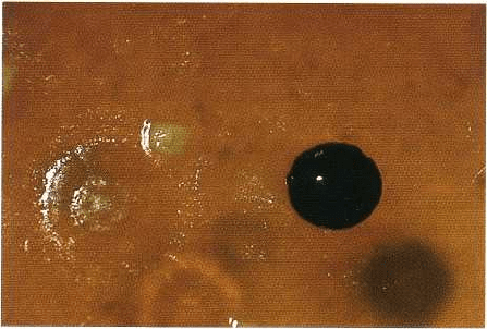
Fig. 4-3. Photograph of part of a primary isolation plate
of a subgingival plaque sample from a subject with
adult periodontitis. The medium and growth condi-
tions were as described in Fig. 4-2. The black-pig-
mented colony is an isolate of Porphyromonas gingivalis.
five for A. actinomycetemcomitans 3-9 months post-
therapy (van Winkelhoff et al. 1992) and 1 of 48 sub-
jects was culture positive 2 years post-therapy (Pavicic
et a1. 1994). There was a significant gain in attachment
level and decrease in probing pocket depth in virtually
all patients after therapy.
Porphyromonas gingivalis
P. gingivalis is a second consensus periodontal patho-
gen. Isolates of this species are Gram-negative, an-
aerobic, non-motile, asaccharolytic rods that usually
exhibit coccal to short rod morphologies. P. gingivalis
is a member of the much investigated "black-pig-
mented Bacteroides" group (Fig. 4-3). Organisms of this
group form brown to black colonies (Oliver & Wherry
1921) on blood agar plates and were initially grouped
into a single species, B. melaninogenicus (Bacterium
melaninogenicum, Burdon (1928)). The black-pig-
mented Bacteroides have a long history of association
with periodontal diseases since the early efforts of
Burdon (1928) through the mixed infection studies of
Macdonald and co-workers (1960) to the current in-
tense interest. In the late 1970s, it was recognized that
the black-pigmented Bacteroides contained species that
were asaccharolytic (eventually P. gingivalis), and
either had an intermediate level of carbohydrate fer-
mentation (which eventually led to a group of species
including P. intermedia) or were highly saccharolytic (
leading to the group that includes Prevotella melanino-
genica).
Early interest in P. gingivalis and other black-pig-
mented Bacteroides arose primarily because of their
essential role in certain experimental mixed infections
(Macdonald et al. 1956, 1963, Socransky & Gibbons
1965) and their production of an unusually large array
of virulence factors (Table 4-2, Haffajee & Socransky
1994, Deshpande & Khan 1999). Members of these
species produce collagenase, an array of proteases (
including those that destroy immunoglobulins),
hemolysins, endotoxin, fatty acids, NH
3
, H
2
S, indole
etc. P. gingivalis can inhibit migration of PMNs across
an epithelial barrier (Madianos et al. 1997) and has
been shown to affect the production or degradation of
cytokines by mammalian cells (Darveau et al. 1998,
Fletcher et al. 1998, Sandros et al. 2000). Studies initi-
ated in the late 1970s and extending to date strength-
ened the association of P. gingivalis with disease and
demonstrated that the species was uncommon and in
low numbers in health or gingivitis but more fre-
quently detected in destructive forms of disease (Table
4-2, Haffajee & Socransky 1994, O'Brien-Simpson et al.
2000). This species has also been shown to be increased
in numbers and/or frequency of detection in deterio-
rating periodontal sites (Dzink et al. 1988, Lopez 2000,
Kamma et al. 2001) or in subjects exhibiting periodon-
tal disease progression (Albandar et al. 1997). The
species has been shown to be reduced in successfully
treated sites but is commonly encountered in sites that
exhibit recurrence of disease or persistence of deep
periodontal pockets post-therapy (Bragd et al. 1987,
Haffajee et al. 1988a, van Winkelhoff et al. 1988, Ber-
glundh et al. 1998, Shiloah et al. 1998, Winkel et al.
1998, Takamatsu et al. 1999, Chaves et al. 2000, Mom
belli et al. 2000). P. gingivalis has been associated with
an increased risk of periodontal disease severity and
progression (Beck et al. 1990, 1992, 1997, Grossi et al.
1994, 1995).
P. gingivalis has been shown to induce elevated
systemic and local immune responses in subjects with
various forms of periodontitis (Table 4-2, Mahanonda
et al. 1991, Haffajee & Socransky 1994, O'Brien-Simp
son et al. 2000). Indeed, there has been a remarkably
intense effort in many laboratories in the last few
years, not only to compare the level of antibody re-
sponse in subjects with and without disease, but to
examine relative avidities of antibody (Lopatin &
Blackburn 1992, Whitney et al. 1992, Mooney et al.
1993), subclass of antibody (Lopatin & Blackburn
1992, Wilton et al. 1992), the effect of treatment (Chen
et al. 1991, Johnson et al. 1993) and the nature of the
antigens which elicit the elevated responses (Ogawa
et al. 1989, Yoshimura et al. 1989, Curtis et al. 1991,
Papaioannou et al. 1991, Duncan et al. 1992, Schifferle
et al. 1993). Noteworthy in this regard are the obser-
vations of Ogawa et al. (1989), which indicate that an
average of approximately 5% of plasma cells in lesions
of advanced periodontitis form antibody to the fim-
briae of P. gingivalis. The consensus of the antibody
studies is that many, but not all, subjects who have
experienced periodontal attachment loss exhibit ele-
vated levels of antibody to antigens of P. gingivalis
suggesting that this species gained access to the un-
MICROBIOLOGY OF PERIODONTAL DISEASE • 117
derlying tissues and may have initiated or contributed
to the observed pathology.
P. gingivalis-like organisms are also strongly related
to destructive periodontal disease in naturally occur-
ring or ligature-induced disease in dogs, sheep or
monkeys (Table 4-2). The species or closely related
organisms were higher in number in lesion sites than
in non-lesion sites in naturally occurring disease.
When disease was induced by ligature in dogs or
monkeys, the level of the species rose at the diseased
sites concomitant with the detection of disease. Of
great interest were the observations of Holt et al. (1988)
who demonstrated that a microbiota suppressed by
systemic administration of rifampin (and without de-
tectable P. gingivalis) would not cause ligature-in-
duced disease, but the reintroduction of P. gingivalis to
the microbiota resulted in initiation and progress of
the lesions. Ligature-induced periodontitis and peri-
implantitis in dogs were also accompanied by a sig-
nificant increase in the detection of P. gingivalis (Nociti
et al. 2001). Like A. actinomycetemcomitans, P.
gingivalis
has been shown to be able to invade human gingival
epithelial cells in vitro (Lamont et al. 1992, Duncan et
al. 1993, Sandros et al. 1993) and buccal epithelial cells
in vivo (Rudney et al. 2001) and has been found in
higher numbers on or in epithelial cells recovered
from the periodontal pocket than in associated plaque
(Dzink et al. 1989). Attachment to and invasion of
epithelial cells appears to be mediated by the P. gingi-
valis fimbriae (Njoroge et al. 1997, Weinberg et al.
1997). Finally, studies in monkeys and gnotobiotic rats
have indicated that immunization with whole organ-
isms or specific antigens affected the progress of the
periodontal lesions. In most instances, periodontal
breakdown was decreased (Evans et al. 1992, Persson
et al. 1994). However, in one study the disease severity
was increased after immunization (Ebersole et al.
1991). The differences in results may have been due to
differences in animal species, the protocol used for
induction of periodontal disease, antigen preparation
or method of immunization. From the viewpoint of
this section, the studies demonstrate that altering the
host–P. gingivalis equilibrium by raising the level of
specific antibodies to P. gingivalis antigens markedly
affected disease outcome. Such data reinforce the im-
portance of this bacterial species in periodontal dis-
ease, at least in the animal model systems employed.
Bacteroides forsythus
The third consensus periodontal pathogen, B.
forsythus, was first described in 1979 (Tanner et al.
1979) as a "fusiform" Bacteroides. This species was
difficult to grow, often requiring 7–14 days for minute
colonies to develop. The organism is a Gram-negative,
anaerobic, spindle-shaped, highly pleomorphic rod.
The growth of the organism was shown to be en-
hanced by co-cultivation with F. n ucleatu in and indeed
commonly occurs with this species in subgingival
sites (Socransky et al. 1988). The species was shown to
have an unusual requirement for N-acetylmuramic
acid (Wyss 1989). Inclusion of this factor in culture
media markedly enhanced growth. The organism was
found in higher numbers in sites of destructive peri-
odontal disease or periodontal abscesses than in gin-
givitis or healthy sites (Lai et al. 1987, Herrera et al.
2000, Papapanou et al. 2000). In addition, B. forsythus
was detected more frequently and in higher numbers
in active periodontal lesions than inactive lesions (
Dzink et al. 1988) (Table 4-3). Further, subjects who
harbored B. forsythus were at greater risk for alveolar
bone loss, attachment loss and tooth loss compared
with subjects in whom this species was not detected (
Machtei et al. 1999). This species has been shown to
produce trypsin-like proteolytic activity (BANA test
positive, Loesche et al. 1992), methylglyoxal (Kashket
et al. 2002) and induce apoptotic cell death (Arakawa
et al. 2000).
Initially, B. forsythus was thought to be a relatively
uncommon subgingival species. However, the studies
of Gmur et al. (1989) using monoclonal antibodies to
enumerate the species directly in plaque samples, sug-
gested the species was more common than previously
found in cultural studies and its levels were strongly
related to increasing pocket depth. Lai et al. (1987)
corroborated these findings using fluorescent-la-
belled polyclonal antisera and demonstrated that B.
forsythus was much higher in subgingival than su-
pragingival plaque samples. Data of Tanner et al. (
1998) suggested that B. forsythus was a major species
found at sites that converted from periodontal health
to disease. B. forsythus was found at higher levels at
sites which showed breakdown after periodontal ther-
apy than sites which remained stable or gained attach-
ment (Shiloah et al. 1998). B. forsythus has also been
shown to be decreased in frequency of detection and
counts after successful periodontal therapy including
SRP (Haffajee et al. 1997, Takamatsu et al. 1999, Darby
et al. 2001), periodontal surgery (Levy et al. 2002), or
systemically administered antibiotics (Winkel et al.
1998, 2001, Feres et al. 2000,). Successful treatment of
peri-implantitis with local delivery of tetracycline was
accompanied by a significant decrease in the fre-
quency of detection of B. forsythus (Mombelli et al.
2001). Ligature-induced periodontitis and peri-im-
plantitis in dogs were accompanied by a significant
increase in the frequency of detection of B. forsythus (
Nociti et al. 2001). Finally, the persistent presence of
B. forsythus at sites in subjects with low severity of
chronic periodontitis indicated a 5.3 times greater
chance of having at least one site in their mouths
losing attachment compared with subjects with occa-
sional or no presence of this species (Tran et al. 2001).
Studies using checkerboard DNA–DNA hybridiza-
tion techniques to examine subgingival plaque sam-
ples confirmed the high levels of B. forsythus detected
using fluorescent-labelled antisera and demonstrated
that B. forsythus was the most common species de-
tected on or in epithelial cells recovered from peri-
odontal pockets (Dibart et al. 1998). It was infre-
quently detected in epithelial cell samples from

118 • CHAPTER 4
Fig. 4-4. Photomicrograph of a sample of subgingival
plaque from subjects with advanced adult periodonti
tis viewed by darkfield microscopy. The sample is
dominated by large spirochetes with the typical cork
-screw appearance.
healthy subjects. Double-labelling experiments dem-
onstrated that B. forsythus was both on and in peri-
odontal pocket epithelial cells indicating the species
ability to invade. Listgarten et al. (1993) found that the
species most frequently detected in "refractory" sub-
jects was B. forsythus. Serum antibody to B. forsythus
has been found to be elevated in a number of perio-
dontitis patients (Taubman et al. 1992) and was often
extremely elevated in a subset of refractory periodon-
tal disease subjects.
The role of this species in periodontal diseases has
been clarified by studies in numerous laboratories
involving non-cultural methods of enumeration such
as DNA probes, PCR or immunologic methods. For
example, Grossi et al. (1994, 1995) considered B.
forsythus to be the most significant microbial risk fac-
tor that distinguished subjects with periodontitis from
those who were periodontally healthy.
Spirochetes
Spirochetes are Gram-negative, anaerobic, helical-
shaped, highly motile microorganisms that are com-
mon in many periodontal pockets (Fig. 4-4). The role
of spirochetes in the pathogenesis of destructive peri-
odontal diseases deserves extended comment.
Clearly, a spirochete has been implicated as the likely
etiologic agent of acute necrotizing ulcerative gingivi-
tis by its presence in large numbers in tissue biopsies
from affected sites (Listgarten & Socransky 1964, List
garten 1965). The role of spirochetes in other forms
of periodontal disease is less clear. The organisms
have been considered as possible periodontal
pathogens since the late 1800s and in the 1980s
enjoyed a resurgence of interest for use as possible
diagnostic indicators of disease activity and/or
therapeutic efficacy (Keyes & Rams 1983, Rams &
Keyes 1983). The major reason for the interest in this
group of organisms has been their increased numbers
in sites with increased pocket depth. Healthy sites
exhibit few, if any, spirochetes, sites of gingivitis
but no attachment loss exhibit low to moderate
levels, while many deep pockets harbor large
numbers of these organisms.
The major difficulty encountered in defining the
role of spirochetes has been the difficulty in distin-
guishing individual species. At least 15 species of
subgingival spirochetes have been described, but in
most studies of plaque samples, spirochetes are com-
bined in a single group or groups based on cell size; i.
e. small, medium or large. Thus, while there may be
pathogens among the spirochetes, their role may have
been obscured by unintentionally pooling their num-
bers with non-pathogenic spirochetes. This would be
similar to combining in a single count, organisms with
coccal morphologies, such as P. gingivalis, Veillonella
parvula and Streptococcus sanguis. In spite of the limi-
tations of combining spirochetes into a single mor-
phogroup, spirochetes have been related with in-
creased risk at a site for the development of gingivitis (
Riviere & DeRouen 1998) and periodontitis (Riviere
et a1.1997). The need to evaluate the role of individual
species of spirochetes in periodontal diseases is rein-
forced by studies of serum antibody responses to dif-
ferent species. When antibody responses to individual
species were examined in subjects with adult or juve-
nile periodontitis or a healthy periodontium, different
responses were observed to different species. Certain
spirochetal species elicited an elevated response in
one or more of the groups with destructive periodon-
tal disease (Mangan et al. 1982, Tew et al. 1985c, Lai et
al. 1986), while others were related to depressed anti-
body responses in certain patient groups (Steinberg &
Gershoff 1968, Tew et al. 1985c). Such data suggest that
pooling spirochete species into a collective group may
obscure meaningful host–parasite interactions.
More recently, specific species of spirochetes have
been related to periodontal breakdown. Treponema
denticola was found to be more common in periodon-
tally diseased than healthy sites, more common in
subgingival than supragingival plaque (Simonson et
al. 1988, Riviere et al. 1992, Albandar et al. 1997, Haf-
fajee et al. 1998, Yuan et al. 2001) and more common
in healthy sites that progressed to gingivitis (Riviere
& DeRouen 1998). T. denticola was shown to decrease
in successfully treated periodontal sites, but not
change or increase in non-responding sites (Simonson
et al. 1992). Cultural studies suggested that T. denticola
and a "large treponeme" were found more frequently
in patients with severe periodontitis than in healthy or
gingivitis sites (Moore et al. 1982). Riviere et al. (
1991a,b,c, 1992) employed a monoclonal antibody
directed against Treponema pallidum, the etiologic
agent of syphilis, to examine supra and subgingival

MICROBIOLOGY OF PERIODONTAL DISEASE • 119
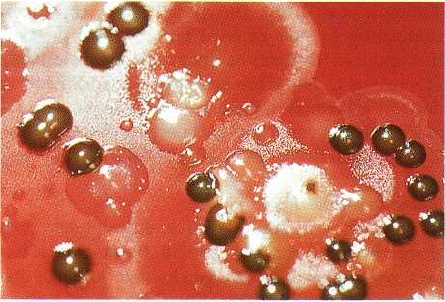
Fig. 4-5. Photograph of part of a primary isolation plate
of a subgingival plaque sample from a subject with
adult periodontitis. The medium and growth condi-
tions were as described in Fig. 4-2. The dark-pig-
mented colonies are isolates of Prevotella intermedia.
plaque samples and/or tissues from healthy, perio-
dontitis and ANUG subjects. This antibody cross-re-
acted with antigens of uncultivated spirochetes in
many of the plaque samples. These "pathogen-related
oral spirochetes" (PROS) were the most frequently
detected spirochetes in supra and subgingival plaques
of periodontitis patients and were the most numerous
spirochetes in periodontitis lesion sites. Their pres-
ence in periodontally healthy sites related to an in-
creased risk of development of periodontitis (Riviere
et al. 1997). The PROS were also detected in plaque
samples from ANUG (Riviere et al. 1991c) and tissue
biopsies from ANUG lesions using immunohisto-
chemical techniques (Riviere et al. 1991a). PROS were
also shown to have the ability to penetrate a tissue
barrier in in vitro systems (Riviere et al. 1991b). This
property was shared with T. pallidum but not with
other cultivated species of oral spirochetes such as T.
denticola, Treponema socranskii, Treponema pectinovorum
or Treponema vincentii. These studies and others sug-
gest that certain species of spirochetes are important
in the pathogenesis of ANUG and certain forms of
periodontitis. Precise evaluation of the role of individ-
ual spirochete species appears to be realistic based on
their detection in plaque samples by immunologic,
PCR or DNA probe techniques. Indeed, enumeration
of even uncultivable spirochete taxa is possible using
oligonucleotide probes (Tanner et al. 1994) or specific
antibody as described above. Studies performed using
such techniques permit better distinction of species of
spirochetes and a clearer understanding of their pos-
sible role in disease.
Prevotella intermedia/Prevotella nigrescens
Table 4-3 summarizes some of the data that suggest a
possible role of other subgingival species in the patho-
genesis of destructive periodontal diseases. At present
the data for other species are more limited, but these
organisms appear to merit further investigation (Zam-
bon 1996). P. intermedia is the second black-pigmented
Bacteroides to receive considerable interest (Fig. 4-5).
The levels of this Gram-negative, short, round-ended
anaerobic rod have been shown to be particularly
elevated in acute necrotizing ulcerative gingivitis (
Loesche et al. 1982), in certain forms of periodontitis
(Tanner et al. 1979, Dzink et al. 1983, Moore et al. 1985,
Maeda et al. 1998, Herrera et al. 2000, Papapanou et
al. 2000), and in progressing sites in chronic periodon-
titis (Tanner et al. 1996, Lopez 2000), and has been
detected by immunohistological methods in the inter-
cellular spaces of periodontal pocket biopsies from
rapidly progressive periodontitis subjects (Hillmann
et al. 1998). Isolates of this species can induce alveolar
bone loss in rats (Yoshida-Minami et al. 1997). Persist
ence of P. intermedia/nigrescens after standard mechani-
cal therapy has been shown to be associated with a
large proportion of sites exhibiting bleeding on prob-
ing (Mombelli et al. 2000). Berglundh et al. (1998)
demonstrated that improved clinical parameters after
the use of mechanical therapy and systemically ad-
ministered amoxicillin and metronidazole were asso-
ciated with a decrease of periodontal pathogens in-
cluding P. intermedia. Successful treatment of peri-im-
plantitis with local delivery of tetracycline also signifi-
cantly decreased the frequency of detection of P. inter-
media/nigrescens (Mombelli et al. 2001).
This species appears to have a number of the viru-
lence properties exhibited by P. gingivalis and was
shown to induce mixed infections on injection in labo-
ratory animals (Hafstrom & Dahlen 1997). It has also
been shown to invade oral epithelial cells in vitro (
Dorn et al. 1998). Elevated serum antibodies to this
species have been observed in some but not all sub-
jects with refractory periodontitis (Haffajee et al.
1988b). Strains of "P. intermedia" that show identical
phenotypic traits have been separated into two spe-
cies, P. intermedia and P. nigrescens (Shah & Gharbia
1992). This distinction makes earlier studies of this "
species" difficult to interpret since data from two
different species may have been inadvertently pooled.
However, new studies which discriminate the species
in subgingival plaque samples might strengthen the
relationship of one or both species to periodontal
disease pathogenesis.
Fusobacterium nucleatum
F. nucleatum is a Gram-negative, anaerobic, spindle-
shaped rod that has been recognized as part of the
subgingival microbiota for over 100 years (Plaut 1894,
Vincent 1899). This species is the most common isolate
found in cultural studies of subgingival plaque sam-
ples comprising approximately 7–10% of total isolates
120 • CHAPTER 4
from different clinical conditions (Dzink et al. 1985,
1988, Moore et al. 1985). F. nucleatum is prevalent in
subjects with periodontitis (Papapanou et al. 2000,
Socransky et al. 2002) and periodontal abscesses (Her
rera et al. 2000). Successful treatment of peri-implan-
titis with local delivery of tetracycline was associated
with a significant reduction in frequency of detection
in several species including F. nucleatum (Mombelli et
al. 2001). Invasion of this species into human gingival
epithelial cells in vitro was accompanied by an in-
creased secretion of IL-8 from the epithelial cells (Han
et al. 2000). The species can induce apoptotic cell death
in mononuclear and polymorphonuclear cells (Jewett
et al. 2001) and cytokine, elastase and oxygen radical
release from leukocytes (Sheikhi et al. 2000).
Although there were differences detected in levels
of this species between active and inactive periodontal
lesions (Dzink et al. 1988), the differences may have
been minimized by the inadvertent pooling of subspe-
cies of F. nucleaturn. Support for this contention may
be derived from the antibody responses in subjects
with different forms of periodontal disease to different
homology groups of F. nucleatum (Tew et al. 1985h). It
is anticipated that a clearer understanding of the role
of F. nucleatum will be achieved when subspecies such
as F. nucleatum ss nucleatum, F. nucleatum ss polymor-
phum, F. nucleatum ss vincentii and F. periodonticum
are
individually evaluated for their association with dis-
ease status and progression.
Carnpylobacter rectus
C. rectus is a Gram-negative, anaerobic, short, motile
vibrio. The organism is unusual in that it utilizes H
2
or formate as its energy source. It was first described
as a member of the "vibrio corroders", a group of short
nondescript rods that formed small convex, "dry
spreading" or "corroding" (pitting) colonies on blood
agar plates. These organisms were eventually shown
to include members of a new genus Wolinella (most
species have been redefined as Campylobacter), and
Eikenella corrodens. C. rectus has been shown to be
present in higher numbers in disease sites as com-
pared with healthy sites (Moore et al. 1983, 1985,
Lippke et al. 1991, Lai et al. 1992, Papapanou et al.
1997, Macuch & Tanner 2000) and it was found in
higher numbers and more frequently in sites exhibit-
ing active periodontal destruction (Dzink et al. 1985,
1988, Tanner & Bouldin 1989, Rams et al. 1993) or
converting from periodontal health to disease (Tanner
et al. 1998). In addition, C. rectus was found less fre-
quently and in lower numbers after successful peri-
odontal therapy (Tanner et al. 1987, Haffajee et al.
1988a, Levy et al. 2002) or treatment of peri-implantitis
with local delivery of tetracycline (Mombelli et al.
2001). C. rectus was also found in combination with
other suspected pathogens in sites of subjects with
refractory periodontal diseases (Haffajee et al. 1988b)
.
Like A. actinomycetemcomitans, C. rectus has been
shown to produce a leukotoxin. These are the only two
oral species known to possess this characteristic
(Gillespie et al. 1992). The species is also capable of
stimulating human gingival fibroblasts to produce IL-
6 and IL-8 (Dongari-Bagtzoglou & Ebersole 1996).
The role of C. rectus has been somewhat difficult to
determine because of the presence in plaque samples
of a number of very closely related organisms such as
Campylobacter showae and Wolinella X (Etoh et al. 1993).
Eikenella corrodens
E. corrodens is a Gram-negative, capnophilic, asac-
charolytic, regular, small rod with blunt ends. It has
been recognized as a pathogen in other forms of dis-
ease, particularly osteomyelitis (Johnson & Pankey
1976), infections of the central nervous system (Em-
merson & Mills 1978, Brill et al. 1982) and root canal
infections (Goodman 1977). This species was found
more frequently in sites of periodontal destruction as
compared with healthy sites (Savitt & Socransky 1984,
Muller et al. 1997, Yuan et al. 2001). In addition, E.
corrodens was found more frequently and in higher
levels in active sites (Dzink et al. 1985, Tanner et al.
1987) and in sites of subjects who responded poorly to
periodontal therapy (Haffajee et al. 1988b). Success-
fully treated sites harbored lower proportions of this
species (Tanner et al. 1987). E. corrodens has also been
found in association with A. actinomycetemcomitans in
some lesions of LJP (Mandell 1984, Mandell et al.
1987). In tissue culture systems, E. corrodens has been
shown to stimulate the production of matrix metallo-
proteinases (Dahan et al. 2001) and IL-6 and IL-8 (
Yumoto et al. 1999). While there is some association
of this species with periodontal disease, to date it has
not been particularly strong (Chen et al. 1989).
Peptostreptococcus micros
P. micros is a Gram-positive, anaerobic, small, asac-
charolytic coccus. It has long been associated with
mixed anaerobic infections in the oral cavity and other
parts of the body (Finegold 1977). Two genotypes can
be distinguished with the smooth genotype being
more frequently associated with periodontitis lesions
than the rough genotype (Kremer et al. 2000). P. micros
has been detected more frequently and in higher num-
bers at sites of periodontal destruction as compared
with gingivitis or healthy sites (Moore et al. 1983,1985,
Herrera et al. 2000, Papapanou et al. 2000, Riggio et al.
2001) and was elevated in actively breaking down
sites (Dzink et al. 1988). The levels and frequency of
detection of the species were decreased at successfully
treated periodontal sites (Haffajee et al. 1988a). Stud-
ies of systemic antibody responses to suspected peri-
odontal pathogens indicated that subjects with severe
generalized periodontitis had elevated antibody lev-
els to this species when compared with healthy sub-
jects or subjects with LJP (Tew et al. 1985a). In a mouse
skin model system, it was shown that P. micros in
combination with either P. intermedia or P. nigrescens
could produce transmissible abscesses (van Dalen et
al. 1998).
MICROBIOLOGY OF PERIODONTAL DISEASE • 121
Selenomonas species
Selenomonas species have been observed in plaque
samples using light microscopy for many decades.
The organisms may be recognized by their curved
shape, tumbling motility and, in good preparations, by
the presence of a tuft of flagella inserted in the
concave side. The Selenomonas spp. are Gram-nega-
tive, curved, saccharolytic rods. The organisms have
been somewhat difficult to grow and speciate. How-
ever, Moore et al. (1987) described six genetically and
phenotypically distinct groups isolated from the hu-
man oral cavity. Selenomonas noxia was found at a
higher proportion of shallow sites (PD < 4 mm) in
chronic periodontitis subjects compared with similar
sites in periodontally healthy subjects (Haffajee et al.
1998). Further, S. noxia was found to be associated with
sites that converted from periodontal health to disease (
Tanner et al. 1998).
Eubacterium species
Certain Eubacterium species have been suggested as
possible periodontal pathogens due to their increased
levels in disease sites, particularly those of severe
periodontitis (Moore et al. 1982, 1985, Uematsu &
Hoshino 1992, Papapanou et al. 2000). E. nodatum,
Eubacterium brachy and Eubacterium timidum are Gram-
positive, strictly anaerobic, small, somewhat
pleomorphic rods. They are often difficult to cultivate,
particularly on primary isolation, and appear to grow
better in roll tubes than on blood agar plates. Some of
these species elicited elevated antibody responses in
subjects with different forms of destructive periodon-
titis (Tew et al. 1985a,b, Vincent et al. 1986, Martin et
al. 1988). The Eubacterium species appear to be prom-
ising candidates as periodontal pathogens; however,
difficulty in their cultivation has slowed assessment
of their contribution. It seems likely that the role of the
Eubacterium species will be clarified when non-cul-
tural methods are routinely employed for their detec-
tion, as discussed for B. forsythus.
The
"milleri" streptococci
Streptococci were frequently implicated as possible
etiologic agents of destructive periodontal diseases in
the early part of the last century. Cultural studies of
the last two decades have also suggested the possibil-
ity that some of the streptococcal species are associ-
ated with and may contribute to disease progression.
At this time, evidence suggests that the "milleri"
streptococci, Streptococcus anginosus, S. constellatus and
S. intermedius might contribute to disease progression
in subsets of periodontal patients. The species was
found to be elevated at sites which demonstrated recent
disease progression (Dzink et al. 1988). Walker and
co-workers (1993) found S. intermedius to be elevated
in a subset of patients with refractory disease at
periodontal sites which exhibited disease progression.
Colombo et al (1998) found that subjects exhibiting a
poor response to SRP and then to periodontal surgery
with systemically administered tetracycline had
higher levels and proportions of S. constellatus, than
subjects who responded well to periodontal therapy.
The data on streptococci are somewhat limited, but a
continued examination of their role in disease seems
warranted.
Other species
Obviously all periodontal pathogens have not yet
been identified. Interest has grown in groups of spe-
cies not commonly found in the subgingival plaque as
initiators or possibly contributors to the pathogenesis
of periodontal disease, particularly in individuals who
have responded poorly to periodontal therapy.
Species not commonly thought to be present in sub-
gingival plaque can be found in a proportion of such
subjects or even in subjects who have not received
periodontal treatment. Emphasis has been placed on
enteric organisms, staphylococcal species as well as
other unusual mouth inhabitants. Slots et al. (1990b)
examined plaque samples from over 3000 chronic pe-
riodontitis patients and found that 14% of these pa-
tients harbored enteric rods and pseudomonads. En-
terobacter cloaceae, K. pneurnoniae, Pseudomonas aerugi-
nosa, Klebsiella oxytoca and Enterobacter agglomerans
comprised more than 50% of the strains isolated. This
group of investigators also examined 24 subjects with
periodontal disease in the Dominican Republic and
found that the prevalence of enteric rods in these
subjects was higher than levels found in subjects in the
US (Slots et al. 1991). In the 16 of 24 subjects in which
this group of organisms was detected, they averaged
23% of the cultivable microbiota. Rams et al. (1990,
1992) identified a number of species of staphylococci
and enterococci in subjects with various forms of peri-
odontal disease. The presence of unusual species in
periodontal lesions suggests the possibility that they
may play a role in the etiology of periodontal diseases.
However, such roles must be evaluated in the same
manner as the species discussed earlier in this section.
It is worth noting that systemically administered
ciprofloxacin improved the treatment response of pa-
tients whose periodontal pockets were heavily in-
fected with enteric rods (Slots et al. 1990a).
More recently, viruses including cytomegalo, Ep-
stein Barr, papilloma and herpes simplex have been
proposed to play a role in the etiology of periodontal
diseases, "possibly by changing the host response to
the local subgingival microbiota (Contreras & Slots
1996, 2000, Parra & Slots 1996, Contreras et al. 1997,
1999a,b, Velazco et al. 1999, Hanookai et al. 2000, Ting
et al. 2000). Human cytomegalo, Epstein Barr and
herpes simplex virus were found more frequently in
deteriorating periodontal sites than in control, stable
periodontitis sites in the same subject (Kamma et al.
2001).
122 • CHAPTER 4
Mixed infections
To this point, attention has been paid to the possible
role of individual species as risk factors for destructive
periodontal diseases. However, microbial complexes
colonizing the subgingival area can provide a spec-
trum of relationships with the host, ranging from
beneficial — the organisms prevent disease, to harmful
— the organisms cause disease. At the pathogenic end
of the spectrum, it is conceivable that different rela-
tionships exist between pathogens. The presence of
two pathogens at a site could have no effect or dimin-
ish the potential pathogenicity of one or other of the
species. Alternatively, pathogenicity could be en-
hanced either in an additive or synergistic fashion. It
seems likely that mixed infections occur in subgingi-
val sites since so many diverse species inhabit this
habitat. Evidence to support this concept has been
derived mainly from studies in animals in which it
was shown that combinations of species were capable
of inducing experimental abscesses, even though the
components of the mixtures could not (Smith 1930,
Proske & Sayers 1934, Cobe 1948, Rosebury et al. 1950,
Macdonald et al. 1956, Socransky & Gibbons 1965). It
is not clear whether the combinations suggested in the
experimental abscess studies are pertinent to human
periodontal diseases. The relationship of microbial
complexes to periodontal diseases will be discussed
in detail below.
THE NATURE OF DENTAL
PLAQUE —
THE BIOFILM WAY OF LIFE
Biofilms colonize a widely diverse set of moist sur-
faces including the oral cavity, the bottom of boats and
docks, and the inside of pipes, as well as rocks in
streams. Infectious disease investigators are interested
in biofilms that colonize a wide array of artificial
devices that have been implanted in the human in-
cluding catheters, hip and voice prostheses and con-
tact lenses. Biofilms consist of one or more communi-
ties of microorganisms, embedded in a glycocalyx,
that are attached to a solid surface. The reason for the
existence of a biofilm is that it allows microorganisms
to stick to and to multiply on surfaces. Thus, attached
bacteria (sessile) growing in a biofilm display a wide
range of characteristics which provide a number of
advantages over single cell (planktonic) bacteria. Ref-
erences to pertinent biofilm literature may be found in
Socransky & Haffajee (2001) and Newman & Wilson (
1999).
The nature of biofilms
Biofilms are fascinating structures. They are the pre-
ferred method of growth for many, perhaps most,
species of bacteria. This method of growth provides a
number of advantages to colonizing species. A major
advantage is the protection that the biofilm provides
to colonizing species from competing microorgan-
isms, from environmental factors such as host defense
mechanisms, and from potentially toxic substances in
the environment, such as lethal chemicals or antibiot-
ics. Biofilms also can facilitate processing and uptake
of nutrients, cross-feeding (one species providing nu-
trients for another), removal of potentially harmful
metabolic products (often by utilization by other bac-
teria) as well as the development of an appropriate
physico chemical environment (e.g. a properly re-
duced oxidation reduction potential).
A crude analogy to the development of a biofilm
might be the development of a city. Successful human
colonization of new environments requires several
important factors including a stable nutrient supply,
an environment conducive to proliferation and an
environment with limited potential hazards. Cities (
like biofilms) develop by an initial "attachment" of
humans to a dwelling site followed by multiplication
of the existing inhabitants and addition of new inhabi-
tants. Cities and biofilms typically spread laterally
and then in a vertical direction, often forming colum-
nar habitation sites. Cities and biofilms offer to their
inhabitants many benefits. These include shared re-
sources and interrelated activities. Inhabitants of cities
or biofilms are capable of "metabolic processes" and
synthetic capabilities that could not be performed by
individuals in an unattached (planktonic) or nomadic
state. An important benefit provided by a city or
biofilm is protection both from other potential colo-
nizers of the same species, from exogenous species
and from sudden harmful changes in the environ-
ment. Individuals in the "climax community" of a
flourishing city/biofilm can facilitate joint activities
and live in a far more stable environment than indi-
viduals who live in isolation. Cities, like biofilms,
require a means to bring in nutrients and raw materi-
als, and to remove waste products. In cities, these are
usually roads, water or sewage pipes; in biofilms they
may be water channels such as those described below.
Cities have maximum practical sizes based on physi-
cal constraints and nutrient/waste limits; so do
biofilms. Cities that are mildly perturbed, e.g. by a
snow storm or a local fire, usually reform a climax
community that is similar to that which was present
in the first place; as do biofilms. However, major per-
turbations in the environment such as prolonged
drought or a radioactive cloud can lay waste to a city.
Major perturbations in the environment such as a toxic
chemical can severely affect the composition or exist-
