Jan Lindhe. Clinical Periodontology
Подождите немного. Документ загружается.

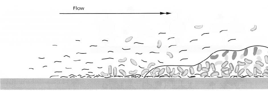
Clean
Molecular
Single
substratum
adsorption
organisms
(Phase 1) (Phase 2)
Multiplication
(
Phase 3)
Sequential adsorption
of organisms
(Phase 4)
Fig. 3-2. Stages in the formation of a biofilm on a clean, hard and non-shedding surface following immersion into a
fluid environment. Phase 1: Molecular adsorption to condition the biofilm formation. Phase 2: Bacterial adhesion
by single organisms. Phase 3: Growth of extracellular matrix production and multiplication of the adhering bacte-
ria. Phase 4: Sequential adsorption of further bacteria to form a more complex and mature biofilm. Adapted from
Marshall (1992).
GENERAL INTRODUCTION TO
PLAQUE FORMATION
Growth and maturation patterns of bacterial plaque
have been studied on natural hard oral surfaces, such
as enamel and dentin, or artificial surfaces, such as
metal or acrylic, using light and electron microscopy
and bacterial culture (Theilade & Theilade 1985). De-
spite differences in surface roughness, free energy and
charge, the most important features of initial plaque
development are similar on all these materials (Sie-
grist et al. 1991).
The ability to adhere to surfaces is a general prop-
erty of almost all bacteria. It depends on an intricate,
sometimes exquisitely specific, series of interactions
between the surface to be colonized, the microbe and
an ambient fluid milieu (Mergenhagen & Rosan 1985).
Immediately upon immersion of a solid substratum
into the fluid media of the oral cavity, or upon cleaning
of a solid surface in the mouth, hydrophobic and
macromolecules begin to adsorb to the surface to form
a conditioning film (Fig. 3-2, Phase 1), termed the
acquired pellicle. This film is composed of a variety of
salivary glycoproteins (mucins) and antibodies. The
conditioning film alters the charge and free energy of
the surface, which in turn increases the efficiency of
bacterial adhesion. Bacteria adhere variably to these
coated surfaces. Some possess specific attachment
structures such as extracellular polymeric substances
and fimbriae, which enable them to attach rapidly
upon contact (Fig. 3-2, Phase 2). Other bacteria require
prolonged exposure to bind firmly. Behaviors of bac-
teria change once they become attached to surfaces.
This includes active cellular growth of previously
starving bacteria and synthesis of new outer mem-
brane components. The bacterial mass increases due
to continued growth of the adhering organisms, adhe-
sion of new bacteria (Fig. 3-2, Phase 4), and synthesis
of extracellular polymers. With increasing thickness,
diffusion into and out of the biofilm becomes more
and more difficult. An oxygen gradient develops as a
result of rapid utilization by the superficial bacterial
layers and poor diffusion of oxygen through the
biofilm matrix. Completely anaerobic conditions
eventually emerge in the deeper layers of the deposits.
Oxygen is an important ecologic determinant because
bacteria vary in their ability to grow and multiply at
different levels of oxygen. Diminishing gradients of
nutrients supplied by the aqueous phase, i.e. the sa-
liva, are also created. Reverse gradients of fermenta-
tion products develop as a result of bacterial metabo-
lism.
Dietary products dissolved in saliva are an impor-
tant source of nutrients for bacteria in the supragingi-
val plaque. Once a deepened periodontal pocket is
formed, however, the nutritional conditions for bacte-
ria change because the penetration of substances dis-
solved in saliva into the pocket is very limited. Within
the deepened pocket, the major nutritional source for
bacterial metabolism comes from the periodontal tis-
sues and blood. Many bacteria found in periodontal
pockets produce hydrolytic enzymes with which they
can break down complex macromolecules from the
host into simple peptides and amino acids. These
enzymes may be a major factor in destructive proc-
esses of periodontal tissues.
Primary colonization is dominated by facultatively
anaerobic Gram-positive cocci. They adsorb onto the
pellicle-coated surfaces within a short time after me-
chanical cleaning. Plaque collected after 24 h consists
mainly of streptococci; S. sanguis is the most promi-
nent of these organisms. In the next phase, Gram-posi-
tive rods, which are present in very low numbers
DENTAL PLAQUE AND CALCULUS • 83
84 • CHAPTER 3
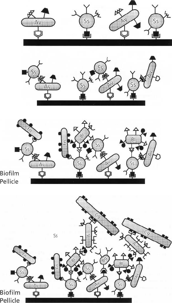
Fig. 3-3. Primary colonization by
predominantly Gram-positive fac-
ultative bacteria. Ss: Streptococcus
sanguis is most dominant. Av: Acti-
uomyces spp. are also found in 24 h
plaque.
Fig. 3-4. Gram-positive facultative
cocci and rods co-aggregate and
multiply.
Biofilm
Pellicle
Biofilm
Pellicle
Fig. 3-5. Surface receptors on the
Gram-positive facultative cocci
and rods allow the subsequent ad-
herence of Gram-negative organ-
isms, which have a poor ability to
directly adhere to the pellicle.
Fn: Fusobacterium nucleatum.
BI: Prevotella intermedia.
Fig. 3-6. The heterogeneity in-
creases as plaque ages and ma-
tures. As a result of ecologic
changes, more Gram-negative
strictly anaerobic bacteria colonize
secondarily and contribute to an
increased pathogenicity of the
biofilm.
initially, gradually increase and eventually outnumber
the streptococci (Fig. 3-3). Gram-positive filaments,
particularly Actinomyces spp., are the predominating
species in this stage of plaque development (Fig. 3-4).
Surface receptors on the deposited Gram-positive
cocci and rods allow subsequent adherence of Gram-
negative organisms with poor ability to attach directly
to pellicle. Veillonella, fusobacteria and other anaerobic
Gram-negative bacteria can attach in this way (Fig. 3-
5). The heterogeneity of plaque thus gradually
increases and, with time, includes large numbers of
Gram-negative organisms. A complex array of
interrelated bacterial species is the result of this
development. Exchange of nutrients between different
species, but also negative interactions, e.g. the
production of bacteriocins, play a role in the establ-
ishment of a stable bacterial community (Fig. 3-6). Due
to the influences of local environmental factors, struc-
turally different types of plaque evolve at different
locations. Protection of the growing plaque from shear
forces and local availability of certain nutrients are
most important. A distinct composition of mature bac-
terial deposits can eventually be recognized at specific
sites and under specific clinical conditions. Examples
are the plaque on smooth enamel surface versus fis-
sure plaque, or the plaque in shallow and less shallow
gingival crevices.
Accumulation of plaque along the gingival margin
leads to an inflammatory reaction of the soft tissues.
The presence of this inflammation has a profound
DENTAL PLAQUE AND CALCULUS • 85
influence on the local ecology The availability of
blood and gingival fluid components promotes
growth of Gram-negative bacterial species with an
increased periodontopathic potential. Bacterial sam-
ples from established gingivitis lesions have increased
numbers of these bacteria. Because of the capability
enzymatically to digest proteins, many of these organ-
isms do not depend upon a direct availability of die-
tary carbohydrates. Such bacteria do not produce ex-
tracellular polymers and develop only loosely adher-
ent plaque in the developing periodontal pocket. Cul-
tivation of samples from advanced periodontal le-
sions reveals a predominance of Gram-negative an-
aerobic rods. Under the microscope, particularly high
numbers of anaerobic uncultivable spirochetes can be
demonstrated. Further details on the microbial ecol-
ogy of subgingival plaque are discussed in Chapter 4.
In summary, immediately following immersion of
hard, non-shedding surfaces into the fluid environ-
ment of the oral cavity, adsorption of macromolecules
will lead to the formation of a biofilrn. Bacterial adhe-
sion to this glycoprotein layer will first involve pri-
mary plaque formers, such as Gram-positive faculta-
tive cocci and rods. Subsequent colonization onto re-
ceptors of these organisms will involve Gram-nega-
tive, strictly anaerobic bacteria, while the primary
plaque formers also multiply to form colonies. The
heterogeneity of the complex biofilm increases with
time, as the ecologic conditions gradually change.
DENTAL PLAQUE AS A BIOFILM
The term biofilrn describes the relatively undefinable
microbial community associated with a tooth surface
or any other hard, non-shedding material (Wilderer &
Charaklis 1989). In the lower levels of most biofilms a
dense layer of microbes is bound together in a poly-
saccharide matrix with other organic and inorganic
materials. On top of this layer is a looser layer, which
is often highly irregular in appearance and may ex-
tend into the surrounding medium. The fluid layer
bordering the biofilm may have a rather "stationary"
sublayer and a fluid layer in motion. Nutrient compo-
nents may penetrate this fluid medium by molecular
diffusion. Steep diffusion gradients, especially for
oxygen, exist in the more compact lower regions of
biofilms. The ubiquity with which anaerobic species
are detected from these areas of biofilms provides
evidence for these gradients (Ritz 1969).
Accumulation of bacteria on solid surfaces is not an
exclusive dental phenomenon. Biofilms are ubiqui-
tous; they form on virtually all surfaces immersed in
natural aqueous environments. Biofilms form particu-
larly fast in flow systems where a regular nutrient
supply is provided to the bacteria. Rapid formation of
visible layers of microorganisms due to extensive bac-
terial growth accompanied by excretion of copious
amounts of extracellular polymers is typical for
biofilms. Biofilms effectively protect bacteria from an
timicrobial agents. Treatment with antimicrobial
sub-stances is often unsuccessful unless the deposits
are mechanically removed. Adhesion-mediated infec-
tions that develop on permanently or temporarily
implanted materials such as intravascular catheters,
vascular prostheses or heart valves are notoriously
resistant to antibiotics and tend to persist until the
device is removed. Similar problems are encountered
in water conduits, wherein potentially pathogenic
bacteria may be protected from chlorination, or on
ship hulls, where biofilms increase frictional resis-
tance and turbulence (Gristina 1987, Marshall 1992).
In summary, dental plaque as a naturally occurring
microbial deposit represents a true biofilrn which con-
sists of bacteria in a matrix composed mainly of ex-
tracellular bacterial polymers and salivary and/or
gingival exudate products.
STRUCTURE OF DENTAL PLAQUE
Supragingival plaque
Supragingival plaque has been examined in a number
of studies by light and electron microscopy to gain
information on its internal structure (Muhlemann &
Schneider 1959, Turesky et al. 1961, Theilade 1964,
Frank & Brendel 1966, Leach & Saxton 1966, Frank &
Houver 1970, Schroeder & De Boever 1970, Theilade
& Theilade 1970, Eastcott & Stallard 1973, Saxton 1973,
Ronstrom et al. 1975, Tinanoff & Gross 1976, Lie 1978).
The introduction of the electron microscope in dental
research was a significant development for studies of
dental plaque, both because the size of many bacteria
approaches the ultimate resolving power of the light
microscope, and because the resins used for embed-
ding allowed for sections thinner than the smallest
bacterial dimension. Hereby the substructure of
plaque could be identified.
In studies of the internal details of plaque, samples
are required in which the deposits are kept in their
original relation to the surface on which they have
formed. This may be accomplished by removing the
deposits with the tooth. If plaque of known age is the
object of study, the tooth surfaces are cleaned at a
predetermined time before removal (McDouga111963,
Frank & Houver 1970, Schroeder & De Boever 1970).
Pieces of natural teeth or artificial surfaces may also
be attached to solid structures in the mouth and re-
moved after a given interval. This method of plaque
collection was already used at the beginning of the last
century by Black (1911). The systematic use of artificial
surfaces for collection of plaque was reintroduced
during the 1950s. Thin plastic foils of Mylar
®
were
attached to mandibular incisor teeth for known peri-
ods, after which they were removed for histologic,
histochemical and electron microscopic examination
of the deposited material (Mandel et al. 1957, Muhle-
86 • CHAPTER 3
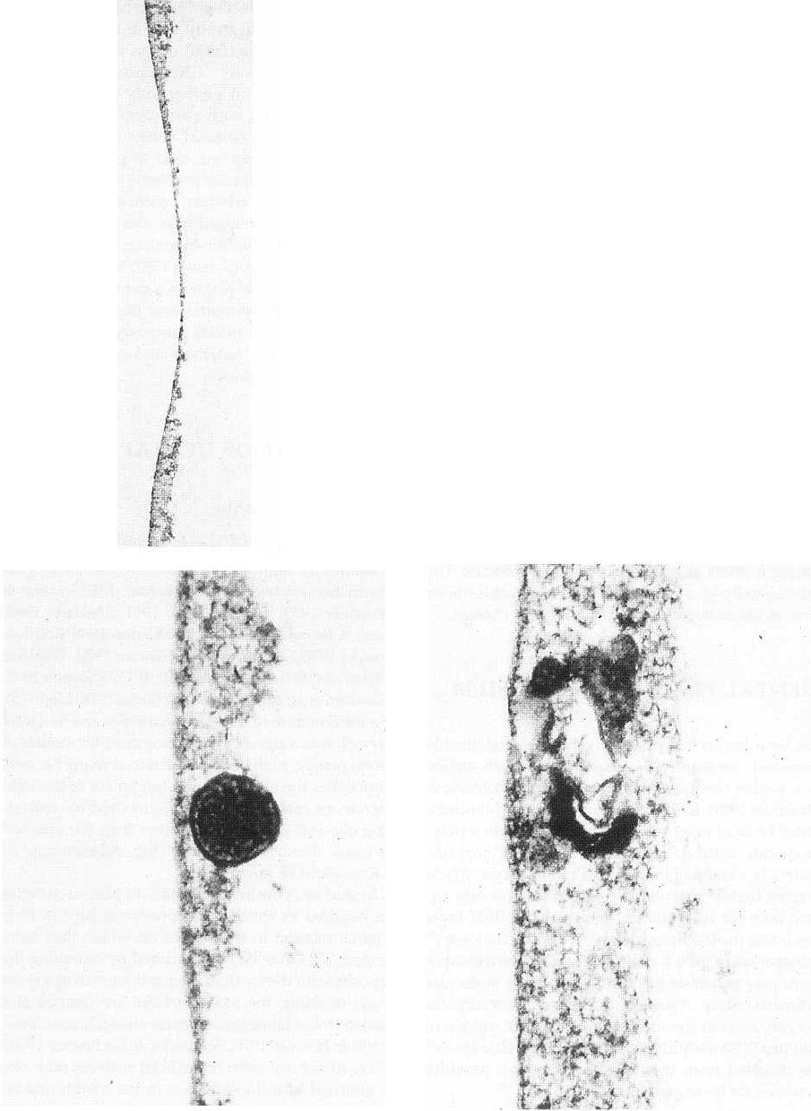
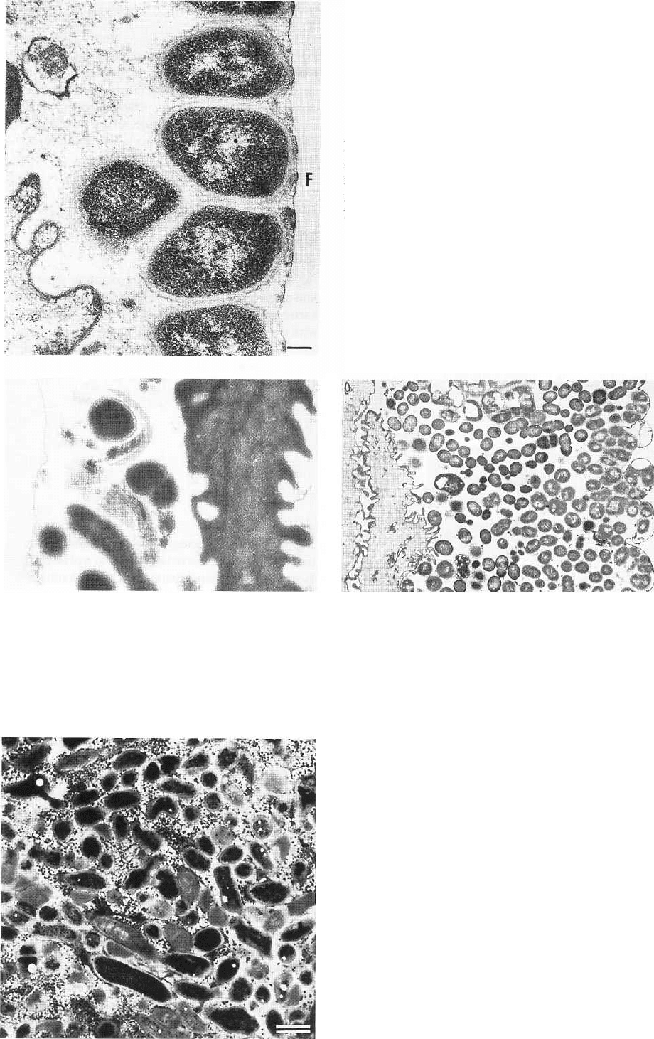
Fig. 3-8. Electron micrographic illustration of a 4-h den-
tal pellicle with a single bacterium included in the film.
The microbe appears attached to the surface. The den-
tal pellicle varies in thickness but has a homogeneous
morphology From Brecx et al. (1981).
mann & Schneider 1959, Zander et al. 1960, Schroeder
1963, Theilade 1964). Other types of plastic materials
Fig. 3-7. Electron micrographic illustration of a 4-h den-
tal pellicle. The pellicle has formed on an artificial sur-
face of plastic, which was painted on to the surface of
the tooth. The plastic surface was exposed to the envi-
ronment for a 4-h period. A thin condensed layer of or-
ganic material is covering the film. The material has a
relatively homogeneous appearance but varies in thick-
ness over the surface. From Brecx et al. (1981).
Fig. 3-9. Electron micrographic illustration of a 4-h den-
tal pellicle, formed on a plastic surface attached to the
buccal surface of a tooth. A condensed layer of organic
material is observed on the surface and cell remnants
are embedded in the film. From Brecx et al. (1981).
such as Westopal
®
, Epon
®
, Araldite
®
, and spray plast
have since been employed for this purpose (Berthold
DENTAL PLAQUE AND CALCULUS • 87
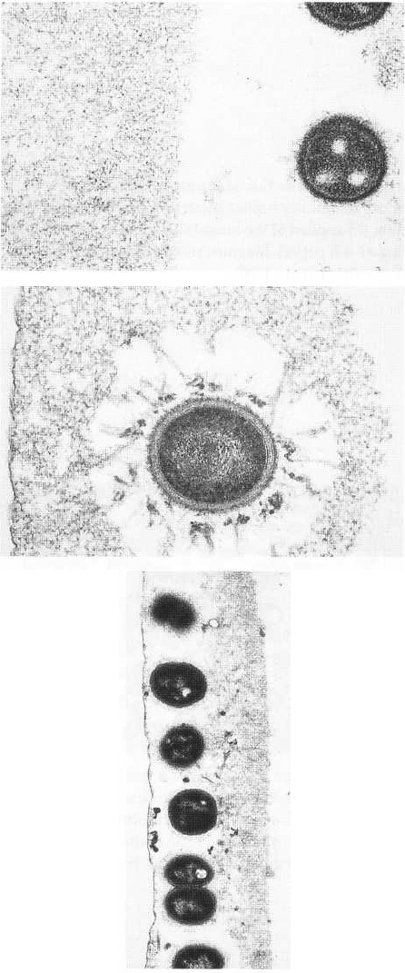
Fig. 3-10. High power electron micrographic illustra-
tion of a 4-h pellicle with bacteria residing in the pelli-
cle at a distance of around one micron from the con-
densed organic material. The pellicle is rather even in
composition and, at the oral side, an irregular con-
densed organic material is seen close to the bacteria.
From Brecx et al. (1981).
et al. 1971, Kandarkar 1973, Lie 1975, Listgarten et al.
1975, Rdnstrom et al. 1975). Results from several such
studies indicate that plaque formed on natural or
artificial surfaces does not differ significantly in struc-
ture or microbiology (Hazen 1960, Berthold et al. 1971,
Nyvad et al. 1982, Theilade et al. 1982a, b), indicating
that at least some of the principal mechanisms in-
volved in plaque formation are unrelated to the nature
of the solid surface colonized. However, there are
small, but important, differences in the chemical com-
position of the first layer of organic material formed
on these artificial surfaces compared with that formed
on natural tooth surfaces (Sonju & R611a 1973, Sonju
& Glantz 1975, Oste et a1.1981). Tooth surfaces, enamel
as well as exposed cementum, are normally covered by
a thin acquired pellicle of glycoproteins (Fig. 3-7). If
removed, e.g. by mechanical instrumentation, it
reforms within minutes. The pellicle is believed to
play an active part in the selective adherence of bacte-
ria to the tooth surface (Fig. 3-8). For details of the
proposed mechanisms, see Chapter 4.
The first cellular material adhering to the pellicle on
the tooth surface or other solid surfaces consists of
coccoid bacteria with numbers of epithelial cells and
polymorphonuclear leukocytes (Fig. 3-9). The bacteria
are encountered either on (Fig. 3-10) or within the
pellicle as single organisms (Fig. 3-11) or as aggregates
of microorganisms (Fig. 3-12). Larger numbers of mi-
Fig. 3-11. High power electron micrographic illustra-
tion of a 4-h pellicle with an embedded bacterium. Th(
bacterium is deposited on the film surface together
with the dental pellicle. Around the bacterium empty
;paces are observed representing the radius of extru-
sions of filaments radiating from the microorganisms.
From Brecx et al. (1981).
Fig. 3-12. Electron micrographic illustration of a 4-h
dental pellicle with bacteria attached to a plastic sur-
face, which had been adhering to a buccal tooth sur-
face and was exposed to the oral environment. A singl
row of bacteria attached to the surface is seen to the
left. On top of the bacteria, a layer of condensed or-
ganic material representing the oral lateral portion of
+ho rlortfnl oollirlo is nn+orl From Rrory o+ a1 (1QR11
88 • CHAPTER 3
Fig. 3-13. Thin section of plaque colony consisting of
morphologically similar bacteria deposited on plastic
film (F) applied to the buccal surface of a premolar dur-
ing an 8-h period. Magnification x 35 000. Bar: 0.2 µm.
From Brecx et al. (1980).
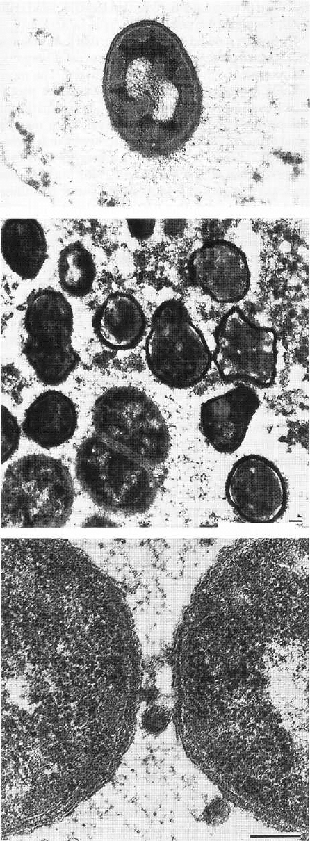
Fig. 3-14. Electron micrographic illustration of early
plaque formation. The film surface on which the pelli-
cle and bacteria adhere is located to the left. Bacteria of
varying morphology are attached to the film. They are
surrounded by organic pellicle material. An epithelial
cell remnant is seen in close vicinity to the microbes.
From Brecx et al. (1981).
Fig. 3-15. Electron micrographic illustration of 24-h den-
tal plaque formed on a plastic film surface attached to the
buccal surface of the tooth. A multilayer bacterial plaque
is noted. A remnant of an epithelial cell has been trapped
in the microbial mass. From Brecx et al. (1981).
Fig. 3-16. Thin section of old plaque stained for the
demonstration of polysaccharides by reacting them
with electron-dense material appearing dark in the il-
lustration. Many bacteria contain large amounts of in-
tracellular polysaccharide, and the intermicrobial ma-
trix contains extracellular polysaccharides. Magnifica-
tion x 7000. Bar: 1µm. From Theilade & Theilade (
1970).
DENTAL PLAQUE AND CALCULUS • 89
Fig. 3-17. High power electron micrographic illustra-
tion of a single bacterium attached to the pellicle by
filaments which extend from the bacterial surface to
the tooth surface. The surface had been exposed to the
oral environment for an 8-h period. From Brecx et al.
(1981)
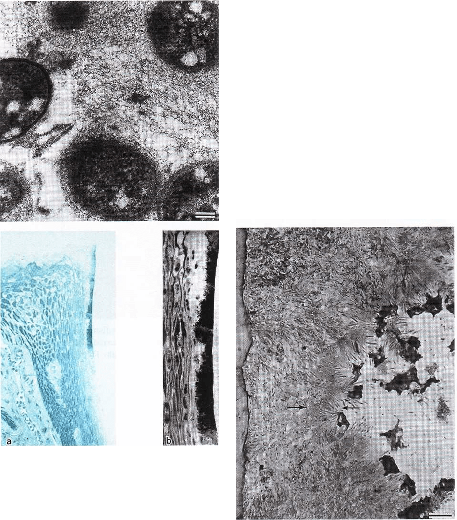
Fig. 3-19. Thin section of plaque with a region predomi-
nated by Gram-negative bacteria. Between them, ves-
icles are surrounded by a trilaminar membrane (two
thin electron-dense layers with an electron-lucent layer
in between). This substructure is also seen in the outer-
most endotoxin containing cell wall layer of the adja-
cent Gram-negative bacteria. Magnification x 110 000.
Bar: 0.1 µm. From Theilade & Theilade (1970).
croorganisms may be carried to the tooth surface by
epithelial cells.
The number of bacteria found on the surface a few
hours after cleaning depends on the procedures applied
to the sample before examination, the reason being that
adherence to the solid surface is initially very weak. If
no special precautions are taken during the preparatory
processing, the early deposits are easily lost (Brecx et
al. 1980). Apparently the adherence
of microorganisms to solid surfaces takes place in two
steps:
1. a reversible state in which the bacteria adhere
loosely, and later
2. an irreversible state, during which their adherence
becomes consolidated (Gibbons & van Houte 1980).
Fig. 3-18. Thin section of plaque with granular or ho-
mogeneous intermicrobial matrix. Magnification x
20 000. Bar: 0.1 µm. From Theilade & Theilade (1970).
90 • CHAPTER 3
Another factor which may modify the number of bac-
teria in early plaque deposits is the presence of gingi-
vitis, which increases the plaque formation rate so that
the more complex bacterial composition is attained
earlier (Saxton 1973, Hillam & Hull 1977, Brecx et al.
1980). Plaque growth may also be initiated by micro-
organisms harbored in minute irregularities in which
they are protected from the natural cleaning of the
tooth surface.
During the first few hours, bacteria that resist de-
tachment from the pellicle may start to proliferate and
form small colonies of morphologically similar organ-
isms (Fig. 3-13). However, since other types of organ-
isms may also proliferate in an adjacent region, the
pellicle becomes easily populated by a mixture of
different microorganisms (Fig. 3-14). In addition,
some organisms seem able to grow between already
established colonies (Fig. 3-15). Finally, it is likely that
clumps of organisms of different species will become
attached to the tooth surface or to the already attached
microorganism, contributing to the complexity of the
plaque composition after a few days. At this time,
different types of organisms may benefit from each
other. One example is the corncob configurations re-
sulting from the growth of cocci on the surface of a
filamentous microorganism (Listgarten et al. 1973).
Another feature of older plaque is the presence of dead
and lysed bacteria which may provide additional nu-
trients to the still viable bacteria in the neighborhood
(Theilade & Theilade 1970).
The material present between the bacteria in dental
plaque is called the intermicrobial matrix and ac-
counts for approximately 25% of the plaque volume.
Three sources may contribute to the intermicrobial
matrix: the plaque microorganisms, the saliva, and the
gingival exudate.
The bacteria may release various metabolic prod-
ucts. Some bacteria may produce various extracellular
carbohydrate polymers, serving as energy storage or
as anchoring material to secure their retention in
plaque (Fig. 3-16). Degenerating or dead bacteria may
also contribute to the intermicrobial matrix. Different
bacterial species often have distinctly different meta-
bolic pathways and capacity to synthesize extracellu-
lar material. The intermicrobial matrix in plaque,
therefore, varies considerably from region to region.
A fibrillar component is often seen in the matrix be-
tween Gram-positive cocci (Fig. 3-17) and is in accord
ance with the fact that several oral streptococci syn-
thesize levans and glucans from dietary sucrose. In
other regions, the matrix appears granular or homo-
geneous (Fig. 3-18). In parts of the plaque with the
presence of Gram-negative organisms, the intermicro-
bial matrix is regularly characterized by the presence
of small vesicles surrounded by a trilaminar mem-
brane, which is similar in structure to that of the outer
envelope of the cell wall of the Gram-negative micro-
organisms (Fig. 3-19). Such vesicles probably contain
endotoxins and proteolytic enzymes, and may also be
involved in adherence between bacteria (Hofstad et al.
1972, Grenier & Mayrand 1987).
It must be remembered, however, that the transmis-
sion electron microscope does not reveal all organic
components of the intermicrobial matrix. The more
soluble constituents may be lost during the proce-
dures required prior to sectioning and examination of
the plaque sample. Biochemical techniques may be
used to identify such compounds (Silverman & Klein-
berg 1967, Krebel et al. 1969, Kleinberg 1970, Hotz et
al. 1972, Rolla et al. 1975, Bowen 1976). Such studies
indicate that proteins and carbohydrates constitute the
bulk of the organic material while lipids appear in
much lower amounts.
The carbohydrates of the matrix have received a
great deal of attention, and at least some of the poly-
saccharides in the plaque matrix are well charac-
terized: fructans (levans) and glucans. Fructans are
synthesized in plaque from dietary sucrose and pro-
vide a storage of energy which may be utilized by
microorganisms in time of low sugar supply. The
glucans are also synthesized from sucrose. One type
of glucan is dextran, which may also serve as energy
storage. Another glucan is mutan, which is not readily
degraded, but acts primarily as a skeleton in the ma-
trix in much the same way as collagen stabilizes the
intercellular substance of connective tissue. It has been
suggested that such carbohydrate polymers may be
responsible for the change from a reversible to an
irreversible adherence of plaque bacteria.
The small amount of lipids in the plaque matrix are
as yet largely uncharacterized. Part of the lipid content
is found in the small extracellular vesicles, which may
contain lipopolysaccharide endotoxins of Gram-nega-
tive bacteria.
Subgingival plaque
Owing to the difficulty of obtaining samples with
subgingival plaque preserved in its original position
between the soft tissues of the gingiva and the hard
tissues of the tooth, there are only a limited number of
studies on the detailed internal structure of human
subgingival plaque (Schroeder 1970, Listgarten et al.
1975, Listgarten 1976, Westergaard et al. 1978). From
these it is evident that in many respects subgingival
plaque resembles the supragingival variety, although
the predominant types of microorganisms found vary
considerably from those residing coronal to the gingi-
val margin.
Between subgingival plaque and the tooth an elec-
tron-dense organic material is interposed, termed a
cuticle (Fig. 3-20). This cuticle probably contains the
remains of the epithelial attachment lamina originally
connecting the junctional epithelium to the tooth, with
the addition of material deposited from the gingival
exudate (Frank & Cimasoni 1970, Lie & Selvig 1975,
Eide et al. 1983). It has also been suggested that the
cuticle represents a secretory product of the adjacent
DENTAL PLAQUE AND CALCULUS • 91
Fig. 3-20. Semithin section of subgingival plaque. An
electron-dense cuticle bordering the enamel space is
visible to the left. Filamentous bacteria are less than in
supragingival plaque. The surface toward the gingival
tissue contains many spirochetes (between arrows).
Various host tissue cells can be seen on the right side.
Magnification x 775. Bar: 10 µm. From Listgarten (
1976).
Fig. 3-21. (a) Light microscopic image of the
dentogingi- val region of a dog with experimental
gingivitis. A thin layer of dentogingival plaque can be
seen, extending from the supragingival region
approximately
1
/2 mm into the gingival sulcus. (b)
Higher magnification of a region of the plaque shown
in (a). The subgingival plaque has a varying thickness
and the epithelial cells are separated from the surface
by a layer of leukocytes. There are also numerous
leukocytes in the superficial portion of the sulcus
epithelium. The apical termina- tion of the plaque is
bordered by leukocytes separating the epithelium from
direct contact with the plaque bac- teria.
epithelial cells (Schroeder & Listgarten 1977). Infor-
mation is lacking concerning its chemical composi-
tion, but its location in the subgingival area makes it
unlikely that salivary constituents contribute to its
formation.
The subgingival plaque structurally resembles su-
pragingival plaque, particularly with respect to
plaque associated with gingivitis without the forma-
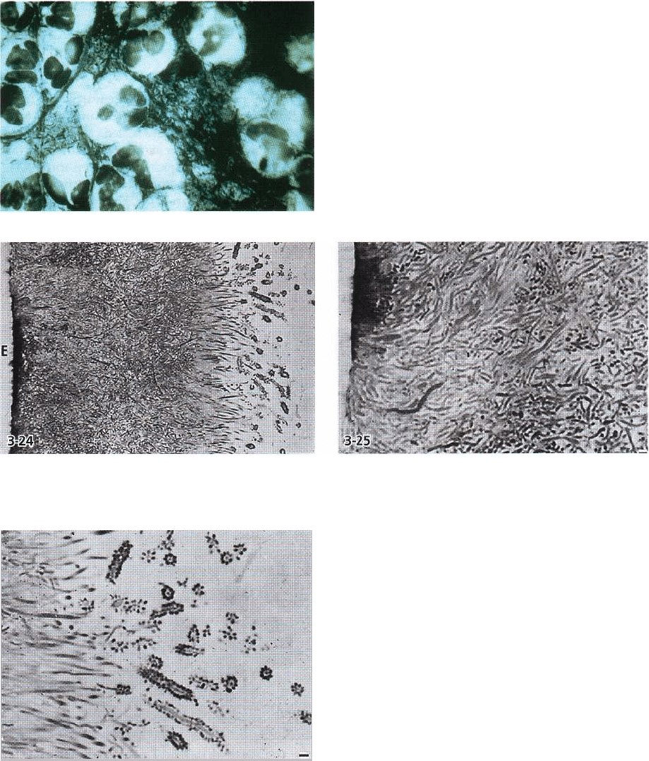
Fig. 3-22. Semithin section of supragingival plaque
with layer of predominantly filamentous bacteria ad-
hering to the enamel (to the left). Lighter staining indi-
cates calcification of part of the plaque close to the
tooth. Magnification x 750. Bar: 10 µm. From Listgarten
(1976).
tion of deep pockets (Fig. 3-21a). A densely packed
accumulation of microorganisms is seen adjacent to the
cuticular material covering the tooth surface (Fig. 3-22).
The bacteria comprise Gram-positive and Gram-
negative cocci, rods and filamentous organ-isms.
Spirochetes and various flagellated bacteria may also be
encountered, especially at the apical extension of the
plaque. The surface layer is often less densely
92 • CHAPTER 3
Fig. 3-23. Light microscopic image of a smear sample
taken from the dentogingival region in a subject who
had abstained from mechanical oral hygiene during 3
weeks. Numerous leukocytes can be observed embed-
ded in a dense accumulation of bacteria.
Fig. 3-24, 3-25. Semithin section of supragingival plaque on enamel (E), which has been dissolved prior to section
ing. Filamentous organisms predominate. At the surface some of these organisms are surrounded by cocci. This
configuration resembles a corncob. Magnification x 750 and x 1400. Bars: 10 µm and 1 µm. From Listgarten (1976).
Fig. 3-26. The corncob formations seen at the plaque
surface in Fig. 3-24 and 3-25. Magnification x 1300. Bar:
1 µm. From Listgarten (1976).
packed and leukocytes are regularly interposed be-
tween the plaque and the epithelial lining of the gin-
gival sulcus (Fig. 3-23).
When a periodontal pocket has formed, the appear-
ance of the subgingival bacterial deposit becomes
much more complex. In this case the tooth surface may
either represent enamel or cementum from which the
periodontal fibers are detached. Plaque accumulation
on the portion of the tooth previously covered by
periodontal tissues does not differ markedly from that
observed in gingivitis (Fig. 3-24). In this layer, filamen-
tous microorganisms dominate (Figs. 3-25, 3-26, 3-27),
but cocci and rods also occur. However, in the deeper
parts of the periodontal pocket, the filamentous or-
ganisms become fewer in number, and in the apical
portion they seem to be virtually absent. Instead, the
dense, tooth-facing part of the bacterial deposit is
dominated by smaller organisms without particular
orientation (Listgarten 1976) (Fig. 3-28).
The surface layers of microorganisms in the peri-
odontal pocket facing the soft tissue are distinctly
different from the adherent layer along the tooth sur-
face, and no definite intermicrobial matrix is apparent
(Figs. 3-28, 3-29). The microorganisms comprise a
larger number of spirochetes and flagellated bacteria.
Gram-negative cocci and rods are also present. The
multitude of spirochetes and flagellated organisms are
motile bacteria and there is no intermicrobial ma-
