Hoque. Advanced Applications of Rapid Prototyping Technology in Modern Engineering
Подождите немного. Документ загружается.


Rapid Prototyping in Correction of Craniofacial Skeletal Deformities
121
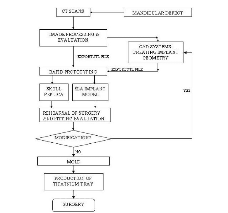
Fig. 2. Computer-aided geometric modeling for the manufacturing of custom implants
1.1 CT data acquisition
CT data acquisition was performed by the spiral volumetric technique (Picker MX8000).
Suitable CT parameters for data acquisition were as follows: zero degree gantry, a resolution
of 512 512 pixel reconstruction matrix image, 1.3 mm slice thickness and a slice
reconstruction interval of 0.6mm.
The scan data are recorded according to the DICOM Norm (a standard of data formatting
and of communication used in medical imagery).
1.2 Medical image processing-segmentation and 3D reconstruction
The 2D image slices from the CT scans were imported into the Materialise’s Interactive
Medical Imaging Control System (Mimics). A thresholding and region growing technique
were used to extract the contour of the skeleton from the CT data. After removing the soft
tissue, a 3-D region-growing technique is then used to isolate the skeletal part of the head
from the CT dataset.

Advanced Applications of Rapid Prototyping Technology in Modern Engineering
122
A 3D image is reconstructed and visualized. The skull data was converted to a mesh based
surface representation (STL) format and was then download to an RP machine to fabricate
the skull replica.
1.3 Design of the custom titanium implant
Customized bone grafting trays were designed using Geomagics studio, version 6.0
(Raindrop Geomagic, Research Triangle Park, NC). Different techniques are applied for
individual cases, including mirroring the non-defect side, implant design from other skull
CT data and geometry modeling. The designs of different implant are to be elaborated in
each case report.
1.4 Rapid prototyping
The CAD model of the skeleton structure and the bone grafting tray were then transferred in
a stereolithography (STL) format, and input to a laser stereolithographic rapid prototyping
system, LPS 600, to manufacture the skull models and customized implants. The model was
sliced into 0.1 mm layer thickness, and then processed through a layer by layer building
process. A physical resin model was thus obtained.
1.5 Rehearsal of surgery and implant fitting evaluation
The RP model makes a clear view about the defect and allow for a surgeon to gain operative
experience and get a clear view of the specific demands required for such an operation.
Preoperative rehearsal of surgery via fitting the physical model of the custom tray with the
patient’s skull replica facilitates the optimal placement for the prosthesis onto the residual
mandible; thereby evaluate the quality of the custom tray. This could reduce the operation
time, and allow for modification of the surgical plans and the implants.
1.6 Production of titanium implant
To obtain a biocompatible titanium tray, the prototyped resin model of the tray was
embedded with a high temperature resistant phosphate investment material. After
successive drying and dipping, the resin model was burn out in an oven, at a temperature of
300-600°C. This led to a casting mold and a titanium tray was cast using this model. The
titanium tray was then subject to post processing: trimming, sandblasting and drilling,
among others.
2. Clinical applications
2.1 Case study: Unilateral mandible defect (fig. 3)
A 24-year-old man with an adamantoma on the left mandibular angle and ramus was
admitted. The surgical plan was to make a block resection treatment to cure the tumor, and
to repair the defect with a rapid prototyped tray.
The CT data of the patient’s skull was acquired and the computer assisted design of the tray
was based on the mirror imaging technique.
Since the defect only involved the left side of the mandible. It was decided to mirror the
undamaged right side onto the left side. To mirror the non-affected side image, a reference
plan is needed. Usually, the center-plane can be established by the landmark of the anatomy
structure, such as maxillary and mandibular adjacent point of central incisors, nasion, nasal

Rapid Prototyping in Correction of Craniofacial Skeletal Deformities
123
septum, the central point of the sella turcica, etc (Zhou et al., 2010). It can also be established
based on the midpoints of the symmetric landmarks, such as the orbital cavity, the condyles,
the temporomandibular joint glenoid fossa, opposite teeth, etc. Generally, cranium and
maxillas and zygomas are more stable than madible, since the latter will displace easily
after unilateral bone resection or overgrowth in one side. Sometimes, to identify
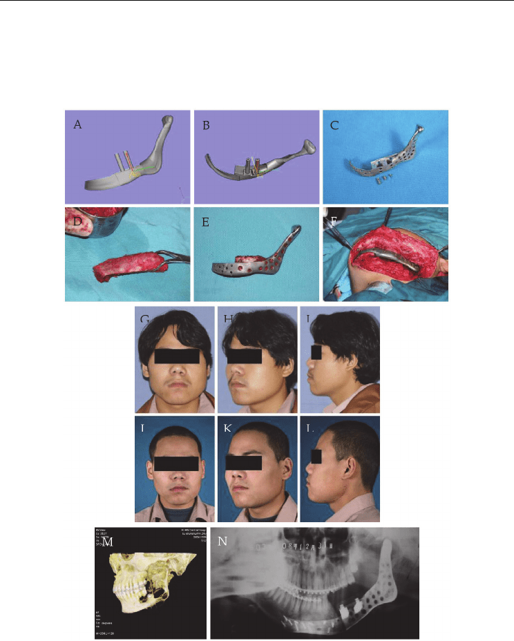
Fig. 3. (A&B) the design of the tray. (C) the titanium tray. (D-F) intraoperative photos, D,
shaping the iliac bone graft, E, marrow-cancellous bone grafts packed in the tray and bone
blocks covered the tray, F, the tray-bone graft complex was fixed onto the mandible to
restore the defect. (G-L) pre- and post-operative facial appearance of the patient. (M)
preoperative 3D view of the skull, tumor on the left mandible. (N) postoperative X-ray view
of the reconstructed mandible.

Advanced Applications of Rapid Prototyping Technology in Modern Engineering
124
the mirror plane is a great challenge. For the deformity only affect a part of the facial or
cranial bone, the unaffected normal part can be used to establish and adjust for the mirror
plane.
A suitable mirror plane could be obtained by trial and error. A reference plane that allows
maximum overlap between the mirrored image and the native image of the normal part of
the skull after mirroring can be considered to be the mirror plane.
After mirroring, the mirrored symmetric structure is considered to be the target contour to
be restored. Then the mirror image was used to design the implant geometry. And
footplates were designed based on the residual mandible ends.
The implant was manufactured by RP process. The SLA model was used to cast the titanium
tray. And the implant was sterilized and prepared for the surgery.
The reconstructive surgery was performed via an extraoral approach. The residual mandible
end was exposed and the bone bed was prepared, the tray was fitted onto the mandible and
highly accurate match was observed. Autologous ilium was harvested from the anterior iliac
crest. Crushed bone marrow-cancellous bone particles were densely packed into the tray and a
cortical-cancellous bone block was placed on the top to cover entire tray. The cortical bone was
drilled and secured on the top of the tray by two dental fixture implants. The tray-bone graft
complex was then fixed onto the mandible with titanium screws. The wound was then closed.
Satisfactory facial appearance and normal occlusion were restored. Over denture was made
to rehabilitate the occlusion.
2.2 Case study: Unilateral cranium defect (fig. 4)
A patient with a huge unilateral cranium defect, involving the left parietal, temporal, frontal
and sphenoid bone, due to traffic accident trauma was admitted for reconstruction.
Using the same mirroring technique, cranium prosthesis was designed and manufactured,
to protect the intracranial contents. An implantation surgery was performed, with taking
good care of the brain. Using the prototyped prosthesis, symmetric cranium was restored,
via a straight approach. The general appearance and radiologic picture demonstrated the
symmetry.
In conclusion, the computer assisted design and rapid prototyping technique facilitate the
reconstructive surgery. By applying the mirroring method, excellent symmetry can be
restored for the asymmetric skeletal defect.
2.3 Case study: Mandibular retraction (fig. 5)
A 28-year-old woman with a mandibular retraction needed chin augmentation. A chin
augmentation of 6 mm was predicted by cephalometric analysis. And an individual
prosthesis was designed and manufactured.
By using the same technology in case study one, a 3D reconstructed CT data was generated
(MIMICS). The defect couldn’t be reconstructed by mirror imaging technique. The patient’s
3D mandible CT data was measured and these measurements data were used to select a
similar mandible. A skull model of a healthy woman with normal mandible contour was
selected and used to design the implant geometry.
The CAD design of the implant was based on the normal mandible data used as a template
to create an anatomically correct mandibular contour. The inner surface of the implant was
based on the anatomic structure of the chin surface, which allow for an easy placement of
the implant onto the chin.

Rapid Prototyping in Correction of Craniofacial Skeletal Deformities
125
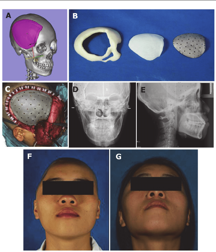
Fig. 4. (A) the design of the cranium prosthesis. (B) the resin model of the cranium and
prosthesis, and the titanium prosthesis. (C) intraoperative photo: the fixation of the
prosthesis to the cranium to repair the defect. (D&E) postoperative X-ray view of the skull.,
F, expose the chin, G, place the prosthesis. (F&G) pre- and post-operative facial appearance
of the patient.
A three-dimensional model was manufactured by using a rapid prototyping machine and
the prototype was used to cast the titanium implant. The prosthesis manufactured using
rapid prototyping technology resulted in simple surgical implantation and better facial

Advanced Applications of Rapid Prototyping Technology in Modern Engineering
126
contour. This technique can also be used to reconstruct segmental defect in the chin region
and to cure hemifacial mocrosomia.
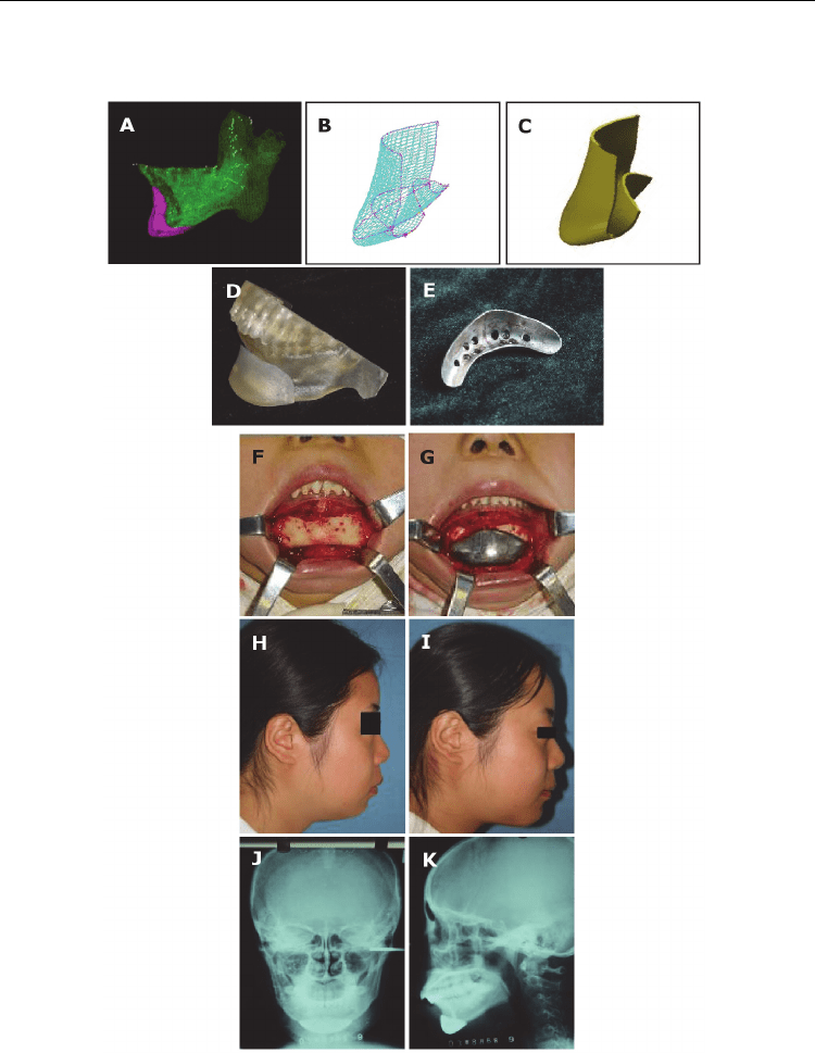
Fig. 5. (A-C) the design of the chin prosthesis. (D) resin model of the prosthesis. (E) the
titanium chin prosthesis. (F&G) intraoperative photos, F, expose the chin, G, place the
prosthesis. (H&I) pre- and post-operative facial appearance of the patient. (J&K)
postoperative X-ray view of the reconstructed mandible.

Rapid Prototyping in Correction of Craniofacial Skeletal Deformities
127
3. Results
We compared the skull model and the CT scan data and found that the physical model’s
dimension was in agreement with the CT scan data and the error was less than 0.3%.
The prototyped models, the skull and the implant, were used to evaluate the design and the
surgical planning. It was found that the physical tray made from virtual data, fitted
perfectly with the exact replica of the patient's skull anatomy. Furthermore, an adequate
symmetry of the jaw was obtained. The surgical planning was accurate and was facilitated
by the RP model.
The custom titanium implants were well fitted in patients. In all cases, the implants were
just inserted and fixed by screws, so that the duration of the surgery was reduced with the
aid of the customized implants.
No complications were observed except that the cancellous bone packed in the grafting tray
was absorbed after a period of time as shows in fig.3N.
4. Conclusions
We introduced the technology of manufacturing individual reconstructive prosthesis for
craniofacial bone defects. This technology involves implant shape design in CAD
environment from CT data, fabrication of the physical model by rapid prototyping process,
creating the mold from the prototype, and then cast of the titanium implant. Clinical studies
demonstrated that this new method can create accurate implant for bone various defects.
We conclude that, with the development of the relative techniques of RP, perfect individual
implants can be manufactured. Also the RP technique facilitates the reconstruction surgery
and makes it more controllable and accurate. Satisfactory aesthetics and functional
rehabilitation of craniofacial deformities can be achieved, that otherwise would remain
difficult.
5. Acknowledgment
We appreciate the assistance by the Institute of Advanced Manufacturing Technology, Xi'an
Jiaotong University, in designing and manufacturing the individual implants.
6. References
Mehta RP. & Deschler DG. (2004). Mandibular reconstruction in 2004: an analysis of
different techniques. Current Opinion in Otolaryngology & Head and Neck Surgery, Vol
12, No.4, (August 2004), pp. 288-2893, ISSN 1068-9508
Boyne PJ. (1973). Methods of osseous reconstruction of the mandible following surgical
resection. Journal of Biomedical Materials Research, Vol 7, No.195, (1973), pp. 195-204,
ISSN 1549-3296
Tideman H., Samman N. & Cheung LK. (1998). Functional reconstruction of the mandible: a
modified titanium mesh system. International Journal of Oral and Maxillofacial
Surgery, Vol.27, No.5, (October 1998), pp. 339-345, ISSN 1399-0020
Samman N., Luk W., Chow T., Cheung L., Tideman H. & Clark R. (1999). Custom-made
titanium mandibular reconstruction tray. Australian Dental Journal, Vol.44, No.3,
(September 1999), pp. 195-199, ISSN 0045-0421

Advanced Applications of Rapid Prototyping Technology in Modern Engineering
128
Eufinger H., Wehmöller M. & Machtens E. (1997). Individual prostheses and resection
templates for mandibular resection and reconstruction. British Journal of Oral and
Maxillofacial Surgery, Vol.35, No.6, (December 1997), pp. 413-418, ISSN 0266-4356
Stojadinovic S., Eufinger H., Wehmöller M. & Machtens E. (1999). One-step resection and
reconstruction of the mandible using computer-aided techniques--experimental
and clinical results. Mund Kiefer Gesichtschir, Vol.3, (May 1999), pp. S151-153, ISSN
1432-9417
Singare S., Dichen L., Bingheng L., Yanpu L., Zhenyu G. & Yaxiong L. (1997). Design and
fabrication of custom mandible titanium tray based on rapid prototyping. Medical
Engineering & Physics, Vol.26, No.8, (October 2004), pp. 671-676, ISSN 1350-4533
Zhou L., Zhao J., Shang H., Liu W., Feng Z., Liu G., Wang J. & Liu Y. (2011). Reconstruction
of Mandibular Defects Using a Custom Made Titanium Tray in Combination with
Autologous Cancellous Bone. Journal of Oral and Maxillofacial Surgery, Vol.69, No.5,
(May 2011), pp. 1508-1518, ISSN 0278-2391
Zhou L., Shang H., He L., Bo B., Liu G., Liu Y., & Zhao J. (2010). Accurate reconstruction of
discontinuous mandible using a reverse engineering/computer-aided
design/rapid prototyping technique: a preliminary clinical study. Journal of Oral
and Maxillofacial Surgery, Vol.68, No.9, (September 2010), pp. 2115-2121, ISSN
0278-2391
9
Application of a Novel Patient - Specific Rapid
Prototyping Template in Orthopedics Surgery
Sheng Lu, Yong-qing Xu and Yuan-zhi Zhang
Department of Orthopedics, Kunming general hospital,
Chengdu military district, PLA, Kunming,
China
1. Introduction
Conventional surgical handwork requires competences such as dexterity or fine motor
skills, which are complemented by visual and tactile feedback. Computer-assisted
orthopaedic surgery aims at improving the perception that a surgeon has of the surgical
field and the operative manipulation. Bony manipulation such as drilling, chiseling, or
sawing can be performed more accurately and implants can be placed more exactly. This
reduces the risk of harming the patient intra-operatively by damaging sensitive structures.
CT scans are very suitable for surgical navigation, especially in orthopaedics. The bones can
be easily distinguished from any other tissue, and can be easily segmented out. The bones
are also the least deformable parts of the body, and therefore the most stable references for
navigation, making it possible for different phases of surgical planning and execution to be
performed well after the patient imaging. Pre-operative planning is typically done in three
orthogonal cross-sectional views made through the CT scan volume.
2. Application of a novel patient - specific rapid prototyping template in
orthopedics surgery
The rapid prototyping template first apply in the hip and knee athroplasty and then apply
in the spine surgery. But the limitation of template design and produce technique, the
authors introduced and validated a novel rapid prototyping templates in the clinical setting.
Report on their experience with spinal pedicle screw placement, [1,2] placement of C2
laminar screws, [3] accurate prosthesis implantation in hip resurfacing arthroplasty, etc
using a novel computer- assisted drill guide template.
2.1 A novel computer- assisted rapid prototyping drill guide template for spinal
pedicle screw placement
Spinal Pedicle screw fixation systems provide three-dimensional (3D) fixation in the spine.
Compared with conventional hook instrumentation, the clinical advantages of such systems
include enhanced correction and stabilization of various deformities, shorter fusion length,
more solid and reliable fixation, and no encroachment into the spinal canal. Therefore,
pedicle screw fixation systems have gained popularity for internal fixation of fractures,
tumors, and deformities of the spine . In spinal pedicle screw insertion, it is important both

Advanced Applications of Rapid Prototyping Technology in Modern Engineering
130
to select the correct size of screw and to place it properly within the pedicle to ensure good
anchoring. Manual placement has a high associated rate of unplanned perforation, which is
the major specific complication of pedicle screw placement and causes a high risk of bone
weakening or lesions of the spinal cord, nerve roots, or blood vessels.
Successful placement of pedicle screws in the cervical spine requires a thorough three-
dimensional understanding of the pedicle morphology in order to accurately identify the
ideal screw axis. Several methods have been explored for precise cervical pedicle screw
placement including anatomic studies, image-guided techniques, computer-assisted surgery
system, and drill templates. These techniques can be broadly classified into five types: (1)
techniques relying on anatomical landmarks and averaged angular dimensions; (2)
techniques with direct exposure of the pedicle, e.g. by laminaminotomy; (3) CT-based
computer assisted surgery (CAS), and (4) fluoroscopy-based CAS techniques. (5) Drill
template techniques.
The principle of image guidance is to register the patient’s pre-operative computed
tomography (CT) scans, thus permitting the surgeon to navigate simultaneously within the
patient and the CT scan volume. Such navigation systems have shown good clinical results.
There are, however, several disadvantages associated with navigation systems. In cases
where screws are to be placed in more than one vertebra, it is necessary to perform a
separate registration step for each vertebra. Intraoperative registration of bone structures
takes up to several minutes, and thus the time taken for the overall procedure is increased
compared with a conventional approach. The navigation equipment often requires
additional personnel to be present during surgery, and this, together with the increased
operating time, leads to a higher risk of intraoperative infection. The navigation equipment
is cumbersome and occupies a lot of space in the operating room. Finally, only a few
hospitals can bear the costs of sensor or robot-based systems. One way to overcome these
drawbacks is the production of personalized templates. These are designed using pre-
operative CT to fit in a unique position on the individual’s bone, and they have carefully
designed holes to guide the drill through a pre-planned trajectory.
2.1.1 A novel patient-specific navigational template for cervical screw placement
Successful placement of pedicle screws in the cervical spine requires a thorough three-
dimensional understanding of the pedicle morphology in order to accurately identify the ideal
screw axis. The accuracy of computer-assisted screw insertion has been demonstrated recently.
The rate of pedicle perforations was 8.6% in the conventional group and 3.0% in the computer-
assisted surgery group in 52 consecutive patients who received posterior cervical or
cervicothoracic instrumentations using pedicle screws. [4] Another group has also reported
similar results, in which the rate of pedicle wall perforation was found to be significantly
lower in the computer-assisted group (1.2%) than in the conventional group (6.7%).[5]
However, despite advances in instrumentation techniques and intra-operative imaging,
successful implementation of posterior cervical instrumentation still remains a challenge.
Considering these difficulties, this study introduces an ingenious, custom-fit navigational
template for the placement of pedicle screws in the cervical spine and further validate it in
the clinical settings. Based on this technique, the trajectory of the cervical pedicle screws
were first identified based on the preoperative CT scan model. The drill template was then
patient-specifically designed so that it can keep in close contact with the postural surface of
the cervical vertebra in order to provide the best stability for drilling.
