Hoque. Advanced Applications of Rapid Prototyping Technology in Modern Engineering
Подождите немного. Документ загружается.

6
Usage of Rapid Prototyping Technique in
Customized Craniomaxillofacial Bone
Tissue Engineering Scaffold
Dong Han
1
, Jiasheng Dong
1*
, De Jun Cao
1
, Zhe-Yuan Yu
1
, Hua Xu
1
,
Gang Chai
1
, Shen Guo-Xiong
1
and Song-Tao Ai
2
1
Department of Plastic and Reconstructive Surgery, Ninth People’s Hospital, Medical
School of Shanghai Jiao Tong University, Shanghai
2
Department of Radiology, Ninth People’s Hospital, Medical School of Shanghai Jiao Tong
University, Shanghai,
People’s Republic of China
1. Introduction
Congenital defect, posttraumatic asymmetries, and depressive deformities in craniofacial
skeleton are common in the craniomaxillofacial surgical practice. In these patients, the injury
or hypoplasty zone and precise shape of the implant that will fill the bone defect must be
determined preoperatively, and an appropriate estimate of the quantity of bone needed
must be made before plastic surgery. A combination of digital medical technology and
tissue engineering has shown great promise for repairing these defects.
Computer-aided design (CAD) and computer-aided manufacturing (CAM) systems can
manipulate three-dimensional (3D) computed tomographic (CT) images of bone, using a
virtual reality force feedback device. Rapid prototyping (RP) is an exceptional innovation in
engineering that has been applied to medicine to build models that provide both visual and
tactile information. CAD/CAM and RP techniques allow the generation of scaffolds for cell
delivery that are custom-made to fit into given bone defects. Craniomaxillofacial bone is
irregular and has a subtle 3D structure, and individualized repair of bone defects is very
important. CAD, CAM, laser scanning and RP technologies have therefore been applied in
craniomaxillofacial surgery.
The scaffolds employed must fit into the anatomical defect and must have sufficient
mechanical integrity as well as a controllable degradation rate. The synthetic biodegradable
polymers poly (lactic acid) (PLA), poly (glycolic acid) (PGA) and poly (lactic-co-glycolic
acid) (PLGA) have attracted much attention in tissue engineering because of their excellent
biocompatibility, ease of fabrication into desired shapes, uniform quality and controllable
degradation timescales compared to natural macromolecules.
The cells for bone tissue engineering must be available in large numbers and must be able to
express the cartilage and bone phenotypes. Bone marrow stem cells (BMSCs) can
differentiate into fibroblastic, osteogenic and adipogenic cells. The method for harvesting
these cells is well established, and BMSC lines can be readily propagated for long periods
without losing their potency. Many BMSCs can be cultured, enabling fabrication of
Advanced Applications of Rapid Prototyping Technology in Modern Engineering
92
transplantable constructs that are composed of appropriate scaffolds with successful BMSC
ingrowth.
Based our previous research work, we developed a novel digital medical support system that
allows us to manipulate 3D CT images and develop the prototype of a custom-made scaffold
for surgical repair of a craniomaxillofacial bone defect in an animal model. In this chapter, we
will describe our workflow of the construction of customized craniomaxillofacial bone tissue
engineering scaffold with an example of dog’s mandibular condyle.
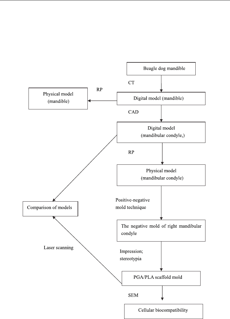
Fig. 1. Flow chart describing the protocol
Usage of Rapid Prototyping Technique
in Customized Craniomaxillofacial Bone Tissue Engineering Scaffold
93
In the pilot study, 12-week-old beagle dogs (male, weighing 10–15 kg) were selected. An
institutional review committee of Shanghai Jiao Tong University School of Medicine
approved all animal study protocols. The animals were examined using 3D CT. For each
dog, a model of the mandibular condyle was produced by RP to guide construction of a
fitted bone substitute scaffold made of PGA/PLA, which was then seeded with BMSCs. The
workflow could be divided into 5 major steps as described in figure 1.
STEP 1: CAD and RP of the dog mandible
The mandibles of beagle dogs were examined using a multi-slice CT scanner (GE Medical
Systems, Light-Speed16, New York, USA). CT scans were carried out with a slice thickness
of 0.625 mm (120 kV, 70 mA). The raw data were exported to Digital Imaging and
Communications in Medicine (DICOM) 3.0 format files. With the help of SimMed (PAMI of
Shanghai Jiaotong University, Shanghai, China), we imported the formatted images and
selected the bone tissue scale (CT threshold, 251–3072) for every slice. A 3D digital model of
the mandible (without soft tissue and cartilage) was then reconstructed slice by slice and
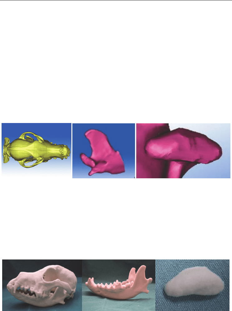
compiled into a solid geometric model in STL format (Figure 2).
(a) (b) (c)
Fig. 2. Virtual model: (a) 3D reconstruction of the skull; (b) mandibular ramus; and (c)
mandibular condyle
The STL files were then imported into Z-Printer 510 (Z Corp Inc., MA, USA). This system
enables 3D printing (3D-P) using resin powder. The process consists of a printer-like
computer system that directs the deposition of layers of sealant alternating with layers of
resin powder (each 0.1 mm thick), resulting in the fabrication from CAD data of 3D models
of the left mandibular condyle, using layer-by-layer manufacturing (Figure 3). Actually, the
detail method of 3D outputting was not important. The keynote procedure here was to have
the first positive solid model that was same as which we had reconstructed in cyber world.
(a) (b) (c)
Fig. 3. (a) Completed RP model: lateral view of the skull. (b) Completed RP model: lateral
view of the mandible. (c) Completed RP model: posterior view of the left mandibular condyle
Advanced Applications of Rapid Prototyping Technology in Modern Engineering
94
STEP 2: Fabrication of a negative mould of the mandibular condyle
The RP model of the mandibular condyle was used as the positive model and was
impressed into semisolid-state gypsum or silicone fibrosum, creating a negative mould,
which was dried at room temperature. The mould was clamshelled by sectioning two sides
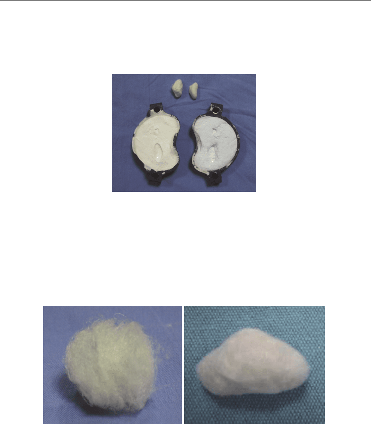
and leaving the bottom intact to allow for easy removal of the positive model (Figure 4).
Fig. 4. Negative mould of the left mandibular condyle. gypsum fibrosum mould, open.
Rapid prototyped PGA/PLA scaffolds in mandibular condyle bone defect reconstruction
STEP 3: Preparation of the PGA/PLA scaffold
Unwoven PGA fibres (60 mg; Albany International Research, Albany, NY, USA) were
layered into the negative mould and 0.6 ml 1.5% PLA (Sigma–Aldrich, St. Louis, MO, USA)
was diluted in dichloromethane solvent and added to maintain the scaffold shape. The
scaffold was then removed from the mould carefully (Figure 5). Then, the geometry
accuracy of the scaffold model was needed to be checked.
(a) (b)
Fig. 5. (a) Unwoven fibres of PGA. (b) PGA/PLA model scaffold of the left mandibular
condyle
STEP 4: Laser scanning of the scaffold model
A 3D laser surface scanning system (VIVID910 3D, Konica Minolta, Tokyo, Japan) with a
Polygon Editing Tool v. 1.03 (Konica Minolta, Tokyo, Japan) was used to acquire the 3D
geometric surfaces of the regenerated left mandibular condyle. The scan was performed
with a 25 mm focus camera at a scan distance of 750 mm. Every model was scanned from
Usage of Rapid Prototyping Technique
in Customized Craniomaxillofacial Bone Tissue Engineering Scaffold
95
five perpendicular angles, and the scanned images from each model were noise-filtered,
smoothed, registered and merged into a single, equidimensional-scale, digital, stereoscopic
model in STL format. The left condyle of the CT-reconstructed STL model was put aside as
the comparison object. Afterwards, the scan result was finely registered to the CT model
using Rapidform2006 (ISUS Ltd, Korea) to match up certain reference points on the
geometric surface, ensuring that the two models had the same coordinates. Then, we
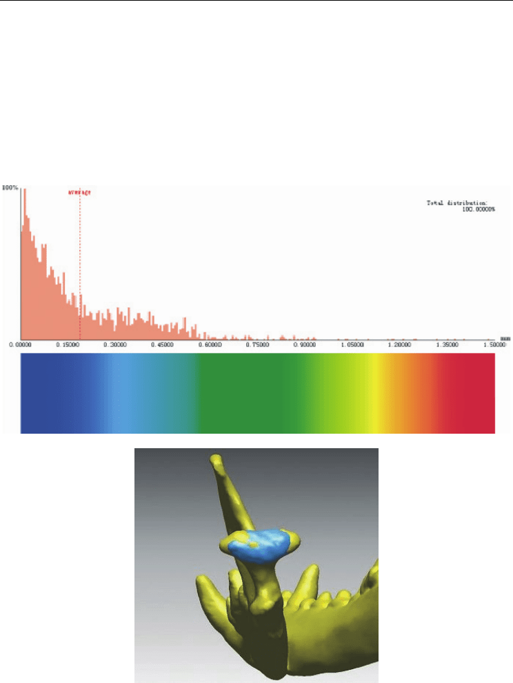
compared the divergence of the two sample datasets, using the Whole Inspection Model tool
in Rapidform2006, which automatically measures the distance (mm) between each
corresponding triangle surface on two registered surfaces and displays the measured
distances as a coloured chart (Figure 6).
(a)
(b)
Fig. 6. (a) Total distribution of scan data. (b) Comparison of the three acquisitions: point
clouds obtained by CT and from the laser scanner

Advanced Applications of Rapid Prototyping Technology in Modern Engineering
96
STEP 5: Cellular biocompatibility with the PGA/PLA scaffold in vitro
The model scaffold was sterilized by soaking in 75% alcohol and washing three times with
phosphate-buffered saline (PBS). This was followed by two washes with Dulbecco’s
modified Eagle’s medium (DMEM; Gibco, USA). Bone marrow aspirates (5 ml) were taken
from the iliac crests of beagle dogs. The aspirates were resuspended in 20 ml DMEM with
0.5 ml heparin sodium. The mixture was centrifuged at 1000 rpm for 5 minutes and the
supernatant removed. The cell pellets were resuspended, loaded onto Ficoll–Paque PLUS
and centrifuged at 1500 rpm for 10 min. Fractions containing low-density mononuclear cells
were harvested with a pipette and washed twice with PBS before culturing. The cells were
cultured in DMEM supplemented with 10% fetal bovine serum. The cultures were
maintained at 37 Ԩ in an atmosphere of 5% CO2. BMSCs at passage 2 (2.5 × 10⁷ cells in 0.5
ml) were evenly dropped onto PGA/PLA scaffolds to form cell/scaffold constructs. These
constructs were then cultured in DMEM supplemented with 10% FBS at 37Ԩ in a 5% CO2
atmosphere. After 7 days of incubation, cell attachment and matrix production on the
polymer were observed via scanning electron microscopy (SEM; JEOL JSM-6360LV, Japan).
Post-processed 3D geometric models from laser scanning demonstrated that the mean error
between the two samples was <0.3 mm; moreover, at error <1 mm, confidence remained at
≥95%. For each tolerance level, the degree of deviation was shown as a percentage (Table 1).
The results indicate that the amount of overlap between the two datasets, expressed as
percentages for tolerance levels 0.1, 0.3, 0.5, 0.8 and 1.00 mm, were 54.01%, 77.88%, 90.26%,
94.83% and 95.65%, respectively. SEM showed that BMSCs were able to adhere to the
scaffold surface and spread and grow along its fibres. Most of these cells had synthesized
and organized abundant extracellular matrices, with which they had created bridges
between one another.
Subjects
(n=8)
1.0mm
(%)
0.8mm
(%)
0.5mm
(%)
0.3mm
(%)
0.1mm
(%)
Mean 95.65 94.83 90.26 77.88 54.01
SD 1.47 2.13 2.78 3.44 3.31
Maximum 97.98 97.08 93.77 82.66 59.63
Minimum 93.45 90.63 85.81 71.28 49.27
Table 1. Degree of deviation (%) at 0.1, 0.3, 0.5, 0.8 and 1.00 mm
In craniomaxillofacial surgery, morphological characteristics of the bone and relevant
mechanisms of injury dictating the complexity and variety of the methods are currently
used to repair the bony defect, of which the most important components are restoration of
tissue volume and reconstruction of the 3D contour and shape.
New technology such as RP has been widely used in craniomaxillofacial reconstructive
surgeries. These technologies have facilitated the transformation of reconstructive surgical
practice from complicated, general, and experience based to simple, accurate, and
digitalized. Because the structure of the craniofacial skeleton is comparatively delicate and
the bone surface is irregular, artificial implant materials cannot be easily molded during
operation. When RP is applied for the preoperative design of the implant, the operation time
will be effectively shortened and the result will be substantially improved. Moreover, a
personalized preoperative design shall make the implant an accurate match for the defect.
Aesthetic needs are both met when the defect is corrected. Rapid prototyping is the
digitalized technology grounded on theories of discrete and accumulative forming that

Usage of Rapid Prototyping Technique
in Customized Craniomaxillofacial Bone Tissue Engineering Scaffold
97
produces the substance layer by layer or point by point. Based on the CT scan data and the
CAD model of the substance, the production for the prototype is accurately conducted. With
the rapid development of the computer software and hardware and further introduction of
mathematics, the CAD/CAM as the importance technology can be applied more widely.
Three-dimensional CT reconstruction was first applied to craniomaxillofacial research and
treatment in 1986. Craniofacial skeleton conditions can be found out clearly through three-
dimensional CT scan. Virtual digital data of CT scan are transferred into a solid three-
dimensional reconstruction model. Therefore, replication of the deformed state can be
interpreted directly, and craniofacial anatomy and periphery relationship can be described
in detail. The accuracy of the model, which has been tested by both domestic and
international scholars, helps physicians to find out the degree and the specificity of the
defect easily, thoroughly, and directly. Thus, the preoperative simulation and the treatment
plan are carried out to reduce both the difficulty and the duration of the surgery. With the
effective communication between the physician and the patient, consensus is reached and
postoperative satisfaction is met. Overall, the three-dimensional reconstruction model is an
irreplaceable tool in the contemporary craniomaxillofacial surgery.
Reconstruction of 3D images from CT data has been used for many years to enhance
interpretation of two-dimensional (2D) CT slices. Construction of CAD/CAM scaffolds
derived from 3D computer-generated images has found various uses in craniofacial and
plastic surgery, and has been particularly valuable when planning complex reconstructive
procedures, such as repairs of large traumatic and complex deformities of the
temporomandibular joint. Collaborations between engineers, surgeons and prosthetic
designers have resulted in efficient exploitation of the available instruments and
technologies .
In a previous study, the unit of craniofacial surgery in our department had applied this
combined technique in the produce of EH (hydroxyapatite granules mixed with an epoxide
acrylate maleic medical resin molding agent) compound artificial bone implants. Between
January 2005 and October 2008, 39 patients with craniomaxillofacial deformities were
treated. All the 39 patients were successfully operated on according to the preoperative
plans. The results after surgery were satisfactory. One patient with temple augmentation
needed secondary surgery to smooth the periphery of the implanted material because of the
prominent edge of the implant that can be felt by the patient. Subcutaneous effusion in 2
patients was treated with suction and compressive bandaging. The recovery was satisfying.
Implants in the 2 patients operated on through intraoral incisions had to be eventually
removed 2 months after the surgeries owing to severe local symptoms of infection such as
swelling and pain. All the other patients showed no complications during uneventful
postoperative follow-ups.
With the application of CAD/CAM and RP, the implant material is perfectly matched for
the skeleton defect. In the discussed case group, the combination of RP and CAD/CAM is
applied in the craniomaxillofacial treatment. It has been proved after surgery that
customized bone implants adhere to the defected area perfectly, and no migration has been
noticed. The adhesion is tight, and the appearance is comparatively symmetrical. As for
defect or depression that is close to the midline of the face, there is no mirror image for
reference. Proper adjustments for the implant production are made during computerized
simulation in accordance with the continuity of the geometric curve of the skeleton,
although the patients’ own opinions are properly taken at the same time. The postsurgical
results were mostly well accepted by the patients. In the EH operation group, 2 patients
Advanced Applications of Rapid Prototyping Technology in Modern Engineering
98
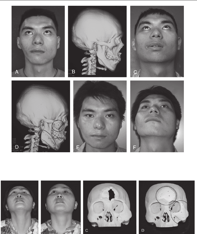
Fiq. 8 A, Preoperative inferior view for patient 1. B, Preoperative three-dimensional CT. C,
One-week postoperative inferior view. D, Postoperative three-dimensional CT. E,
Anteroposterior view of the same patient after 1 year. F, Inferior view after 1 year.
Fig. 9. A, Preoperative inferior view of patient 2. B, Postoperative inferior view (3 mo). C,
Three-dimensional skull model. D, Preoperatively made EH compound artificial bone
implant.
with temple augmentation were operated on. Usually, a lipoinjection or a Medpor implant is
chosen for the reconstructive surgery. However, after sufficient communication with the
patients, computerized reconstruction was applied for the design of the EH compound
artificial bone implants to meet the patients’ demands. The surgical procedures were easy

Usage of Rapid Prototyping Technique
in Customized Craniomaxillofacial Bone Tissue Engineering Scaffold
99
and safe. The implants were placed subperiosteumly. No foreign body rejection or extrusion
was noticed during the follow-up.
While studies have investigated the suitability of various materials in the construction of
CAD/CAM scaffolds, histological studies focusing on BMSC seeding in scaffolds have
demonstrated that PGA/PLA is one of the best materials available for the regeneration of
new bone and cartilage. However, sculpting the external volume of the scaffold and
generating strategies for its RP using PGA/PLA are significant challenges.
We have described a new method to tailor-make PGA/PLA mandibular condylar scaffolds
for bone and cartilage regeneration that ensures excellent morphological alteration and
accuracy. A physical replica of the mandible was generated from 3D CT data using
CAD/CAM and RP, and was used as the positive model in the negative mould technique to
prefabricate a PGA/PLA scaffold in the shape of the mandibular condyle. To evaluate the
accuracy of the copy thus obtained, a laser scanning system was used. In general, if the
difference could be seen in <90% of the face after simulated implant placement, this was
doomed to be a faithful reproduction; in addition, when aligned, maps of the merged scans
showed that, on average, 90% of the created composite facial scans correlated to the original
with an error up to 0.85 mm, which was considered to be clinically acceptable. As for bone
reconstruction of craniomaxillofacial features, the tolerance levels reached in our results are
more acceptable. A comparison between the copy and the original morphology confirmed
the efficacy of this method for accurate reconstruction of craniomaxillofacial features.
Synthetic absorbable implants made of materials such as elastic PGA/PLA can be readily
adapted to the shape of a given defect. Because of their resilience, these materials may not
sufficiently resist static or dynamic stresses. While each material has its advantages and
disadvantages, it must at least accomplish its specified objectives. PGA and PLA are
degradable, and their degradation product is weakly acidic. The acid–base neutralization
reaction that occurs locally during degradation can prevent aseptic inflammation, maintain
acid–base balance and promote osteogenesis. Zhou demonstrated that physiological repair
of defects in articular cartilage and the corresponding subchondral bone was achievable
using autologous BMSCs and PGA/PLA polymers. This study shows that BMSCs dispersed
throughout a PGA/PLA scaffold in in vitro culture, proving that PGA/PLA have excellent
cellular compatibility. BMSCs also have multi-lineage differentiation potential, particularly
for osteogenic and chondrogenic differentiation, making them ideal seeding cells for tissue
engineering.
In summary, our digital medical system enabled the fabrication of a customized PGA/PLA
scaffold that very accurately recreated the original anatomical form and also had excellent
cellular compatibility. Furthermore, the cost of equipment for this procedure was not high,
since RP machines and laser scanners are available at low cost. This digital medical support
system may be useful for craniomaxillofacial surgeons when reconstructing complex bony
injuries and abnormalities.
2. References
[1] Ciocca L, De Crescenzio F, Fantini M, et al. CAD/CAM and rapid prototyped scaffold
construction for bone regenerative medicine and surgical transfer of virtual
planning: a pilot study. Comput Med Imaging Graph 2009; 33(1): 58–62.

Advanced Applications of Rapid Prototyping Technology in Modern Engineering
100
[2] Klein M, Glatzer C. Individual CAD/CAM fabricated glass–bioceramic implants in
reconstructive surgery of the bony orbital floor. Plast Reconstr Surg 2006; 117(2):
565–570.
[3] Yeong WY, Chua CK, Leong KF, et al. Rapid prototyping in tissue engineering:
challenges and potential. Trends Biotechnol 2004;22(12): 643–652.
[4] Dean D, Min KJ, Bond A. Computer aided design of large-format prefabricated cranial
plates. J Craniofac Surg 2003; 14(6): 819–832.
[5] Peltola SM, Melchels FP, Grijpma DW, et al. A review of rapid prototyping techniques
for tissue engineering purposes. AnnMed 2008; 40(4): 268–280.
[6] Ciocca L, Mingucci R, Gassino G, et al. CAD/CAM ear model and virtual construction of
the mold. J Prosthet Dent 2007; 98(5):339–343.
[7] Zhou G, Liu W, Cui L, et al. Repair of porcine articular osteochondral defects in non-
weightbearing areas with autologous bone marrow stromal cells. Tissue Eng 2006;
12(11):3209–3221.
[8] Xu H, Han D, Dong JS, Shen GX, Chai G, Yu ZY, Lang WJ, Ai ST. Rapid prototyped
PGA/PLA scaffolds in the reconstruction of mandibular condyle bone defects. Int J
Med Robot. 2010; 6(1):66-72.
[9] Rosen JM, Long SA, McGrath DM, et al. Simulation in plastic surgery training and
education: the path forward. Plast Reconstr Surg 2009; 123(2): 729–738; discussion,
739–740.
[10] Hollister SJ. Porous scaffold design for tissue engineering. Nat Mater 2005; 4(7): 518–
524.
[11] Hutmacher DW, Sittinger M, Risbud MV. Scaffold-based tissue engineering: rationale
for computer-aided design and solid free-form fabrication systems. Trends
Biotechnol 2004; 22(7): 354–362 [review].
[12] Moroni L, de Wijn JR, van Blitterswijk CA. Integrating novel technologies to fabricate
smart scaffolds. J Biomater Sci Polym Ed 2008; 19(5): 543–572.
[13] Wang L, Detamore MS. Tissue engineering the mandibular condyle. Tissue Eng 2007;
13(8): 1955–1971.
[14] Hutmacher DW. Scaffolds in tissue engineering bone and cartilage. Biomaterials 2000;
21(24): 2529–2543.
[15] Kau CH, Richmond S, Zhurov AI, et al. Reliability of measuring facial morphology
with a three-dimensional laser scanning system. Am J Orthod Dentofacial Orthop
2005; 128(4): 424–430.
[16] Yu D, Li Q, Mu X, et al. Bone regeneration of critical calvarial defect in goat model by
PLGA/TCP/rhBMP-2 scaffolds prepared by low-temperature rapid-prototyping
technology. Int J Oral Maxillofac Surg 2008; 37(10): 929–934.
[17] Cao W, Miyamoto Y. Direct slicing from AutoCad solid models for rapid prototyping.
Int J Adv Manuf Technol 2003;21:739-742
[18] Muller A, Krishnan KG, Uhl E, et al. The application of rapid prototyping techniques
in cranial reconstruction and preoperative planning in neurosurgery. J Craniofac
Surg 2003;14:899-914.
[19] Mavili ME, Canter HI, Saglam-Aydinatay B, et al. Use of three-dimensional medical
modeling methods for precise planning of orthognathic surgery. J Neurosurg Sci
2008;52:113-116
