Hoque. Advanced Applications of Rapid Prototyping Technology in Modern Engineering
Подождите немного. Документ загружается.

Application of a Novel Patient - Specific Rapid Prototyping Template in Orthopedics Surgery
141
(a) (b) (c) (d)
(e) (f)
(g) (h)

Advanced Applications of Rapid Prototyping Technology in Modern Engineering
142
(i) (j)
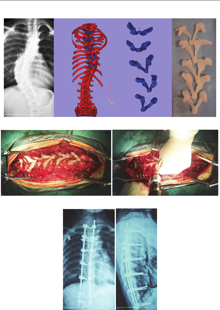
Fig. 8. A 12-year-old male with a 62° pre-operative curve; thoracic pedicle screw was
inserted using the drill template;
a: AP X-ray; b: 3-D model of the spine; c: Pedicle screw trajectory and design of the
navigational template; d: RP model of the navigational template, e: The navigational
template fits the posterior part of the thoracic perfectly in the operation; f: Drill the trajectory
by a power drill. g,h, I,j: X ray and CT scan show that the screws were fully contained within
the pedicle and body; corrected to 11° one year after surgery, giving 82.3% correction of the
major curve.
motion along all axes. The addition of transarticular screw fixation, according to Magerl and
Seemann, offers a better biomechanical stability. But, Magerl screw fixation must be very
precise point of placement to make screw fixation in the lateral mass of atlas, this can result
in a significant risk to the vertebral artery. The size of the C2 pedicle can limit the ability to
safely place these screws, particularly taking into account the anomalous position of the
vertebral artery in relation to the isthmus of C2 in up to 20% of the population. Leonard and
Wright [7] described a new technique of C2 laminar screw for rigid screw fixation of the axis
and incorporation into atlantoaxial fixation or subaxial cervical constructs, and subsequent
cases have shown good clinical results with this technique. C2 laminar screws are appealing
due to the reduced risk of injury to the vertebral artery and biomechanical stability. The
present method of C2 laminar screw placement relies on anatomical landmarks for screw
placement. Placement of C2 laminar screws using drill template has not been described in
the literature. A novel computer- assisted drill guide template for placement of C2 laminar
screws, Which designed to simplify and shorten the surgical act and at the same time further
enhance the accuracy of screw positions in the C2 laminar.
Before the operation, a spiral three-dimensional (3-D) CT scan (LightSpeed VCT, GE, USA)
was performed on cervical spine of each patient with 0.625-mm slice thickness and 0.35-mm
in-plane resolution. The images were stored in DICOM format, and transferred to a
workstation running MIMICS 10.01 software (Materialise company, Belgium) to generate a 3-
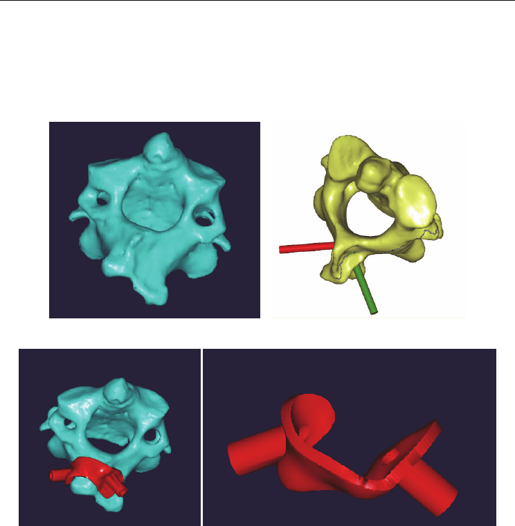
D reconstruction model for the desired C2 vertebrae (Fig. 9a). The 3-D vertebral model was
then exported in STL format, and opened in a workstation running Reverse Engineering (RE)
software UG imageware12.0 (EDS, USA)for determining the optimal screw size and
orientation. A screw with a diameter of 4 mm was placed virtually into the 3-D spinal model
on both sides. The virtual screw’s entry point and the trajectory was placed centered on the
lamina without violating the cortex and two screws will not interfere with each other. (Fig. 9b).
The optimal screw size was determined according to the size of laminea as well. Afterwards,
a navigational template was constructed with one drill guide on either side. The template
Application of a Novel Patient - Specific Rapid Prototyping Template in Orthopedics Surgery
143
surface was created to be the inverse of C2 spinous process and laminar, thus potentially
enabling a fit in a lock-and-key fashion similar to a physical casting of the vertebral surface,
and specifically avoided overlap onto adjacent segments (Fig. 9c). The inner diameter of the
hollow cylinder was created to accommodate the preplanned trajectory for drilling. Once
these had been done, a drill template was constructed with a surface designed to be the
inverse of the vertebral surface (Fig. 9d).
(a) (b)
(c) (d)
Fig. 9. The design of the virtual navigational template
a: 3-D model of C2 vertebra; b: virtual display of the planned laminar screw trajectory; c:
navigational template fit the vertebral perfectly; d: The virtual 3-D model of navigational
template
The computer model was then exported in STL format. The biomodel of the C2 vertebra as
well as its corresponding navigational template were both produced by acrylate resin (Somos
14120, DSM Desotech Inc, USA) using stereolithography rapid prototyping (RP) technique.
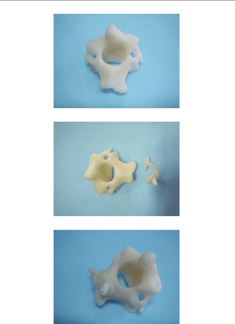
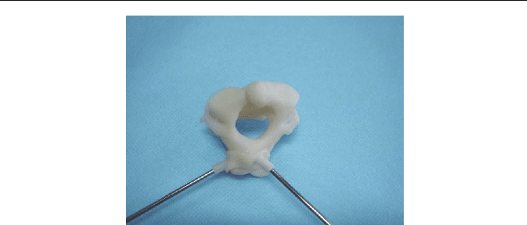
The accuracy of the navigational template was before surgery examined by visual inspection.
The biomodel and its coresponding template were placed together, and a standard electric
power drill was used to drill screw trajectory into the biomodel at the predefined placement,
and visual inspection was taken for any violation of C2 laminar (Fig. 10).
Advanced Applications of Rapid Prototyping Technology in Modern Engineering
144
(a)
(b)
(c)
Application of a Novel Patient - Specific Rapid Prototyping Template in Orthopedics Surgery
145
(d)
Fig. 10. The accuracy of the navigational template was examined by visual inspection
a: RP model of C2 vertebra; b: RP model of C2 vertebra and navigational template; c:
navigational template fit RP model of vertebra perfectly; d: insert the K wire by navigational
template; the accuracy of the navigational template was examined by visual inspection.
From June 2006 to Septempber 2008, 9 patients (four males, five females, age 17–53 years)
with basilar invagination requiring the posterior instrumentation were performed
occipitocervical fusion surgery by C2 laminar screw fixation. There are eight cases with
occipitalization of the atlas, six cases accompanied by C2-C3 vertebral fusion. In three
patients a transoral surgery was performed firstly, followed by posterior surgery. The
anatomy of C2 pedicle was observed preoperative X ray and CT scan, if the pedicle is very
narrow, thin, or vertebral artery high riding, it would be inappropriate placement of pedicle
screw, the laminea screw fixation can be choosed.
Tracheal intubation using general anesthesia, patients are placed in the prone position with the
head and cervical spine maintained in the neutral position using the Mayfield head holder.
The spinous process, laminae and lateral masses of of C2 are then exposed as needed. Then the
drill template was placed on the spinous process and laminae of C2. Template and the
corresponding spinous process were fitted well. The high-speed drill is used along the
navigational channel to drill the trajectory of laminar screw. Using a hand drill, the trajectory
of laminar screw is carefully drilled to a depth of preoperation plan. A 4.0 mm screw is
carefully inserted along the same trajectory. Using the same technique as above, a 4.0mm
screw is placed into the other lamina. After screw placement, all exposed laminar surfaces are
decorticated with the high-speed drill. Autologous iliac crest bone grafts are wedged under the
rods between the occipital bone and the spinous process and lamina of C2.
Using the virtual 3-D model, the optimal entry point for the bore can be choosed, thus
determining the entry point and direction for the C2 laminar screw. The drill template was
created to fit the postural surface of C2 spinous process very well. The accuracy of the drill
template was before operation examined by drilling K wire trajectory into the vertebra
biomodel. Each navigational template fits its corresponding vertebral biomodel perfectly,
and K wire was found to be inserted through the drill hole into the C2 lamina, no violation
was found by visual inspection.
Nine patients with basilar invagination were performed occipitocervical fusion surgery, the
bilateral cross laminar screw fixation was preformed in eight patients, unilateral laminar

Advanced Applications of Rapid Prototyping Technology in Modern Engineering
146
screw fixation and other side of pedicle screw fixation was preformed in a patient. Average
follow-up was 9 months (range 4–13 months). Preoperative and postoperative functional
comparisons were made using the ASIA grading scale for all cases. Seven Of the patients
(77.8%) improved at least one ASIA grade. Two patients have not improve. No patient
suffered neurological deterioration as a result of the procedure.
(a) (b) (c)
(d) (e) (f)
(g) (h) (i)
Fig. 11. The navigational template applied in the operation of a 42- year- old women with
basilar invagination, occipitalization of the atlas and C2- C3 vertebral fusion.
a, b: Pre-operative CT and T1 sagittal MRI displaying ventral brainstem compression; c:
Preoperation CT show the pedicle of C2 is narrow and can not accommodate the 3.5 mm
pedicle screw; d: Navigational template fit the spinous process and lamina of C2 perfectly;
e: The high-speed drill is used along the navigational channel to drill the trajectory of
laminar screw; f: Insert the cervical pedicle screw by navigational template; g, h, i:
Postoperative CT showing accurate placement of C2 laminar screws

Application of a Novel Patient - Specific Rapid Prototyping Template in Orthopedics Surgery
147
Seventeen C2 laminar screws were inserted using drill emplate. No screw inserted
complications such as spinal cord, nerves, vertebral artery injury appear in this group. The
mean operative time between fixation of the template to the lamina and placement of the
screw was 1 to 2 minutes. Operation time reduced through use of the navigational template.
No additional computer assistance was needed during surgery, and fluoroscopy was used
only once, after all the C2 laminar screws had been inserted. The method thus significantly
reduced radiation exposure for the members of the surgical team.
It takes about 16 hours to manufacture the RP model, and the price of each RP model of the
vertebra and navigational template is about $20. Postoperative CT scans showed that the
individual template has a higher precision. No laminar screw misplacement occurred using
the individual template. (Fig. 11)
2.2 Design and primary application of computer-assisted, patient-specificnavigational
templates in metal-on-metal hip resurfacing arthroplasty
Total hip resurfacing arthroplasty, viewed by many as representing an evolution from the
mold arthroplasty procedure of Smith-Petersen, has been considered an alternative to total
hip arthroplasty for adult patients with osteoarthritis of the hip or congenital hip dysplasia.
It has been performed with a variety of materials, designs, surgical approaches, techniques,
and fixation methods [1]. Many advantages of hip resurfacing arthroplasty have been
suggested, including bone conservation [2-4], improved function as a consequence of
retention of the femoral head and neck and more precise biomechanical restoration [5],
decreased morbidity for revision arthroplasty [6], reduced dislocation rates [7,8], and
normal femoral loading and reduced stress shielding [9]. In hip resurfacing arthroplasty,
prosthesis location is key to postoperative joint stability. In conventional hip arthroplasty,
the neck-shaft angle is determined by the design of the prosthesis itself, whereas in hip
resurfacing, it is determined by valgus or varus placement of the femoral prosthesis. When
the prosthesis is inserted by conventional positioning, some positioning devices are needed;
however, accuracy
cannot always be achieved with current devices. Successful positioning
thus depends largely on how experienced the surgeon is. With the development of
computer-aided design and computer-aided engineering, the trend in medical technology
has been toward individualization. We conducted a study in which we introduced and
validated novel locating navigation templates in the clinical setting. The templates' designs
are based on reverse engineering. The patient-specific design allows close contact with
acetabular and femoral features to provide better stability and function than is possible with
conventional positioning devices.
Three-dimensional (3-D) computed tomography (CT) pelvic scan image data were obtained
from 10 healthy volunteers who underwent a spiral 3-D CT scan (Light- Speed VCT; GE,
Fairfield, CT) using a 0.625-mm slice thickness and 0.35-mm in-plane resolution. Data were
transferred via a digital imaging and communications in medicine network into a computer
workstation. Three dimensional models of the hips were reconstructed using Amira
software (version 3.1; TGS, San Diego, CA) and saved in stereolithography format. The 3-D
models were then imported into Imageware software (version 12.0; EDS, Palo Alto, CA).
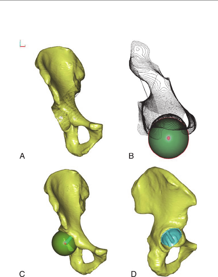
First, data for the transaction planes (1-mm thickness) of the acetabular and femoral head
surfaces were extracted; and the correctly fitting globe was produced. The globe center was
considered to be the rotation center of both the acetabulum and the femoral head. Second,
we presumed an ideal acetabular location of about 45° abduction and 18° anteversion and
Advanced Applications of Rapid Prototyping Technology in Modern Engineering
148
an optimum location for the femoral head implant of 140° abduction along the axis of the
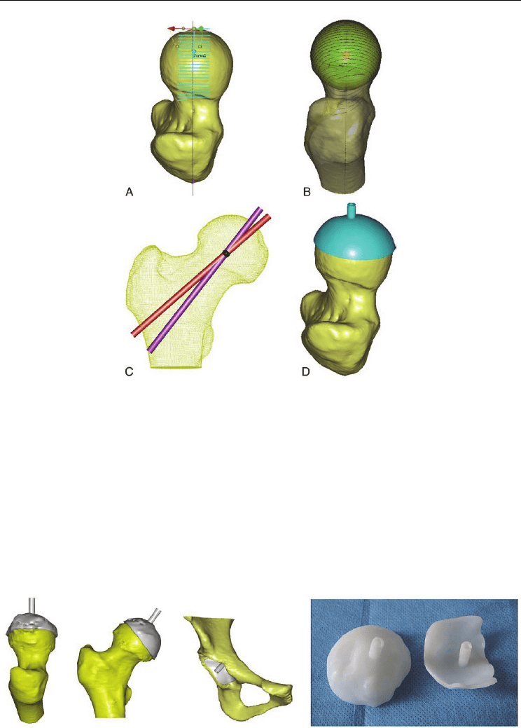
femoral neck. Finally, we designed specific navigation templates according to the
anatomical features of the acetabular contour and the femoral head (Figs. 12 and 13).
Fig. 12. Determination of the rotation center and the design process for a navigation
template for the acetabulum: (A) preparing a 3-D reconstruction of the hip and dissection of
acetabular surface, (B) constructing the rotation center of the acetabulum (small ball), (C)
determining the navigation channel, and (D) producing the navigation template.
Application of a Novel Patient - Specific Rapid Prototyping Template in Orthopedics Surgery
149
Fig. 13. The design process for a femoral head navigation template: (A) determining the
axial ray on the femoral neck and dissecting the femoral head, (B) constructing the center of
femoral head (small ball), (C) determining the navigation channel, and (D) producing the
navigation template.
20 patients (11 men and 9 women) with pathologic changes in one hip who were 24 to 37
years old were scheduled for total hip resurfacing arthroplasty. They were randomly
assigned to undergo either conventional implantation of a prosthesis (control group) or
implantation of a prosthesis guided by navigation templates (NT group). The navigation
templates were made as described in the preceding section. Navigation template models
were exported in stereolithography format and formed from acrylate resin (Somos
14120;DSMDesotech Inc, Stanley, NC) using the stereolithography rapid prototyping (RP)
technique (Fig. 14).
Fig. 14. Template models of the acetabulum and femoral head.
Advanced Applications of Rapid Prototyping Technology in Modern Engineering
150
Duration of surgery and intraoperative blood loss for the 2 groups were compared.
Deviation between the ideal abduction angles and the actual angles of the implanted
acetabular cup was calculated, as was deviation of cup anteversion angles. The deviation
between the neck-shaft angle and the actual implanted short stem-shaft angle (SSA) and that
between the anteversion angle of the femoral neck and the anteversion angle of the actual
implanted short stem were measured. Angle deviations in the NT group were compared
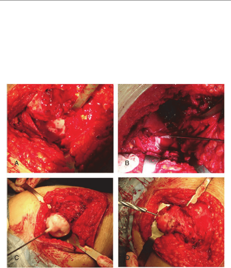
with those in the control group. In the NT group, after dislocation of the hip and insertion of
the template into the acetabulum, a 3.2-mm–diameter guide pin was placed in the template
hole. We followed the guide pin with the acetabular reamer to drill and shape the prosthetic
socket at 45° abduction and 18° anteversion.
Fig. 15. Surgical procedure in the navigation template group: (A) locating the acetabulum,
(B) inserting a guide pin, (C) matching the navigation template and inserting a Kirschner
wire, and (D) enlargement canals.
When the femoral head was dislocated and correctly exposed, we matched the locating
template to the surface of the femoral head as well as possible. We then inserted a Kirschner
wire into the femoral neck according to the template. We evaluated the axial location of the
femoral component (shown by the Kirschner wire) with anteroposterior (AP) and lateral
position photographs obtained during surgery by a C arm machine (Fig. 15). We obtained
