Hammond C. The Basics of Crystallography and Diffraction
Подождите немного. Документ загружается.


214 The diffraction of X-rays
A
A
B
B
B
B
reference plane
C
C
hkl
reflection
hkl
reflection
(a) (b)
Fig. 9.9. Representation of anomalous scattering of atom labelled B: (a) hkl reflection, B displaced to
B
; and (b)
¯
h
¯
k
¯
l reflection, B displaced to B
.
from the ‘top’or ‘underside’of the reflecting planes. This is the condition for anomalous
scattering. Figure 9.9 (due to W.L. Bragg) shows the geometry involved for just three
atoms, A, B and C, one of which, B, scatters anomalously.
For the hkl reflection, Fig. 9.9(a), B scatters as if it were situated at B
and for the
¯
h
¯
k
¯
l
reflection, Fig. 9.9(b), B scatters as if it were at B
. The resultant amplitudes, obtained
by combining the effects of all the atoms, will clearly be different.
The effect may also be represented in the Argand diagram; the alteration of phase of
an anomalously scattering atom is equivalent to combining the normal f of the atom with
a vector f at right angles to it as shown in Fig. 9.8(b), where the resultant structure
factors F
hkl
and F
¯
h
¯
k
¯
l
which are now different, are shown by the dashed lines.
Anomalous scattering (or absorption) can be ‘put to use’to distinguish centrosymmet-
ric and non-centrosymmetric point groups. In particular it can be used to distinguish the
right and left-handed (dextro and laevo) crystals in the enantiomorphic point groups—
e.g. tartaric acid (Fig. 4.6, page 107). In so doing a salt is synthesized which contains an
anomalously scattering atom for the X-ray wavelength used. The first such experiments
were carriedout by J. M.Bijvoet
∗
using crystalsof sodium rubidium tartrate. It isveryfor-
tunate (even chances!) that the dextro- and laevo-configurations thus determined agree
with those of chemical convention—otherwise all the dexro- and laevo-conventions
would have had to be interchanged.
To summarize, these simple examples show how the amplitude, F
hkl
, and hence the
intensity, I
hkl
, of the reflected X-ray beam from a set of hkl planes can be calculated from
the simple ‘structure factor’ equation on p. 209; all we need to know are the positions
of the atoms in the unit cell (the x
n
y
n
z
n
values) and their atomic scattering factors,
f
n
. The great importance of the equation is that it can be applied, as it were, ‘the other
way round’: by measuring the intensities of the reflections from several sets of planes
(the more the better), the positions of the atoms in the unit cell can be determined.
This is the basis of crystal structure determination, which has developed and expanded
since the pioneering work of the Braggs, so that, at the time of writing, some 400 000
different crystal structures are known. Many of these are very complex, for example
protein crystals in which the motif may consist of several thousand atoms. Again, it
∗
Denotes biographical notes available in Appendix 3.
9.3 The broadening of diffracted beams 215
should be emphasized that the procedures are invariably not straightforward because the
phase information in going from the F
hkl
to the (measured) I
hkl
values is lost, e.g. as in
Example 4, Fig. 9.7. This is called the phase problem in crystal structure determination,
which may be understood with reference to Fig. 9.3(b). All F
hkl
vectors with the same
modulus or amplitude will give the same observed intensity I
hkl
; the value of the phase
angle , which is an essential piece of information in the vector-phase diagram, is lost.
In short, we do not know in which direction the vector F
hkl
‘points’, e.g. as in Fig. 9.7.
In some cases (as in Example 4), the problem is simply solved if we are able to arrange
the origin to coincide with a centre of symmetry in the crystal in which case, as shown
in Example 3, Fig. 9.5, the phase angle φ is zero and the structure factor F
hkl
is a real
number with no imaginary component. However, in the many cases where the crystal
does not possess a centre of symmetry, we must resort to more subtle procedures, the
details of which are beyond the scope of this book. One method is to arrange a heavy
atom (possibly substituted in the crystal structure for a light atom) to be at the origin.
Then, in terms of our vector-phase diagram (Fig. 9.3(b)), f
0
is so large that it dominates
the contributions of all the other atoms such that the phase angles for all the F
hkl
values
are small and therefore can more easily be guessed at. In all cases the structure factor
equation is expressed as it were in a ‘converse’ form (or transform of that on p. 209 in
which atomic positions (expressed as electron (X-ray scattering) density) are expressed
in terms of the F
hkl
values of the reflections.
The notion of electron density provides a much more realistic representation of
atomic structure. Atoms, which are detected by X-rays from the scattering of their
constituent electrons, have a finite size and the atomic coordinates essentially represent
those positions where the amount of scattering (the electron density) is the highest. In
our two-dimensional plan views (Section 1.8) we may therefore represent the atoms as
hills—a contour map of electron density; the ‘higher the hill’ the greater the atomic scat-
tering factor of the atom. These ideas, which involve the application of Fourier analysis,
are introduced in Chapter 13, but it is a subject of great complexity which is covered in
more detail in those books on crystal structure determination which are listed in Further
Reading.
9.3 The broadening of diffracted beams:
reciprocal lattice points and nodes
In Chapter 8 we treated diffraction in a purely geometrical way, incident and reflected
beams being represented by single lines implying perfectly narrow, parallel beams and
reflections only at the Bragg angles. Of course, in practice, such ‘ideal’ conditions do
not occur; X-ray beams have finite breadth and are not perfectly parallel to an extent
depending upon the particular experimental set-up. Such instrumental factors give rise
to broadening of the reflected X-ray beams: the reflections peak at the Bragg angles and
decrease to zero on either side. However, broadening is not solely due to such instru-
mental factors but much more importantly also arises from the crystallite size, perfection
and state of strain in the specimen itself. The measurement of such broadening (having
accounted for the contribution of the instrumental factor) can then provide information
on such specimen conditions.
216 The diffraction of X-rays
We now consider the effects of crystal size on the broadening and peak intensity of
the reflected beams, which lead to the Scherrer equation (Section 9.3.1) and the notion
of integrated intensity (Section 9.3.2). In Section 9.3.3 we consider the imperfection (or
mosaic structure) of real crystals. The effect of lattice strain on broadening is covered in
Section 10.3.4.
9.3.1 The Scherrer equation: reciprocal lattice points and nodes
In Section 7.4 we found that when the number N of lines in a grating, or the number
of apertures in a net, were limited, the principal diffraction maxima were broadened
and surrounded by much fainter subsidiary maxima. These phenomena are shown in
Fig. 7.3(d) and diagrammatically in Fig. 7.9. Precisely the same considerations apply to
X-ray (and electron) diffraction from ‘real’ crystals in which the number of reflecting
planes is limited: and the broadening and occurrence of the subsidiary maxima can be
derived by similar arguments. This, in turn, leads us to modify our concept of reciprocal
lattice points, which are not geometrical points, but which have finite size and shape,
reciprocally related, as we shall see, to the size and shape of the crystal. There is, as
far as I know, no common term to express the fact that reciprocal lattice points do have
a finite size and shape, except in special cases such as the reciprocal lattice streaks or
‘rel-rods’ which occur in the case of thin plate-like crystals. Reciprocal lattice nodes
seems to be the closest approximation to a common term.
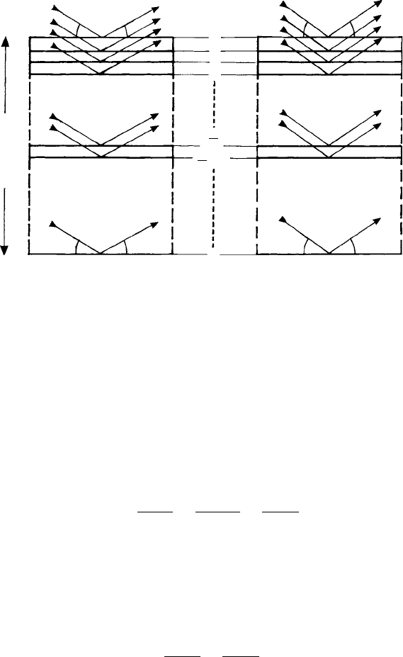
The broadening of the reflected beams from a crystal of finite extent is derived as
follows. Consider a crystal of thickness or dimension t perpendicular to the reflecting
planes, d
hkl
, of interest. If there are m planes then md
hkl
= t. Consider an incident
beam bathing the whole crystal and incident at the exact Bragg angle for first order
reflection (Fig. 9.10 (a)). For the first two planes labelled 0 and 1, the path difference
λ = 2d
hkl
sin θ; for planes 0 and 2 the path difference is 2λ = 4d
hkl
sin θ and so on—
constructive interference between all the planes occurs right through the crystal. Now
consider the interference conditions for an incident and reflected beam deviated a small
angle δθ from the exact Bragg angle (Fig. 9.7(b)). For planes 0 and 1 the path difference
will be very close to λ as before and there will be constructive interference. However, for
planes 0 and 2, 0 and 3, etc. the ‘extra’path difference will deviate increasingly from 2λ,
3λ etc.—and when it is an additional half-wavelength destructive interference between
the pair of planes will result.
The condition for destructive interference for the whole crystal is obtained by notion-
ally ‘pairing’ reflections in the same way as we did for the destructive interference of
Huygens’wavelets across a wide slit (Fig. 7.8). Consider the constructive and destructive
interference condition between planes 0 and (m/2) (halfway down through the crystal).
At the exact Bragg angle θ (Fig. 9.10 (a)), the condition for constructive interference is
(m/2)λ = (m/2)2d
hkl
sin θ. The condition for destructive interference at angle (θ + δθ)
is given by (m/2)λ + λ/2 = (m/2)2d
hkl
sin(θ + δθ). Now this is also the condition
for destructive interference between the next pair of planes 1 and (m/2) + 1—and so
on through the crystal. This equation gives us, in short, the condition for destructive
interference for the whole crystal and the angular range δθ (each side of the exact Bragg
angle) of the reflected beam.

9.3 The broadening of diffracted beams 217
0
2
m
m
t = md
hkl
(u + du)
(u + du)
(u + du)
u
u
uu
1
2
3
2
+ 1
m
Fig. 9.10. Bragg reflection from a crystal of thickness t (measured perpendicular to the particular set
of reflecting planes shown). The whole crystal is bathed in an X-ray beam (a) at the exact Bragg angle
θ and (b) at a small deviation from the exact Bragg angle, i.e. angle (θ +δθ). The arrows represent the
incident and reflected beams from successive planes 0,1,2,3 … (m/2) (half-way down) and … m (the
lowest plane).
Expanding the sine term and making the approximations cos δθ = 1and sin δθ ≈ δθ
gives:
(m/2)λ + λ/2 = (m/2)2d
hkl
sin θ +(m/2)2d
hkl
cos θδθ.
Cancelling the terms (m/2)λ and (m/2)2d
hkl
sin θ and substituting md
hkl
= t gives
2δθ =
λ
t cos θ
=
2d sin θ
t cos θ
=
2 tan θ
m
.
This is the basis of the Scherrer
∗
equation which relates the broadening of an X-ray beam
to the crystal size t or number of reflecting planes m. The broadening is usually expressed
as β, the breadth of the beam at half the maximum peak intensity and in which the angles
are measured relative to the direct beam. As indicated in Fig. 9.11, β is approximately
equal to 2δθ, hence
β =
λ
t cos θ
=
2 tan θ
m
.
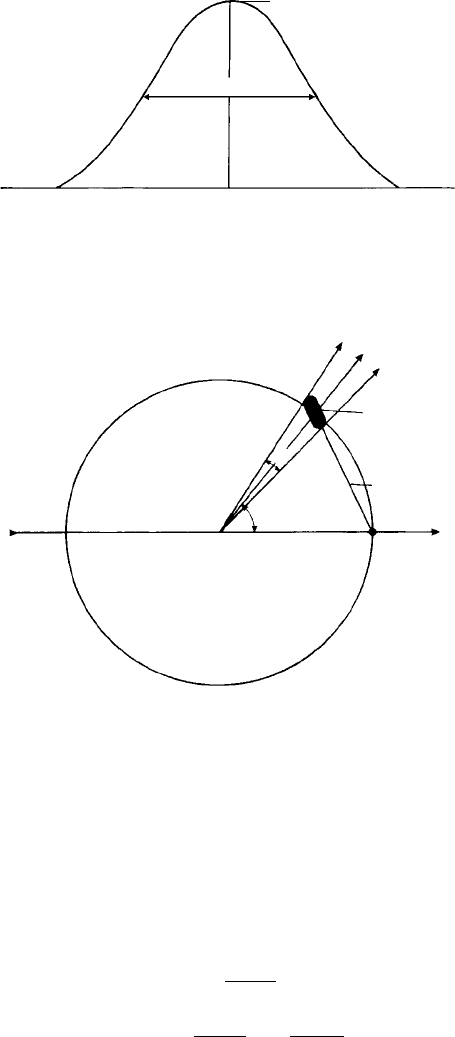
The broadening can be represented in the Ewald reflecting sphere construction in terms
of the extension of the reciprocal lattice point to a node of finite size. Figure 9.12 shows
∗
Denotes biographical notes available in Appendix 3.

218 The diffraction of X-rays
2(u – du)2(u + du)
2 du ≈ b
2u
I
max
Fig. 9.11. A schematic diagram of a broadened Bragg peak arising from a crystal of finite thickness.
The breadth at half the maximum peak intensity, β, is approximately equal to 2δθ. Note that the angular
‘2θ’ scale is measured in relation to the direct and reflected beams.
Reciprocal
lattice node
Incident
X-ray
beam
2b
2u
d*
hkl
Fig. 9.12. The Ewald reflecting sphere construction for a broadened reflected beam, β, which
corresponds to an extension 1/t of the reciprocal lattice node.
a diffracted beam (angle 2θ to the direct beam) which is broadened over an angular
range 2β ≈ 4δθ. This is expressed by extending the reciprocal lattice point into a node
of finite length which, as the crystal rotates, intersects the reflecting sphere over this
angular range. Let δ(d
∗
hkl
) represent the extension of the reciprocal lattice point about its
mean position. Now since
d
∗
hkl
= d
∗
hkl
=
2 sin θ
λ
;
δ(d
∗
hkl
) = δ
2 sin θ
λ
=
2 cos θ
λ
δθ.

9.3 The broadening of diffracted beams 219
Substituting for δθ from above gives:
δ(d
∗
hkl
) =
2 cos θ
λ
·
λ
2t cos θ
=
1
t
i.e. the extension of the reciprocal lattice node is simply the reciprocal of the crystal
dimension perpendicular to the reflecting planes. This applies to all the other directions
in a crystal with the result that the shape of the reciprocal lattice node is reciprocally
related to the shape of the crystal. For example, in the case of thin, plate-like crystals
(e.g. twins or stacking faults), the reciprocal lattice node is a rod or ‘streak’perpendicular
to the plane of the plate.
The extension of the reciprocal lattice node δ
d
∗
hkl
may also be expressed as a ratio
or proportion of d
∗
hkl
, i.e. since
δ
d
∗
hkl
=
1
t
=
1
md
hkl
=
d
∗
hkl
m
,
then
δ
d
∗
hkl
d
∗
hkl
=
1
m
,
i.e. the ratio is simply equal to the reciprocal of the number of reflecting planes, m.
The above is a simplified treatment, both of the Scherrer equation and the extension
of reciprocal lattice points into nodes. The Scherrer equation is normally applied to the
broadening from polycrystalline (powder) specimens and includes a correction factor
K (not significantly different from unity) to account for particle shapes.
4
Hence the
Scherrer equation is normally written:
β =
Kλ
t cos θ
=
Kλ sec θ
t
.
Finally, it should be noted that the reciprocal lattice nodes are also surrounded by sub-
sidiary nodes (or satellites, maxima, or fringes) just as in the case of light diffraction
from gratings with a finite number of lines (Section 7.4, Fig. 7.9). In most situations these
subsidiary nodes are very weak because the number of diffracting planes contributing to
the beam is large. However, in the case of X-ray diffraction from specimens consisting
of a limited number of diffracting planes, superlattice repeat distances or multilayers,
the subsidiary nodes (or satellites, maxima or fringes) are observable and are important
in the characterization of the specimen, as described in Section 9.6.
4
Note that the observed breadth at half the maximum peak intensity, β
OBS
, includes additional sources of
broadening arising from the experimental set up and instrumentation, β
INST
, which must be ’subtracted’ from
β
OBS
in order than β can be determined (see Section 10.3.3).

220 The diffraction of X-rays
9.3.2 Integrated intensity and its importance
In Section 9.3.1, we have seen the effect of crystal thickness on broadening. What
is its effect on the peak intensity, I
max
(Fig. 9.11)? To answer this question we will
carry out a ‘thought experiment’. Let us suppose that in our crystal, of thickness t,
there are a total of n unit cells each of which contributes a scattering amplitude |F
hkl
|.
The total scattered amplitude is therefore n|F
hkl
| and the total scattered intensity, I
max
,
is proportional to n
2
|F
hkl
|
2
. Now let us separate the crystal into two halves, each of
thickness t/2. The X-ray peaks from each crystal are twice the breadth of that for the
single crystal (half the thickness, twice the breadth). The total scattered amplitude from
each crystal is n/2
|
F
hkl
|
and hence the peak intensity is proportional to n
2
/4
|
F
hkl
|
2
.
Added together, the two crystals give a peak intensity, I
max
= n/2
|
F
hkl
|
2
, i.e. half that
of the single crystal but a peak breadth twice that of the single crystal. What is constant
and independent of the ‘state of division’—or crystallite size in the specimen—is the
area under the diffraction peak. This quantity, usually measured in arbitrary units, is
called the integrated intensity of the reflection or just the integrated reflection. It is not a
measurement of intensity, but rather a measurement of the total energy of the reflection.
Furthermore, except for situations in which dynamical interactions need to be taken
into account (i.e. large, perfect crystals in which the reflected beams are comparable in
intensity with the direct beam—see Section 9.1), the integrated intensity is a measure of
|F
hkl
| (and of course crystal volume).
9.3.3 Crystal size and perfection: mosaic structure and
coherence length
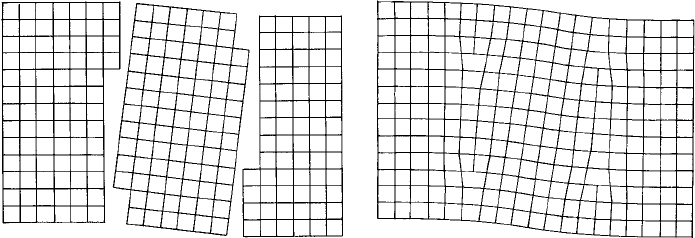
Except for rather special cases there is rarely a continuity of structure throughout the
whole volume of a ‘single’ crystal, but rather is separated into ‘blocks’ of slightly vary-
ing misorientation (Fig. 9.13(a))—a situation recognized in the early days of X-ray
diffraction from the discrepancy between the intensities predicted for ‘perfect’ crystals
(a) (b)
T
T
T
T
Fig. 9.13. Representation of a single crystal: (a) divided into three mosaic blocks; and (b) the
boundaries as ‘walls’ of edge dislocations.
9.4 Fixed θ, varying λ X-ray techniques 221
and those actually observed. Ewald termed this a ‘mosaic’ structure—but of course
the nature of the mosaic blocks and the boundaries were unknown. It is now evident,
from transmission electron microscopy, that crystals contain dislocations which may be
distributed uniformly throughout the structure, or arrange themselves (as shown in the
simple case in Fig. 9.13(b)), into sub-grain boundaries. However, it remains the case
that the relationship between sub-grain size, as measured by electron microscopy, and
coherence length or mosaic size, as measured by X-ray broadening, is by no means
clear.
In an X-ray experiment, as a crystal is ‘swept’ through its diffracting condition, the
individual crystallites of the mosaic structure reflect at slightly different angles and the
total envelope of the diffraction profile, or integrated intensity, is the sum of all the
separate reflections.
9.4 Fixed θ, varying λ X-ray techniques: the Laue method
X-ray single crystal techniques may be classified into two groups depending upon the
way in which Bragg’s law is satisfied experimentally. There are two variables in Bragg’s
law—θ and λ—and a series of fixed values, d
hkl
. In order to satisfy Bragg’s law for any
of the d-values, either λ must be varied with θ fixed, or θ must be varied with λ fixed.
The former case has only one significant representative—the (original) Laue method and
its variants, whereas there are many methods based upon the latter case.
The geometry of the Laue method, in terms of the reflecting sphere construction, has
already been explained in Section 8.4. Now we need to consider the practical applications
of the technique. The important point to emphasize is that each set of reflecting planes
with Laue indices hkl (see Section 8.4) gives rise to just one reflected beam. Of all the
white X-radiation falling upon it, a lattice plane with Miller indices (hkl) reflects only
that wavelength (or ‘colour’) for which Bragg’s law is satisfied, a reflecting plane of half
the spacing with Laue indices 2h 2k 2l reflects a wavelength of half this value, and so
on. In other words the reflections from planes such as, for example, (111) (Miller indices
for lattice planes) 222, 333, 444, etc. (Laue indices for the parallel reflecting planes) are
all superimposed.
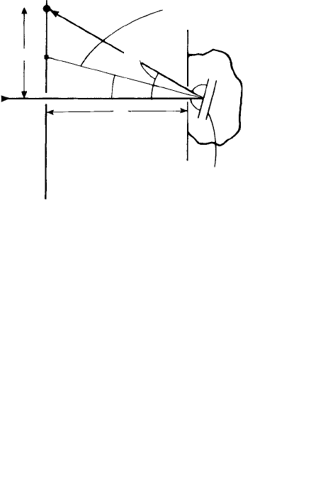
The usual practice is to record the back-reflected beams since thick specimens which
are opaque to the transmission of X-rays through them can be examined. The set-up
showing just one reflected beam in the plane of the paper is shown in Fig. 9.14(a). In
analysing the film it is necessary to determine the projection of the normal (or reciprocal
lattice vector) of each of the reflecting planes on to the film from each reflection S and
then to plot these on a stereographic projection. By measuring the angles between the
normals, and then comparing them with lists of angles such as given for cubic crystals
in Section A4.4 (Appendix 4), it is possible to identify the reflections. In practice such
manual procedures (involving the use of the Greninger net
5
) have largely been replaced
5
Greninger nets are prepared for a particular specimen-film distance (e.g. 30 mm), on which are drawn a
series of calibrated hyperbolae (for the back reflection case). By measuring the distance between two spots
along a hyperbola the angle between the normals, or the reciprocal lattice vectors of the two planes giving rise
to these spots, can be obtained.

222 The diffraction of X-rays
(b)
(a)
Reflection on film
Film
(90°– u)
(180°– 2u)
u
u
x
Lattice planes (hkl) in a single
crystal in the specimen
Projection of normal to lattice
planes (or reciprocal
lattice vector d*
hkl
)
S
r
Fig. 9.14. (a) The geometry of the back-reflection Laue method for a particular set of hkl planes in a
single crystal. The wavelength of the reflected beam is that for which Bragg’s law is satisfied for the
particular fixed θ and d
hkl
value, (b)A back-reflection Laue photograph of a single crystal of aluminium
oriented with a
100
direction nearly parallel to the incident X-ray beam showing the reflections S
from many (hkl) planes. They lie on a series of intersecting hyperbolae (close to straight lines in this
photograph), each hyperbola corresponding to reflections from planes in a single zone, [uvw]. Note the
four-fold symmetry of the intersecting zones indicating the
100
crystal orientation. (Photograph by
courtesy of Prof. G. W. Lorimer.)
by computer programs which determine the orientation of the crystal using as input
data the positions of spots on the film, film-specimen distance and (assumed) crystal
structure.
Hence (back reflection) Laue photographs may be used to determine or check the ori-
entation of single crystals and the orientation relationships between crystals. An example
of a Laue photograph is shown in Fig. 9.14(b).

9.5 Fixed λ, varying θ X-ray techniques 223
Figure 9.14(a) should be compared with Fig. 8.7. Consider for example the 201 recip-
rocal lattice point which is situated in the ‘nest’ of spheres representing the wavelength
range of the incident ‘white’X-radiation. Bragg’s law is satisfied for the particular wave-
length represented by the sphere which passes through the 201 reciprocal lattice point
and the direction of the reflected beam (indicated by the arrow) is from the centre of this
sphere (which can be found by construction) and the 201 reciprocal lattice point. This
beam makes angle (180
◦
− 2θ ) with the incident beam and the reciprocal lattice vector
d
∗
201
makes angle (90
◦
− θ ) with the incident beam.
9.5 Fixed λ, varying θ X-ray techniques:
oscillation, rotation and precession methods
In Section 8.4, Fig. 8.6, we showed the Ewald reflecting sphere construction for the case
where the incident X-ray beam was incident along the a
∗
reciprocal lattice vector (from
the left). Figure 8.6(a) shows the h0l section of the reciprocal lattice through the centre
of the sphere and the origin of the reciprocal lattice and Fig. 8.6(b) shows the h1l section
of the reciprocal lattice and the smaller (non-diametral) section of the Ewald sphere. For
the particular wavelength and particular incident beam direction drawn in Fig. 8.6, only
two planes—201 and 21
1 satisfy Bragg’s law. Now, instead of varying λ, as in the Laue
case (Fig. 8.7), let us change the direction of the incident beam such that it is no longer
incident along the a
∗
reciprocal lattice vector direction. This variation in the Bragg angle
is accomplished in practice by moving the crystal.
9.5.1 The oscillation method
Consider the crystal, Fig. 8.6, oscillated say ±10
◦
about an axis parallel to the b
∗
reciprocal lattice vector (or y-axis), i.e. perpendicular to the plane of the paper. As the
crystal is (slowly) oscillated, the angles between the incident beam and hkl planes vary
and whenever Bragg’s law is satisfied for a particular plane ‘out shoots’ momentarily a
reflected beam. The oscillation of the crystal can be represented by an oscillation of the
reciprocal lattice about the origin: imagine a pencil fixed to the crystal perpendicular
to the page with its point at the origin of Fig. 8.6(a). As you oscillate the pencil the
reciprocal lattice oscillates about the origin but the Ewald sphere remains fixed, centred
along the direction of the incident beam from the left: whenever a reciprocal lattice point
intersects the sphere ‘out shoots’ a reflected beam.
The relative movement of the sphere and reciprocal lattice is most easily represented,
not by drawing the whole reciprocal lattice in its different positions, but by drawing the
Ewald sphere at the extreme limits of oscillation say 10
◦
in one direction and 10
◦
in the
other. These limits are shown in Fig. 9.15 for (a) the h0l and (b) the h1l reciprocal lattice
sections. The shaded regions, called ‘lunes’because of their shape, represent the regions
of reciprocal space through which the surface of the sphere passes as it is oscillated.
Only those reciprocal lattice points which lie within these shaded regions give rise to
reflected beams.
There are several ways of recording the reflected beams. The simplest is to arrange a
cylindrically shaped film coaxially around the crystal (with a hole to allow the exit of the
