Glusker J.P., Trueblood K.N. Crystal Structure Analysis: A Primer
Подождите немного. Документ загружается.

58 Experimental measurements
Normal to diffraction
plane = diffraction
Vector
X-ray tube
Detector
Crystal
q
q
q
Diffracted beam
Incident beam
Direct beam
Bragg reflection
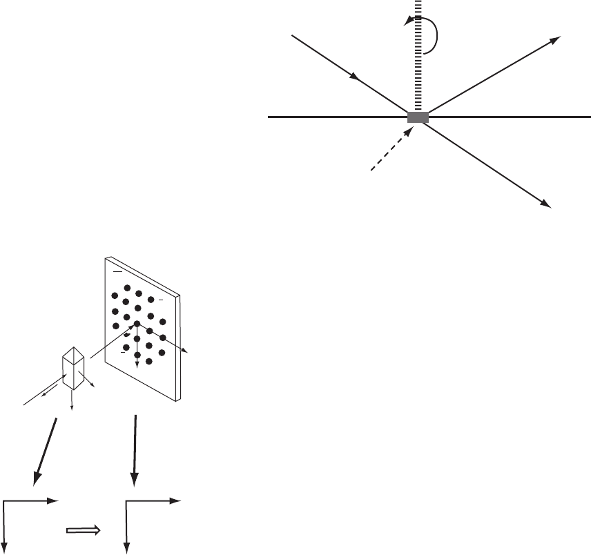
Fig. 4.7 Source, crystal, and detector.
Diagram of the relative arrangement of the X-ray source, the detector, and the crystal and
their relationship to the diffraction vector. All are in the same plane.
resulting diffraction pattern is recorded on photographic film placed
around the crystal. If the axis of rotation or oscillation is perpendicular
to the X-ray beam, the resulting photograph contains lines (layers) of
Bragg reflections (see Figure 4.10). As can be seen in this figure, many
of the Bragg reflections overlap each other, so that indexing them may
difficult. Therefore the Weissenberg camera was invented, in which
the camera is moved as the crystal is rotated or oscillated. Only one
layer from an oscillation photograph is selected, by the positioning
of a metal screen with a slit in it, between the film and the X-ray
source (Weissenberg, 1924). The crystal is oscillated back and forth,
Crystal
Detection
device
Reciprocal
lattice
Diffraction
pattern
c
h
k
b
*
a*
a*
b
*
k
hk0
hk0
hk0
hk0
h
X rays
Fig. 4.8 The relation between the crystal
orientation and the diffraction pattern.
The relative orientation of the reciprocal
lattice of a crystal (expressed here as a
∗
and b
∗
), and its indexed X-ray diffraction
pattern (expressed here as h and k). Note
the relationship of a
∗
to h and b
∗
to k.From
the positions of diffracted beams on the
detection device it is possible to deduce
the dimensions of the reciprocal lattice
and hence of the crystal lattice; hence the
indices h, k,andl of each Bragg reflection.
while the slit ensures that only one layer of Bragg reflections (for
example, a specific value for the h index) is recorded on the film. At
the same time the camera moves in a direction parallel to the axis of
crystal oscillation. The most important feature is that the motion of
the camera is coupled to the oscillation of the crystal, which helps in
interpreting the photograph. Bragg reflections on a Weissenberg pho-
tograph can therefore be more readily indexed than on an oscillation
photograph.
An even more useful type of X-ray diffraction photograph is pro-
duced by a precession camera (Figures 3.8a and 4.11) (Buerger, 1964).
It gives an undistorted view of one selected plane of the recipro-
cal lattice. This makes it particularly useful for measuring unit-cell
dimensions and assigning a space group to the crystal. Here the cam-
era motion is more complicated in order that the recorded image
of the diffraction pattern may be simple. In fact, direct measure-
ment of all reciprocal lattice parameters is possible from a series of
precession photographs, with an appropriate scale factor taken into
Equipment for diffraction studies 59
Unit cell
Crystal lattice
Reciprocal lattice
000
l /d
100
= a*
l /d
001
= c*
001
101
0
0
1
2
3
4
1
2
3
4
1
100
b
c
a
x
z
h
d
100
d
001
0
(b)
(a)
Unit in reciprocal
lattice
b*
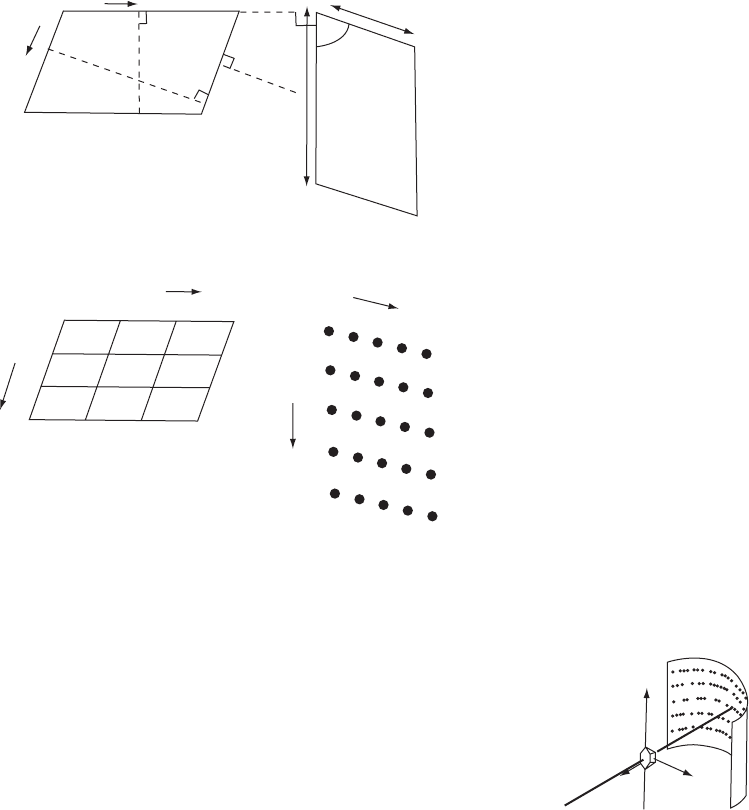
Fig. 4.9 The reciprocal lattice.
(a) The relationship between the unit cell of a crystal and its reciprocal lattice. (b) Indexing
of a reciprocal lattice.
consideration. An axis of the crystal perpendicular to the required
Crystal
mounted
on fiber
with c*
vertical
Photographic
film
X-ray beam
a
b*
c*
Fig. 4.10 Layer lines.
An X-ray diffraction photograph is
obtained from a crystal mounted with
the reciprocal lattice axis, c
∗
,vertical.On
oscillation about this vertical axis the
diffraction pattern shows layer lines, each
with a constant value for the index l along
them.
reciprocal lattice plane is inclined by an angle Ï (typically 30
◦
)tothe
direct incident X-ray beam, and this then precesses (like the motion
of a toy spinning top) about the incident X-ray beam. The flat film
holder, which has an annular screen that isolates a single plane of
the reciprocal lattice, follows the precession motion, ensuring that
the film is always parallel to the selected reciprocal lattice plane
of the crystal being photographed. It does this in such a way that the
direct beam always hits the center of the film. The photograph that
results from this complicated set of motions is simple to interpret.
This method is very useful for triclinic crystals and for macromolecular
crystals.
Generally, crystal symmetry, crystal lattice constants, and diffraction
data are currently measured with a diffractometer (Figure 4.12). The
incident radiation may be X rays from a sealed tube, a rotating anode,
or a synchrotron source, or it may be a neutron beam. A diffractometer
requires a collimated incident beam and a beamstop to collect that part
of the direct beam that has passed undeflected through the crystal. The
60 Experimental measurements
037
X rays
a
b⬚
c⬚
k
l
012
012
008 004 008
017
029
031
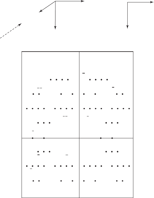
PARTIALLY INDEXED PRECESSION PHOTOGRAPH
033
012
012
037
040
Fig. 4.11 Indexing a precession photograph.
The indexing of Bragg reflections on a precession photograph. Note the systematic
absences—0k0 with k odd and 00l with l odd. By convention the positive direction of
a is toward the X-ray source.
detection system is an image plate or a charge-coupled device, rarely
photographic film. Many modern diffractometers do not require any
orientation of the crystal, only centering of the crystal, so that no matter
how the instrument is oriented the crystal is always centered in the
incident beam. A goniometer head can, however, be used to align the
crystal, if required. Protein crystals, mounted with mother liquor in a
capillary, are also put in a centering device. While both imaging with
film and digital signaling are employed for the detection of diffracted
radiation, they operate in different ways. A film records light as the
result of a series of chemical reactions, while charge-coupled devices
convert light (caused when X-ray photons hit a phosphor) directly into
a digital signal.
Equipment for diffraction studies 61
X-ray
source
omega (angle of
chi circle with
respect to
incident beam)
phi (f)
(a)
(b)
phi (f)
detection
device
2 theta (2
q)
omega (
w)
omega (
w)
block
omega (
w)
2 theta (2
q)
2 theta (2
q) circle
attached to detector
chi (
c)
circle
incident
diffracted
beam
X-ray beam
direct
beam
X-ray
source
phi (
f)
phi
(
f)
detection
device
2 theta (2
q)
omega (
w)
2 theta (2
q)
2 theta (2θ)
kappa (k)
block
kappa (k)
incident
diffracted
beam
X-ray beam
direct beam
kappa (k)
2q = angle between directions of
incident and diffracted beams
= angle detector has to be rotated
to intercept diffracted beam
f = spindle axis of gonoimeter head
w = angle between diffracted vector
and plane of c-circle
c = angle between f axis (gonio. head)
and diffractometer axis
(equatorial plane)
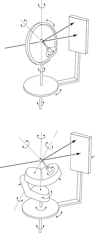
Fig. 4.12 An automatic diffractometer.
(a) A four-circle diffractometer. The crystal is mounted on a goniometer head, for which the spindle axis is ˆ. The goniometer head is
attached to the ˜ circle. The angle ˜ is the angle between the ˆ axis of the goniometer head and the base of the diffractometer. The ˜
circle can be rotated about the ˘ axis, where ˘ is the angle between the diffraction vector and the plane of the ˜ circle. The detector is
moved on the 2Ë circle, where 2Ë is the angle between the incident and diffracted X-ray beams. The detection device can be an image
plate or a charge-coupled device. The setup for serial measurement is shown here. (b) A diffractometer with kappa (Í) geometry. The
omega block rotates about the base plate while the kappa block rotates about the omega block as shown. This simulates the chi circle
motions in the instrument in (a) but avoids clashes.
62 Experimental measurements
There are several types of diffractometers. Some move a detector to
measure each Bragg reflection sequentially, and some employ a flat
detection device, an “area detector,” that measures a large number of
Bragg reflections at one time. The source of radiation is usually fixed
in space and, in a sequentially measuring diffractometer, the required
angular settings for the crystal and detector with respect to the incident
beam are calculated in advance once a few Bragg reflections have been
located and identified. This type of diffractometer is composed of sev-
eral mechanical circles that rotate the crystal or the detection system
with respect to the X-ray beam, as shown in Figure 4.12a. In this “four-
circle diffractometer” the crystal can be rotated around three axes (˜, ˆ,
and ˘) independently, and the detector can be rotated about a fourth
angle (2Ë, concentric with, but independent of, ˘), in the equatorial
plane parallel to the base of the instrument. The crystal is mounted on
a goniometer head and can be rotated about the vertical ˆ axis (phi)
of this mounting (see Figure 4.12a). The goniometer head is mounted
on the ˜ circle, which tilts the crystal about the horizontal ˜ axis (chi).
The 2Ë circle is attached to the detector device. This is concentric with
the ˘ circle that rotates the sample. The ˜ circle is mounted on top of the
˘ circle, and the ˆ circle is mounted on top of the ˜ circle. Usually the
entire instrument is controlled by a computer and the data collection
is then done automatically. There are also diffractometers that utilize
the kappa (Í) geometry (Figure 4.12b). This type of diffractometer was
designed specifically to reduce mechanical clashes during data collec-
tion. The ˘, ˆ, and 2Ë circles remain, but the ˜ circle is replaced by a
Í block that sits on the ˘ block (which replaces the ˘ circle) and this
controls the orientation of the crystal and its goniometer head.
If the measurement is to be sequential, the intensity of a Bragg
reflection is measured with the detector and recorded, together with
measurements of the background intensity near the Bragg reflection,
and then a new set of angles is calculated and another intensity mea-
surement made. One normally advances incrementally through the
Miller indices, hkl. In this way a systematic scan of all desired Bragg
reflections is done completely automatically. Alternatively, if the crystal
is stationary and white radiation is used, an image plate or charge-
coupled device will be positioned to receive and record as many as
possible of the diffracted beams. For this Laue diffraction, the inci-
dent radiation is white radiation with a range of wavelengths. It has
proved useful for studies of enzyme reactions (Hajdu et al., 1987).
For example, a crystal of the enzyme glycogen phosphorylase b was
mounted in a flow cell and substrate solution was passed over it. Laue
photographs (stationary crystal, white radiation) were taken with syn-
chrotron white radiation (over 10,000 Bragg reflections per second) at
a series of times after initiation of the biochemical reaction. A com-
parison of electron-density maps from the various data sets showed
the course of the reaction as a substrate was converted to product (by
phosphorylation).
Detection systems 63
Detection systems
The intensities of the diffracted beams are measured by intercepting
the beams with a detecting material or device that is sensitive to X
rays. The intensity at the peak of the diffraction spot is measured, or,
better, the peak profile is scanned. Measurements of background counts
are also made, or calculated from the profile of the peak, and used to
correct the recorded intensities. Measurements may be done electroni-
cally or photographically and may concentrate on one diffracted beam
at a time (as is often done with a diffractometer) or on many diffracted
beams at the same instant (as with electronic analogues of photographic
film).
The simplest detection device for X rays is photographic film. This
contains silver halide in an emulsion on its surface. When the film
is developed, black metallic silver is deposited at the positions at
which the diffracted beams hit the photographic film. The darkness
of each spot so formed is a measure of the intensity of the dif-
fracted beam. These intensities can be measured with a film scan-
ner. Film is not used much nowadays, because of the development
of electronic detection devices (with superior detection capabilities)
and current problems in obtaining photographic film suitable for X-ray
studies.
Electronic detectors of X rays that have an appreciable area for detec-
tion of the diffraction pattern, and offer the possibility of resolving
and individually measuring the intensities of diffraction maxima at
different points across this area, are now preferred. They consist of scin-
tillation counters, television-enhanced scanning devices, image plates,
and charge-coupled devices, and are the equivalent of electronic film.
Position-sensitive detectors can measure the position at which a Bragg
reflection hits the detection device. These various devices represent the
development of improved ways of recording a diffraction pattern elec-
tronically in a computer-readable manner, and image plates and charge-
coupled devices are the current instruments of choice for this. Whereas
photographic film records photons through a series of chemical reac-
tions, charge-coupled devices convert light directly into a digital signal.
Scintillation counters make use of the ability of certain substances to
emit visible light by fluorescence when X rays hit them. The intensity
of the emitted light is measured by a photomultiplier tube. Similarly,
television area detectors contain a phosphor that produces visible light
when hit by X rays. The photon signal is intensified and then detected
by a television photocathode. These methods of detection are now less
used than image plates and charge-coupled devices. Neutrons, which
lack any charge, and readily penetrate materials, are detected by gas or
scintillator detectors; these are similar to the X ray detectors described
above (Wilson, 2000).
An image plate is a storage phosphor on which a latent image is
formed when X rays hit it. It contains plastic sheets with powdered
64 Experimental measurements
phosphor crystals, doped with divalent europium ions, on their
surfaces. When X rays hit these sheets, the divalent europium ions
are converted to metastable trivalent ions and the electrons that are
liberated are stored ready for release when scanned by a laser beam
of visible light. When trivalent europium ions are encountered, blue
light (wavelength 3900 Å) is emitted that can be scanned and converted
to a digital image. This latent image has to be read; it is exposed to
laser light, which causes the emission of light of a different wavelength,
which is then detected. The image plate can then be erased, ready for
the next use, while the data from the scanning of the latent image, which
are in a computer-readable form, are then ready for use in structure
determination. The location of the direct beam is evident on the image,
and from the positions of diffracted beams it is possible to determine
the direction, as well as the intensity, of each Bragg reflection. Neutrons
can only be detected if they have undergone some reaction that results
in the emission of energetic charged particles; this means that a con-
verter must be used. Neutron image plates contain elements such as
gadolinium (which has a very high neutron, but not proton, capture
cross-section, or stopping power) that absorb neutrons and act as a
converter to enable the neutrons to emit electromagnetic radiation (such
as gamma rays), which can be detected like the X rays in the description
above.
Charge-coupled devices are used widely in X-ray diffraction equip-
ment. They are two-dimensional grids of radiation-sensitive semi-
conductor capacitors that have the capability of transferring charge
between their neighbors. They acquire a charge when hit by a photon,
and electron–hole pairs are generated by the photoelectric effect. The
total charge that is built up is a measure of the number of photons that
have been detected (the radiation intensity), and it is collected in an
array of electrodes. The charge and position of each pixel are transferred
as a result of a differential voltage across the electrodes, and the data
are read and digitized by a computer (see Ladd and Palmer, 2003). This
gives an immediate computer listing of the intensity and position on the
detection device, and therefore this device is closer to a direct detector
than is an image plate.
When white radiation is incident on a crystal, as in the Laue method,
it is necessary to know the wavelength of the radiation that causes
a particular Bragg reflection. The time-of-flight neutron diffraction
technique depends on the fact that neutrons with different energies
(wavelengths) travel at different speeds. Therefore a measurement of
the time of flight will reveal the wavelength of the diffracted beam
(generally selected from a multiwavelength incident beam). The instant
at which the diffracted beam hits the crystal and then impacts on the
detection system is measured and recorded. This, with the known dis-
tance traveled, gives the velocity of the neutron and hence its wave-
length. Therefore the wavelength of each diffracted neutron can be
measured.
Preparing measured I(hkl) for subsequent analysis 65
Preparing measured I(hkl) for subsequent
analysis
Since the intensity I(hkl) of any radiation propagated as a wave is
proportional to the square of its amplitude, |F (hkl)| the intensity of
the diffracted beam corresponding to the diffraction maximum for each
set of planes hkl is proportional to |F (hkl)|
2
. Modifications to I(hkl)are
necessary in order to correct for the geometry of measurement. Weak
Bragg reflections are measured carefully, rather than being ignored. Of
the various correction factors that are used, the Lorentz factor takes
into account the time that it takes for a Bragg reflection, represented
as a reciprocal lattice point with a finite size, to cross the surface of the
sphere of reflection; the longer the time, the higher the intensity. The
Lorentz factor equalizes the time taken to measure each Bragg reflec-
tion. The polarization factor depends on the state of polarization of the
incident X-ray beam; X rays are polarized on scattering, with reduction
of the intensity of the Bragg reflection. Corrections for absorption of
X rays by the crystal are also made; ideally, the path lengths through
the crystal of many component waves of each diffracted beam are com-
puted, and the diminution in intensity resulting from absorption can
then be determined. Semiempirical absorption corrections, based on the
intensity variation as certain intense Bragg reflections are scanned while
the crystal is rotated, are more generally used. If a crystal is strongly
absorbing for the radiation used, it may be shaped (with a scalpel
or razor blade) until it is approximately spherical so that absorption
corrections may be more uniform. Generally it is better to avoid using
a crystal larger than the primary beam, although this may be necessary
for protein crystals that are damaged by the X-ray beam, so that one can
move the crystal to an undamaged area during data collection. The aim
is to keep the amount of matter exposed to radiation independent of the
crystal orientation.
It is then possible to determine the absolute value (without phase) of
the structure factor F (hkl) from these measurements, as follows:
I(hkl)=k
1
{Î
3
V
c
Lp Abs/˘V
2
}|F (hkl)|
2
=K{Lp Abs}|F (hkl)|
2
=k
2
|F (hkl)|
2
(4.3)
where k
1
, k
2
, and K are constants, V
c
is the volume of the crystal that
is bathed in the incident beam, V is the volume of the unit cell, Lp
consists of the Lorentz and polarization factors, Abs is an absorption
correction, and ˘ is the angular velocity of the crystal. Thus, values of
k
2
|F (hkl)|
2
and hence of k
2
1/2
|F (hkl)| are immediately available once
intensity measurements have been made. The values of Lp and Abs
contain only known quantities and therefore can readily be computed
for each Bragg reflection.
66 Experimental measurements
0
2
1
0
−1
−2
0.1 0.2 0.3 0.4 0.5
B = 0.95 Å
2
0.6 0.7 0.8 0.9 1.0
sin
2
q/l
2
ln(〈F
2
〉 / 〈 f
2
〉)
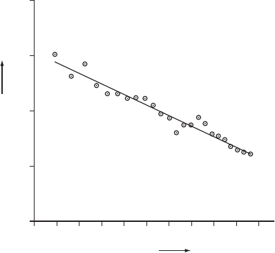
Fig. 4.13 Wilson plot.
A Wilson plot of sin
2
Ë/Î
2
versus the logarithm of a function of the measured structure
factors, F (hkl )=F . The slope gives an overall measure of the displacement factors. The
intercept gives the scale factor necessary to obtain intensities on an absolute scale.
If the value of I(hkl), corrected for Lp and Abs, is called I
corr
, we can
say
I
corr
= I(hkl)/{Lp Abs} =K|F (hkl)|
2
=K|F
novib
|
2
exp(−2B
iso
sin
2
Ë/Î
2
)
(4.4)
where |F
novib
| is the value of |F (hkl)| for a structure composed of non-
vibrating point atoms. The application of the Lp correction involves no
knowledge of the structure. An estimation of Abs can be made from a
knowledge of the shape, orientation, and composition of the crystal.
The value of |F(hkl)| so derived contains information on the atomic
displacement factors, B. Thus F = |F
novib
|exp(−B
iso
sin
2
Ë/Î
2
)(seeTrue-
blood et al., 1996). It is possible to derive B
iso
and K in Eqn. (4.4) from
the experimental data by a “Wilson plot” (Wilson, 1942). It is assumed
that, to a first approximation, the average intensity of Bragg reflections
at a certain value of 2Ë depends only on the atoms present in the cell, not
on their positions—that is, that the arrangement of atoms in the crystal
structure is random. By comparison of the averages of the observed
intensities in ranges (shells) of sin
2
Ë/Î
2
with the theoretical values for
a unit cell with the same atomic contents, approximate values for K
and B
iso
can be found from the Wilson plot (Figure 4.13). Values of the
resulting scale factor K can then be used for preparation of a full list
of values of |F (hkl )| on an approximately absolute scale (relative to the
scattering by one electron) for all Bragg reflections measured. The value
of B
iso
obtained from this graph will indicate the extent of disorder from
unit cell to unit cell in the crystal structure.
Summary 67
The reader should note that the intensity, I(hkl), is a simple function
of the structure amplitude |F |. However, an inspection of Eqn. (4.4)
shows that each value of |F(hkl)|, and hence of the intensity, I(hkl), of the
diffracted beams contains, with few exceptions, a contribution from every
atom in the unit cell. The unraveling of these contributions makes the
structure solution complicated.
Summary
The diffraction of a crystal by X rays results from the constructive and
destructive interference of the X rays that have been scattered by each
individual atom in the structure. Three types of experimental diffrac-
tion data may be obtained:
(1) The angle of scattering (2Ë, the angular deviation from the direct
undeviated beam), which is used to measure the spacings of the
reciprocal lattice and hence the spacings of the crystal lattice.
These spacings can be used to derive the size and shape of the
unit cell.
(2) The orders of diffraction (hkl) of each diffracted beam.
(3) The intensities of the diffracted beams, I(hkl), which may be
analyzed to give the positions of the atoms within the unit cell.
These atomic positions are usually expressed as fractions of the
unit-cell edges.