Glusker J.P., Trueblood K.N. Crystal Structure Analysis: A Primer
Подождите немного. Документ загружается.

28 Diffraction
IN PHASE, HIGH INTENSITY
PARTIALLY OUT OF PHASE, LOWER INTENSITY
OUT OF PHASE, NO INTENSITY
A = 1.4, I = 2.0.
l/4 = phase difference
1.4
A = 2.0, I = 4.0 in phase
0 = phase difference
(a)
(b)
(c)
Resultant wave
2.0
A = 0.0, I = 0.0 out of phase
l/2 = phase difference
λ
Amplitude = 1.0
0.0
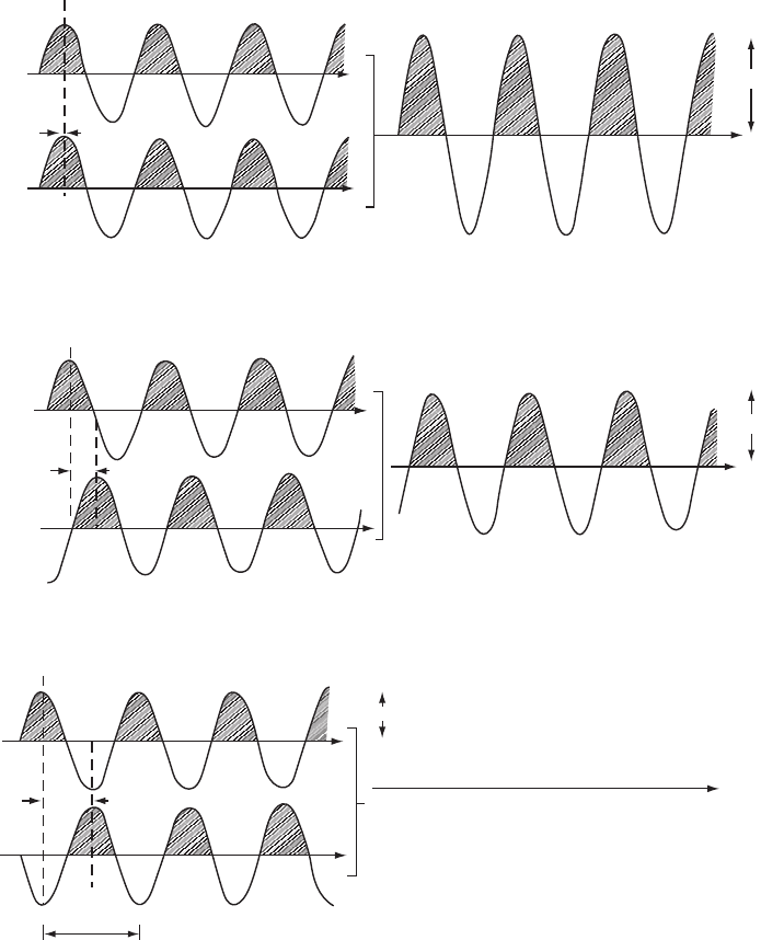
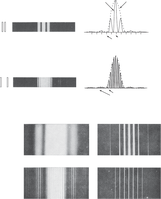
Fig. 3.2 Interference of two waves. Summation of waves.
Three examples are shown of what happens when two parallel waves of the same wavelength and equal amplitude add. In each
example, the two separate waves are shown on the left and their sum or resultant wave on the right. The different examples are
characterized by varying phase differences. The relative phase of a wave is the position of a crest relative to some arbitrary point (see
Figure 1.2). This position (relative phase) is usually expressed as a fraction of the wavelength, and often this fraction is multiplied by
360
◦
or 2π radians, so that the phase will be given as an angle. Thus a phase difference of Î/4 may be given as 1/4, 90
◦
,or2/π radians.
The resultant wave has the same wavelength, Î, as the original two waves. The intensity, I , of the resultant wave is proportional to the
square of its amplitude, A, obtained on wave summation.

Diffraction of visible light by single slits 29
Figure 3.1a, the slit is narrow and the diffraction pattern is wide, while
in the other, Figure 3.1b, the slit has a greater width but the diffrac-
tion pattern is narrower. This implies that there is a reciprocal relation
between the angular spread of the scattering or diffraction pattern in
a particular direction and the corresponding dimension of the object
causing the scattering. The smaller the object, the larger the angular
spread of the diffraction pattern. What is actually involved is the ratio of
Î (the wavelength of the radiation used) to the minimum dimension, a,
of the scattering object (for example, the width of the slit); the larger
the value of Î/a (wavelength divided by slit width), the greater the
spread of the pattern. Therefore Figure 3.1b might equally well be a
view of the same slit as in Figure 3.1a, illuminated with radiation of
wavelength about 2.2 times shorter than that used in Figure 3.1a. It
is, in fact, possible to produce this change of scale by any change in
a and Î whose combined effect is to decrease the value of Î/a by a
factor of 2.2.
The phenomenon of interference between two waves traveling in
the same direction and the importance of phase differences between
these two parallel waves are illustrated in Figure 3.2. The amplitude
of the wave resulting from the interaction of two separate waves trav-
eling in the same direction with the same wavelength, and a con-
stant phase difference, depends markedly on the size of this phase
difference. Figure 3.2 shows how such waves may be summed
‡
for
‡
The displacements from the mean (zero),
parallel to the vertical axis (the ordinates),
are directly summed at many points along
the horizontal axis (the abscissae) to give
the resultant wave.
three examples of different phase differences (zero, a quarter, and
half a wavelength). The intensity of the resulting beam is propor-
tional to the square of the amplitude of the summed waves in each
case.
The variations in intensity seen in Figure 3.1 arise from the inter-
ference of the secondary wavelets generated within the slit, as shown
in Figure 3.3. In the direction of the direct beam, the waves scattered
by the slit are totally in phase and reinforce one another to give max-
imum intensity. However, at other scattering angles, as illustrated in
Figure 3.3, the relative phases of the waves cause interference between
waves traveling in the same direction so that the intensity falls off as a
function of scattering angle; this leads to an overall intensity contour
of the diffraction peak, and we term this “the envelope.” At most
scattering angles the different scattered waves are neither completely in
phase nor completely out of phase, so that there is partial reinforcement
and thus an intermediate intensity of the diffracted beam. The result is
illustrated in the single-slit diffraction pattern (the envelope) shown on
the right of Figure 3.3.
(a) Phase difference zero. In this case there is total reinforcement, and the waves are said to be “in phase” or to show “constructive
interference”. If the original waves are of unit amplitude, the resultant wave has amplitude 2, intensity 4.
(b) Phase difference Î/4. Partial reinforcement occurs in this case to give a resultant wave of amplitude 1.4, intensity 2.
(c) Phase difference Î/2. The waves are now completely “out of phase” and there is destructive interference, which gives no resultant
wave (that is, a wave with amplitude 0, intensity 0).
30 Diffraction
Direct beam
resultant (A)
Resultant (B)
Resultant (C)
A
B
C
C
C
B
B
A
Envelope
Intensity
DIFFRACTION BY
ONE SLIT
DIFFRACTION BEAM PROFILE
(THE ENVELOPE)
Fig. 3.3 Diffraction by a single slit.
Diffraction from a single slit is diagrammed by the superposition of waves generated within the area of the slit. The variation in
intensity with increasing angle is shown by the different amplitudes of the resultant waves (A, B, and C) at different angles. Left-hand
side: diffraction by a single slit; right-hand side: diffracted beam profile (the envelope), showing the location of A, B, and C on this
envelope.
Diffraction of light by regular arrays of slits
In order to consider what happens when a crystal that has a periodic
internal structure diffracts radiation, we now describe diffraction by
a series of equidistantly aligned slits. Reinforcement of the diffracted
beam occurs at angles at which the path difference between two par-
allel waves is an integral number of wavelengths; for example, when
the two waves are out of phase by three wavelengths (n = 3), there
will be reinforcement at a specific scattering angle and the wave will
be described as the “third order of diffraction” (see Figure 3.4). As
shown in Figure 3.5, the diffraction pattern of a single slit is modified
by interference effects when increasing numbers of slits are placed
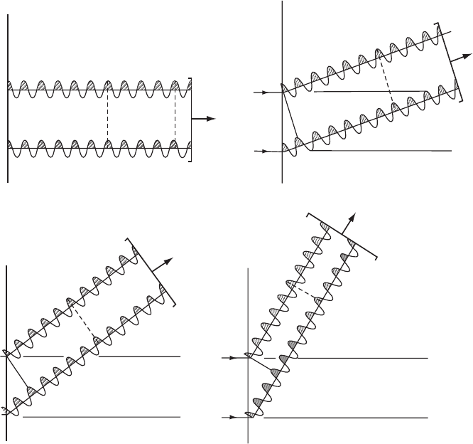
Diffraction of light by regular arrays of slits 31
Third order 300
(Three wavelengths
path difference)
Direct beam
(Zero path difference)
First order 100
(One wavelength
path difference)
Second order 200
(Two wavelengths
path difference)
000
100
300
200
Fig. 3.4 Orders of diffraction.
First, second, third, and higher orders of diffraction are obtained as scattered waves differ by one, two, three, and more wavelengths.
Readers should satisfy themselves that with a smaller spacing a between scattering objects, the angle at which a given order of
diffraction occurs is proportionally increased.
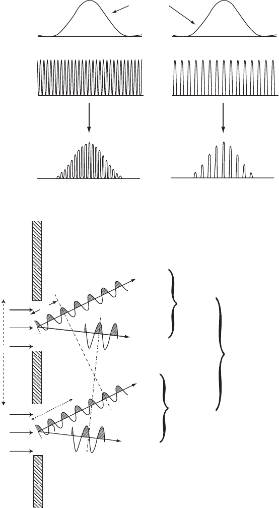
side by side in a regular manner to form a one-dimensional grating.
Sometimes rays proceeding in a specific direction after scattering are
in phase and sometimes they are not. The important point to note is
that the diffraction pattern from a grating of slits is a sampling of the single-
slit pattern in narrow regions that are representative of the spacings between
the slits (see Figure 3.5). With even as few as 20 slits in the “grating”
(see Figure 3.6), the small subsidiary maxima vanish almost completely
and the lines in the diffraction pattern are sharp. The overall diffraction
pattern of a series of slits is thus composed of an “envelope” and a series
of “sampling regions” within the envelope. This envelope represents
the diffraction pattern of a single slit (see Figure 3.3). The “sampling
regions” result from interference of waves scattered from equivalent
points in different slits; the spacing of these sampling regions in the
diffraction pattern (see Figure 3.6) is inversely related to the spacing of
the slits.
32 Diffraction
Larger spacing
between slits
Sampling regions
(a)
Envelope
++
Smaller spacing
between slits
SAMPLING
REGIONS
(D
1
and D
2
)
(E
1
and E
2
)
3l
D
2
D
1
E
2
E
1
(b)
ENVELOPE
(D
1
, E
1
)
(D
2
, E
2
)
DIFFRACTION BY TWO SLITS
In
phase
(third order)
Partially
out of
phase
ENVELOPE
q
d
2l
Fig. 3.5 Diffraction by two slits.

The reciprocal lattice 33
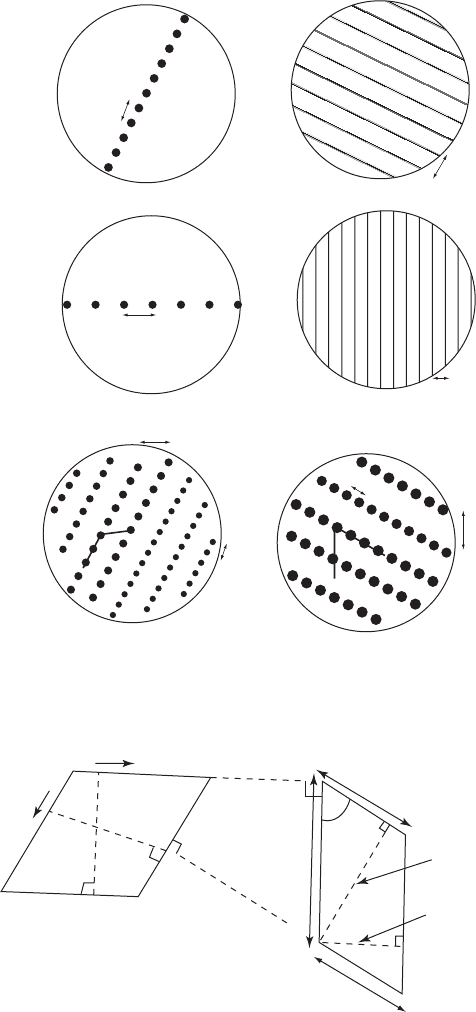
Figure 3.7 shows schematically how a two-dimensional regular
arrangement of simple scattering objects, in this case holes in an opaque
sheet (drawn as black spots on the left), produces a two-dimensional
diffraction pattern (drawn as lines or spots on the right). Each of the
one-dimensional gratings in Figures 3.7a and b produces (in two dimen-
sions) a pattern of scattered light (the diffraction pattern) consisting of
lines (representing the maxima of light, as seen on the right). These
lines are perpendicular to the direction of the original grating because
interference effects between light scattered from adjacent holes reduce
the scattered light intensity effectively to zero in all directions except
that perpendicular to the repeat direction of the original grating. Hence
lines of diffracted light are formed. In Figure 3.7c, a combination of
both kinds of one-dimensional gratings that were shown in (a) and (b)
are present at once. This gives a regular two-dimensional grating. The
lattice of the diffraction pattern in Figure 3.7c is necessarily, then, the
“reciprocal” of the lattice of the original scattering objects (the crystal
lattice) shown on its left; see Figure 3.7d. This will now be described.
The reciprocal lattice
In addition to the lattice of the crystal structure in real or crys-
tal space (discussed earlier), there is a second lattice, related to the
first, that is of importance in diffraction experiments and in many
other aspects of solid state physics. This is the reciprocal lattice, intro-
duced by Josiah Willard Gibbs in 1884, long before X-ray diffraction
was known (Gibbs, 1901; Ewald, 1921). Its definition in terms of the
crystal lattice vectors is shown in Appendix 3. In the reciprocal lat-
tice a point, (hkl), is drawn at a distance 1/d
hkl
from the origin (the
direct beam, (000)), and in the direction of the perpendicular distance
between the (hkl) crystal lattice planes (Figure 3.7d). The relation-
ship between these two important lattices (the crystal and reciprocal
lattices) is a particularly simple one if the fundamental translations
of the crystal lattice are all perpendicular to one another; then the
(a) An overview of the envelope profile (equivalent to diffraction by a single slit or
an atomic arrangement) and the sampling regions (equivalent to the diffraction
of a series of equidistant slits or a crystal lattice). The envelope is accessed at the
sampling regions only.
(b) When diffraction occurs from two slits, there are two effects to consider:
(1) The variation in intensity with angle as a result of interference of the waves
generated within each slit separately. Interference between D
1
and E
1
and
between D
2
and E
2
gives the “envelope,” as obtained for a single slit (see also
Figure 3.3). This is the equivalent of diffraction by a single slit.
(2) The interference of scattered waves at a given angle with those at the same
angle from the adjacent slit (D
1
with D
2
from the next slit, E
1
with E
2
from the
next slit, etc.). At angles of constructive interference, when the two resultant
waves are in phase, “sampling” of the “envelope” occurs, as shown in part (a).
At certain other angles, no diffraction is observed. This sampling is the result
of the distance between the two slits.
34 Diffraction
Diffraction pattern of two slits
Photometer
trace
Envelope
Photometer
trace
Sampling regions
Sampling
region
Sampling regions
Wide spacing (6a) between slits (each width a). Narrow spacing between sampling regions.
d = 2a
d = 2a
d = 6a
d = 6a
Photograph
Slits
Slits
Photograph
Narrow spacing (2a) between slits (each width a). Wide spacing between sampling regions
(a)
Varying numbers of slits
1 slit
(b)
5 slits
20 slits2 slits
Fig. 3.6 Diffraction patterns from equidistant parallel slits.
(a) The effect of varying the distance, d, between two slits of constant width, a, is shown. On the left is a diagram of the slits with
spacings of 2a and 6a, respectively, between them. In the center is shown a photograph of the diffraction pattern. On the right,
a photometer tracing of the diffraction pattern for the combination of the two slits is drawn as a solid line, and the diffraction
pattern for a single slit, referred to in the text as the “envelope,” is drawn as a dashed line. The envelope in both cases has the
same shape because it represents the diffraction pattern of a single slit of the same width. The regions of the “envelope” that are
sampled are indicated by short vertical lines at the lower edge of the drawings on the right. When there is a relatively narrow
spacing between the slits (d =2a), the distance between sampling regions is relatively large, as shown in the upper diagram.
When there is a relatively wide spacing between the slits (d =6a), the distance between sampling regions has decreased; that is,
there is an inverse relationship of the spacing of the sampling regions to the spacing of the slits.

Diffraction of X rays by atoms in crystals 35
fundamental translations of the reciprocal lattice are parallel to those of
the crystal lattice, and the lengths of these translations are inversely
proportional to the lengths of the corresponding translations of the
crystal lattice. With nonorthogonal axes, the relationships between
the crystal lattice and the reciprocal lattice are not hard to visualize geo-
metrically; a two-dimensional example is given in Figure 3.7d. As we
shall see shortly, the fundamental importance of the reciprocal lattice in
crystal diffraction arises from the fact that if a structure is arranged on a
given lattice, then its diffraction pattern is necessarily arranged on the lattice
that is reciprocal to the first.
§
§
This may be stated alternatively as fol-
lows. The diffraction pattern of a molecu-
lar crystal is the product of the diffraction
pattern of the molecule (also called the
molecular transform) with the diffraction
pattern of the crystal lattice (which is also
a lattice, the reciprocal lattice, described
above). The result is a sampling of the
molecular transform at each of the recipro-
cal lattice points. The diffraction pattern of
a single molecule is too weak to be observ-
able. However, when it is reinforced in a
crystal (containing many billions of mole-
cules in a regular array) it can be readily
observed, but only at the reciprocal lattice
points.
Diffraction of X rays by atoms in crystals
It is a principle of optics that the diffraction pattern of a mask with
very small holes in it is approximately equivalent to the diffraction
pattern of the “negative” of the mask—that is, an array of small dots
at the positions of the holes, each dot surrounded by empty space. This
equivalence is discussed lucidly by Richard Feynman (Feynman et al.,
1963). In a crystal, the electrons in the atoms act, by scattering, as sources
of X rays, just as the wavefront in the slits in a grating may be regarded
as sources of visible light. There is thus an analogy between atoms in a
crystal, arranged in a regular array, and slits in a grating, arranged in
a regular array. In diffraction of X rays by crystals, as of visible light
by slits in a grating, the intensities of the diffraction maxima show a
variation in different directions and also vary significantly with angle
of scattering.
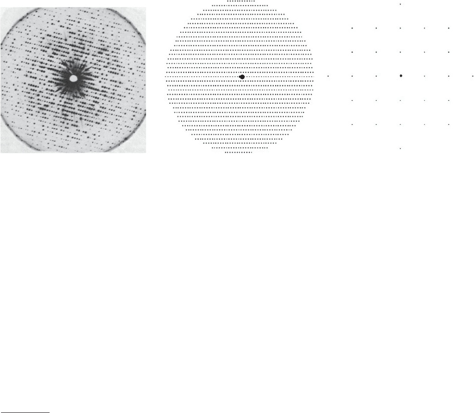
Most unit cells contain a complex assembly of atoms, and each atom
is comparable in linear dimensions to the wavelength of the X rays
or neutrons used. Figure 3.8a shows a typical X-ray diffraction photo-
graph, taken by the “precession method,” which records the reciprocal
lattice without distortion. Considerable variation in intensity of the
individual diffracted beams is evident; this is a result of the arrange-
ment of atoms (and their accompanying electron density) in the struc-
ture. The analogy with Figures 3.3, 3.5, and 3.6 holds; that is, the X-
ray photograph is merely a scaled-up sampling of the diffraction pattern of
the contents of a single unit cell. The “envelope,” which is shown by the
(b) Diffraction patterns are shown for gratings containing 1, 2, 5, and 20 equidistant slits, illuminated by parallel radiation of the
same wavelength. The diffraction pattern for a grating composed of 20 (or more) slits consists only of sharp lines, the intervening
minor maxima having disappeared; similarly, the diffraction pattern for a crystal composed of many unit cells contains sharp
diffraction maxima.
Summary of key points:
(1) The size and shape of the envelope are determined by the diffraction pattern of a single slit.
(2) The positions of the regions in which the envelope is sampled are determined by the spacing between the slits.
From Fundamentals of Optics by Francis A. Jenkins and Harvey E. White, 3rd edition (1957) (Figures 16E and 17A). Copyright © 1957,
McGraw-Hill Book Company. Used with permission of McGraw-Hill Book Company.
ORIGINAL
GRATING
(a)
(b)
(c)
(d)
a
a*
a
reciprocal space
real space
CRYSTAL
LATTICE
RECIPROCAL
LATTICE
010
000
110
a*
b*
a
b*
b*
k/b
K/b
K/a
k/a
0
b
100
1.0
1.0
d
100
d
010
b
b
g
g*
g
g*
DIFFRACTION
PATTERN
Fig. 3.7 Diagrams of diffraction patterns from one- and two-dimensional arrays. Relation
between the crystal lattice and reciprocal lattice.
Diffraction of X rays by atoms in crystals 37
(b)
(a)
Fig. 3.8 X-ray diffraction photographs taken by the precession method.
(a) The precession method gives an undistorted representation of one layer of the reciprocal lattice. An X-ray precession photograph
of a crystal of myoglobin is shown here. The direct X-ray beam, which might otherwise “fog” the film, has been intercepted, hence
the white hole in the middle of the photograph. The radial streaks, found for very intense Bragg reflections, occur because the X
rays are not truly monochromatic (one wavelength) but contain background radiation of varying wavelength but lower intensity.
As a result the spot appears somewhat smeared out (that is, for each Bragg reflection, sin Ë/Î is constant but since Î varies for
the background “white radiation,” sin Ë must also vary, giving rise to a streak rather than a spot on the film). Note the regularity
of the positions of spots in this photograph but the wide variation in intensity (from a very black spot to one that is almost or
apparently absent). The positions of the spots (diffracted beams) give information on unit cell dimensions; the intensities of the
spots give information on the arrangement of atoms in that unit cell.
Photograph courtesy Dr. J. C. Kendrew.
(b) A comparison of diagrams of the diffraction patterns of myoglobin (large unit cell, monoclinic, a = 64.5 Å, b = 30.9 Å not shown,
c = 34.7 Å, β = 106.0
◦
) on the left and potassium chloride (small unit cell, cubic, a = 6.29 Å) on the right. The larger the unit cell,
the nearer together the diffraction spots if the wavelength of the radiation is the same for both. Variations in Bragg reflection
intensities are not shown in these diagrams. Note that many Bragg reflections are measured when the unit cell is large.
variation in intensities of the individual diffracted-beam spots, is the
diffraction pattern of the scattering matter (the electrons of the atoms)
in a single unit cell. The “sampling regions,” which are the positions of
the diffracted-beam spots, are arranged on a lattice that is “reciprocal”
to the crystal lattice. Measurements of the distances between these will
lead to the dimensions of the unit cell, and they sample the diffraction
(a, b, c) On the left is shown the grating used and, on the right, the corresponding
diffraction pattern (such as might be obtained by holding the original grating in front of a
point source of light). a and b are direct lattice vectors in the crystal or grating, and a
∗
and
b
∗
are vectors in the diffraction pattern (a and b are the spacings of the original gratings,
and a
∗
and b
∗
are spacings in the diffraction pattern). The reciprocal relationships of a and
b to the spacings of certain rows in the diffraction pattern are shown. These are diagrams,
and no intensity variation is indicated. The black dots on the left-hand side represent
holes that cause diffraction, giving the pattern on the right-hand side, in which black
lines or spots represent appreciable intensity for diffracted light.
Adapted from H. Lipson and W. Cochran. The Crystalline State. Volume III. The Determi-
nation of Crystal Structures. Cornell University Press: Ithaca, New York; G. Bell and Sons:
London (1966) (Lipson and Cochran, 1966).
(d) The relationships of a and b in the crystal lattice to a
∗
and b
∗
in the corresponding
reciprocal lattice are shown.