Glusker J.P., Trueblood K.N. Crystal Structure Analysis: A Primer
Подождите немного. Документ загружается.


xx Symbols used in this book
F
P
, F
PH1
, F
PH2
,
F
H1
, F
H2
, F
M
,
F
M
, F
R
, F
T
, F
T
Structure factors for a given value of hkl for a protein (P), two heavy-atom derivatives
(PH1andPH2), the parts of F due to certain atoms (M, M
, H1andH2) and the rest
of the molecule (R), and for the total structure (T and T
).
F Structure factor when represented as a vector.
F
novib
Value of F for a structure containing only nonvibrating atoms.
F
+
, F
−
Values of F(hkl)andF (
¯
h
¯
k
¯
l) when anomalous-dispersion effects are measurable.
f (hkl), f , f
j
Atomic scattering factor, also called atomic form factor, for the hkl reflection relative to
the scattering by a single electron. The subscript j denotes atom j.
f
, f
When an anomalous scatterer is present the value of f is replaced by ( f + f
)+if
.
G(r) Radial distribution function.
G, H Values of A and B with the scattering factor contribution ( f + f
+ f
) removed (see
Chapter 10).
H Reciprocal lattice vector.
H, K Indices of two Bragg reflections. H = h, k, l; K = h
, k
, l
.
hkl, −h, −k, −l,
¯
h
¯
k
¯
l, hkil
Indices of the Bragg reflection from a set of parallel planes; also the coordinates of a
reciprocal lattice point. If h, k,orl are negative they are represented as −h, −k, −l or
¯
h
¯
k
¯
l. In hexagonal systems a fourth index, i = −(h + k), may be used (see Appendix 2).
(hkl) Indices of a crystal face, or of a single plane, or of a set of parallel planes.
I Body-centered lattice.
I(hkl), I Intensity (on an arbitrary scale) for each reflection.
I
corr
Value of I corrected for Lp and Abs.
i An “imaginary number,” i =
√
−1
i, j Any integers.
Lp Lorentz and polarization factors. These are factors that are used to correct values of
I for the geometric conditions of their measurement
l The distance between two points in the unit cell (e.g., bond length).
l A direction cosine.
M Molecular weight of a compound.
M
1
,M
2
,M,M
Atoms or groups of atoms that are interchanged during the preparation of an isomor-
phous pair of crystals. Heavy atoms substituted in a protein, P.
m Mirror planes.
N The number of X-ray reflections observed for a structure.
N
Avog.
Avogadro’s number. The number of molecules in the molecular weight in grams, 6.02 ×
10
23
.
n Any integer. Used for n-fold rotation axes. Also used as a general constant.
n
r
Screw axis designations, where n and r are integers (2, 3, 4, 6 and 1, ..., (n − 1),
respectively).
P, PH1, PH2 Protein (P), also heavy-atom derivatives PH1andPH2.
P(uvw), P,
P
s
(uvw)
The Patterson function, evaluated at points of u, v, w in the unit cell. The P
s
(uvw)
function is used with anomalous-dispersion data (see Chapter 10).

Symbols used in this book xxi
P, A, B, C, F , I Lattice symbols. Primitive (P), centered on one set of faces (A, B, C), or all faces (F )of
the unit cell, or body-centered (I).
P
+
Probability that a triple product is positive (see Eqns. 8.6 and 8.7).
p, q Path differences.
Q The quantity minimized in a least-squares calculation.
R Discrepancy index R =
|
(
|
F
o
|
−
|
F
c
|
)
|
|
F
o
|
. Also called R factor, R value, or residual.
R/S System of Cahn and Ingold for describing the absolute configuration of a chiral
molecule.
r The distance on a radial distribution function.
s(hkl) The sign of the reflection hkl for a centrosymmetric structure.
t Crystal thickness.
U
11
, U
ii
, U
ij
Anisotropic vibration parameters.
u
2
Mean square amplitude of atomic vibration.
u, v, w The coordinates of any one of a series of systematically spaced points, expressed as
fractions of a, b,andc, in the unit cell for a Patterson (or similar) function.
V
c
, V, V
∗
The unit-cell volume in direct and reciprocal space.
V
M
Matthews coefficient, volume on Å
3
per dalton of protein.
w(hkl) The weight of an observation in a least-squares refinement.
X, Y, Z Cartesian coordinates for atomic positions.
x, y, z; x
j
, y
j
, z
j
;
x, y, z, u
Atomic coordinates as fractions of a, b,andc. The subscript j denotes the atom under
consideration. If the system is hexagonal a fourth coordinate, u, may be added (see
Appendix 2).
x
1
, x
2
, x
j
, x
r
Displacements of a wave at a given point. The waves are each designated 1, 2, j; r is
the resultant wave from the summation of several waves.
x, y, z Coordinates of any one of a series of systematically spaced points, expressed as frac-
tions of a, b, c filling the unit cell at regular intervals.
Z Number of molecules in a unit cell.
Z
i
, Z
j
The atomic number (total number of diffracting electrons) of atoms i and j.
·, ‚, „ Interaxial angles between b and c, a and c,anda and b, respectively (alpha, beta,
gamma).
·
∗
, ‚
∗
, „
∗
Interaxial angles in reciprocal space.
·(hkl), ·, ·
M
, ·
P
,
·
H
Phase angle of the structure factor for the reflection hkl. · =tan
−1
(B/ A).
·
1
, ·
2
, ·
j
, ·
r
Phases of waves 1, 2, j,andr, the resultant of the summation of waves, relative to an
arbitrary origin.
ƒ|F | The difference in the amplitudes of the observed and calculated structure factors, |F
o
|−
|F
c
| (delta |F |).
ƒÒ Difference electron density.
‰ Interbond angle.
‰
ij
An index that is 1 when i = j and 0 elsewhere; i and j are integers (delta).
xxii Symbols used in this book
ε Epsilon factor used in calculating normalized structure factors (see Glossary).
Ë, Ë
hkl
The glancing angle (complement of the angle of incidence) of the X-ray beam to the
“reflecting plane.” 2Ë is the deviation of the diffracted beam from the direct X-ray
beam (two theta).
Í A device for aligning the crystal and detector in a diffractometer that utilizes Í geomet-
ry (Figure 4.12) (kappa).
Î Wavelength, usually that of the radiation used in the diffraction experiment (lambda).
Ï/Ò Mass absorption coefficient. Ï, linear absorption coefficient; Ò, density.
Ò(xyz), Ò
obs
, Ò
calc
Electron density, expressed as number of electrons per unit volume, at the point x, y, z
in the unit cell (rho).
Summation sign (sigma).
1
,
2
Listing of triple products of normalized structure factors (see Chapter 8).
Ù Torsion angle.
ˆ An angular variable, proportional to the time, for a traveling wave. It is of the form
2πÌt, where Ì is a frequency and t is the time (phi).
ˆ Angle on spindle axis of goniometer head. See diffractometer (Figure 4.12).
ˆ
H
The phase angle of the structure factor of the Bragg reflection H.
˜ Angle between ˆ axis and diffractometer axis (see Figure 4.12) (chi).
¯ Angle incident beam makes with lattice rows (see Appendix 4) (psi).
˘ Angle between diffraction vector and plane of ˜ circle on diffractometer (Figure 4.12)
(omega).
The mean value of a quantity.
1, 2, 3, 4, 6 Rotation axes.
¯
1,
¯
2,
¯
3,
¯
4,
¯
6 Rotatory-inversion axes.
2
1
,4
1
,4
2
,4
3
Screw axes n
r
.

CRYSTALS AND
DIFFRACTION
Part
I
This page intentionally left blank

Introduction
1
Much of our present knowledge of the architecture of molecules has
been obtained from studies of the diffraction of X rays or neutrons by
crystals. X rays
*
are scattered by the electrons of atoms and ions, and the
*
“X ray” for a noun, “X-ray” for an adjec-
tive.
interference between the X rays scattered by the different atoms or ions
in a crystal can result in a diffraction pattern. Similarly, neutrons are
scattered by the nuclei of atoms. Measurements on a crystal diffraction
pattern can lead to information on the arrangement of atoms or ions
within the crystal. This is the experimental technique to be described in
this book.
X-ray diffraction was first used to establish the three-dimensional
arrangement of atoms in a crystal by William Lawrence Bragg in 1913
(Bragg, 1913), shortly after Wilhelm Conrad Röntgen had discovered
X rays and Max von Laue had shown in 1912 that these X rays could
be diffracted by crystals (Röntgen, 1895; Friedrich et al., 1912). Later, in
1927 and 1936 respectively, it was also shown that electrons and neu-
trons could be diffracted by crystals (Davisson and Germer, 1927; von
Halban and Preiswerk, 1936; Mitchell and Powers, 1936). Bragg found
from X-ray diffraction studies that, in crystals of sodium chloride, each
sodium is surrounded by six equidistant chlorines and each chlorine by
six equidistant sodiums. No discrete molecules of NaCl were found and
therefore Bragg surmised that the crystal consisted of sodium ions and
chloride ions rather than individual (noncharged) atoms (Bragg, 1913);
this had been predicted earlier by William Barlow and William Jackson
Pope (Barlow and Pope, 1907), but had not, prior to the research of the
Braggs, been demonstrated experimentally. A decade and a half later, in
1928, Kathleen Lonsdale used X-ray diffraction methods to show that
the benzene ring is a flat regular hexagon in which all carbon–carbon
bonds are equal in length, rather than a ring structure that contains
alternating single and double bonds (Lonsdale, 1928). Her experimental
result, later confirmed by spectroscopic studies (Stoicheff, 1954), was of
great significance in chemistry. Since then X-ray and neutron diffraction
have served to establish detailed features of the molecular structure of
every kind of crystalline chemical species, from the simplest to those
containing many thousands of atoms.
We address ourselves here to those concerned with or interested in
structural aspects of chemistry and biology who wish to know how
3
4 Introduction
crystal diffraction methods can be made to reveal the underlying three-
dimensional structure within a crystal and how the results of such
structure determinations may be critically assessed. In order to explain
why molecular structure can be determined by single-crystal diffraction
of X rays or neutrons, we shall try to answer several questions: Why use
crystals and not liquids or gases? Why use X rays or neutrons and not
other types of radiation? What experimental measurements are needed?
What are the stages of a typical structure determination? How are the
structures of macromolecules such as proteins and viruses determined?
Why is the process of structure analysis sometimes lengthy and com-
plex? Why is it necessary to “refine” the approximate structure that is
first obtained? How can one assess the reliability of a crystal structure
analysis?
This book should be regarded not as an account of “how to do it” or of
practical procedural details, but rather as an effort to explain “why it is
possible to do it.” We aim to give an account of the underlying physical
principles and of the kinds of experiments and methods of handling
the experimental data that make this approach to molecular structure
determination such a powerful and fruitful one. Practitioners are urged
to look elsewhere for details.
The primary aim of a crystal structure analysis by X-ray or neutron
diffraction is to obtain a detailed three-dimensional picture of the con-
tents of the crystal at the atomic level, as if one had viewed it through
an extremely powerful microscope. Once this information is available,
and the positions of the individual atoms are therefore known precisely,
one can calculate interatomic distances, bond angles, and other features
of the molecular geometry that are of interest, such as the planarity of a
particular group of atoms, the angles between planes, and conformation
angles around bonds. Frequently the resulting three-dimensional repre-
sentation of the atomic contents of the crystal establishes a structural
formula and geometrical details hitherto completely unknown. Such
information is of great interest to chemists, biochemists, and molecular
biologists who are interested in the relation of structural features to
chemical and biological effects. Furthermore, precise molecular dimen-
sions (and information about molecular packing, molecular motion in
the crystal, and molecular charge distribution) may be obtained by this
method. These results expand our understanding of electronic struc-
ture, molecular strain, and the interactions between molecules.
Atoms and molecules are very small and therefore an extensive
magnification is required to visualize them. The usual way to view
a very small object is to use a lens, or, if even higher magnification
is required, an optical or electron microscope. Light scattered by the
object that we are viewing is recombined by the lens system of the
microscope to give an image of the scattering matter, appropriately
magnified, as shown in Figure 1.1a. This will be discussed and illus-
trated later, in Chapter 3. What is important is how the various scattered
light waves interact with each other, that is, the overall relationship
between the relative phases
**
of the various scattered waves (defined
**
Relative phases (discussed in Chapter 3)
describe the relationships between the
various locations of peaks and troughs of
a series of sinusoidal wave motions. They
are described as “relative” phases because
they are measured with respect to a fixed
point in space, such as but not necessarily
the selected origin of the unit cell.
Introduction 5
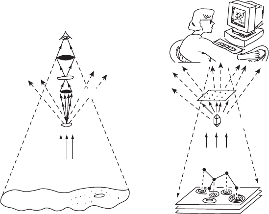
Objective
lens
Eyepiece lens
Detector
Crystallographer
Crystal
Object
Visible light
Enlarged image
(a) (b)
Electron-density map
MICROSCOPE X-RAY DIFFRACTION
Object
Molecule
X rays
Detection device
(electronic
film)
Computer
Fig. 1.1 Analogies between light microscopy and X-ray diffraction.Analogies between the two methods of using scattered radiation for
determining structure are shown here—optical microscopy on the left, X-ray diffraction on the right. The sample that is under study in
both instruments scatters some of the incident radiation and this gives a diffraction pattern.
(a) In the ordinary optical microscope there are two lenses. The lower objective lens gathers light that has been scattered by the
object under study and focuses and magnifies it. The eyepiece or ocular lens, which is the one we look through, increases this
magnification. There is no need to record a diffraction pattern because the light that is scattered by the object under examination
is focused by these lenses and gives a magnified image of that object. The closer the objective lens is to the object, the wider
the angle through which scattered radiation is caught by this lens and focused to form a high-resolution image. The rest of the
radiation is lost to the surroundings.
(b) With X rays the diffraction pattern has to be recorded electronically or photographically, because X rays cannot (at this time) be
focused by any known lens system. Therefore the recombination of the diffracted beams (which is done by an objective lens in
the optical microscope) must, when X rays are used, be done mathematically by a crystallographer with the aid of a computer. As
stressed later (Chapter 5), this recombination cannot be done directly, because the phase relations among the different diffracted
beams cannot usually be measured directly. However, once these phases have been derived, deduced, guessed, or measured
indirectly, an image can be constructed of the scattering matter that caused diffraction—the electron density in the crystal.
in Figure 1.2); this is because, when two scattered waves proceed in
the same direction, the intensity of the combined wave will depend on
the difference in the phases of the two scattered beams. If they are “in
phase” they will enhance each other and give an intense beam, but if
they are “out of phase” they will destroy each other and there will be no
apparent diffracted beam. Generally it is found that such enhancement
or destruction is only partial, so that the diffracted beams have some
intensity and the diffraction pattern that is obtained contains diffracted
beams that have differing intensities—some are weak and some are
intense.
In an optical microscope, that is, a microscope that uses light that
is visible to the human eye, the radiation scattered by the object is
6 Introduction
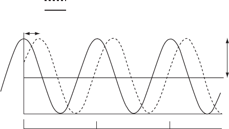
Origin
Phase of
relative to
or to the origin
0º 360º
Wavelength or 360º as unit
Amplitude
|F(hkl)|
720º
Fig. 1.2 A sinusoidal wave.
A sinusoidal wave, showing its amplitude, phase relative to the origin, and wavelength.
Sine and cosine functions are sinusoidal waves with different phases [cos x =sin(x + π/2)
when the distance traveled is measured in radians]. Shown is a cosine wave (black line),
which has a peak at the wave origin. This wave origin coincides with the origin in space
that has been selected by the crystallographer. A second wave (dashed line) has its peak in
a different location. The distance between these two peaks defines their “relative phase.”
recombined by the lens system (the objective lens) so that a magni-
fied image of the object under study is obtained (Figure1.1a). Light
flows through and beyond the lens system of the microscope in such
a way that the relationships between the phases of the scattered waves
are maintained, even after these waves have been recombined by the
second lens (the eyepiece lens). In a similar way, X rays are scattered
by the electrons in atoms and ions (Figure 1.1b), but, in contrast to the
situation with visible light, these scattered X rays cannot be focused
by any presently known experimental techniques. This is because no
electric or magnetic field or material has yet been found that can refract
X rays sufficiently to give a practicable X-ray lens. Therefore an X-ray
microscope cannot yet be used to view atoms (which have dimensions
too small to permit them to be visible with an ordinary light micro-
scope). Much research on a possible X-ray lens is currently in progress
(see Shapiro et al., 2005; Sayre, 2008). The information obtained from
an X-ray diffraction experiment, however, is three-dimensional, and
therefore the great usefulness of this method will doubtless continue
after an X-ray lens can be made.
Since a lens system cannot be used to recombine scattered X rays to
obtain images at atomic resolution, some other technique must be used
if one wishes to view molecules. In practice, the diffracted (scattered)
X rays or neutrons are intercepted and measured by a detecting sys-
tem, but this means that the relationships between the phases of the
scattered waves are lost; only the intensities (not the relative phases)
of the diffracted waves can be measured. If the values of the phases
of the diffracted beams were known, it would be possible to combine
them with the experimental measurements of the diffraction pattern
Introduction 7
and simulate the recombination of the scattered radiation—just as if a lens
had done it—by an appropriate, though complicated, calculation (done
by a crystallographer and a computer in Figure 1.1b). Then we would
have an electron-density map, that is, an image of the material that
had scattered the X rays. This mathematical calculation, the Fourier
synthesis of the pattern of scattered or “diffracted” radiation (Fourier,
1822; Porter, 1906; Bragg, 1915), is a method for summing sinusoidal
waves in order to obtain a representation of the material that scat-
tered the radiation. Such a Fourier synthesis is a fundamental step in
crystal structure determination by diffraction methods and is a central
subject of our discussion (described in detail in Chapters 5, 6, 8, and
9). The difficult part of correctly summing these sinusoidal waves is
termed the “phase problem,” that is, finding where the peaks of each
sinusoidal wave should lie with respect to the others in the summa-
tion. Any of several methods (to be described) can be used to over-
come this difficulty and determine the phases of the various diffracted
beams with respect to each other. When the correct phases are known
(that is, derived, deduced, guessed, or measured indirectly), the three-
dimensional structure of the atomic contents of the crystal (and hence
of the molecules or ions that it contains) will be revealed as a result of a
Fourier synthesis.
Why make the effort to carry out a crystal structure analysis? The
reason is that when the method is successful, it is unique in provid-
ing an unambiguous and complete three-dimensional representation of
the atoms in the crystal. This three-dimensionality is incredibly useful
because chemical and biological reactions occur in three dimensions,
not two; surface and internal structures of molecules, plus informa-
tion on their interactions with other molecules, are revealed by this
powerful technology. Other experimental methods can also provide
structural information. For example, large molecules, such as those of
viruses, can also be visualized by use of an electron microscope, but
individual atoms deep inside each virus molecule cannot currently
be distinguished. Newer technologies such as field ion microscopy
and scanning tunneling microscopy (or atomic force microscopy) are
now providing views of molecules on the surfaces of materials, but
they also do not provide the detailed and precise information about
the internal structure of larger molecules that X-ray and neutron dif-
fraction studies do. Infrared and microwave spectroscopic techniques
give quantitative structural information for simple molecules. High-
field nuclear magnetic resonance (NMR), the main alternative method
currently used for structure determination, can also provide distances
between identified atoms and can be used to study fairly large mole-
cules. No other method can, however, give the entire detailed three-
dimensional picture that X-ray and neutron diffraction techniques can
produce.
Crystal diffraction methods do, however, have their limitations,
chiefly connected with obtaining samples with the highly regular long-
range three-dimensional order characteristic of the ideal crystalline