Glusker J.P., Trueblood K.N. Crystal Structure Analysis: A Primer
Подождите немного. Документ загружается.


18 Crystals
CRYSTAL LATTICE
CRYSTAL STRUCTURE
STRUCTURAL MOTIF
Convolution
Fig. 2.6 The crystal lattice and choices of unit cells.
The generation of a two-dimensional “crystal structure” from a crystal lattice and a
structural motif (an apple in this example). The crystal lattice is obtained from the crystal
structure by replacing each complete repeating unit by a point. The replacement of
each point in the crystal lattice by an apple would lead to a two-dimensional crystal
structure. This crystal structure may be described alternatively as the convolution of an
apple and the crystal lattice. There are many ways in which unit cells may be chosen in the
repeating pattern of apples. Some possible alternative choices are shaded, each having
the same area despite varying shape. This can be verified by noting that the total content
of any chosen unit cell in this figure is one apple. Infinite repetition in two dimensions of
any one of these choices for unit cell will reproduce the entire pattern.
Wolff and Gruber, 1991), that is, to select the three shortest noncoplanar
vectors in the crystal lattice. This may help in establishing whether
two crystals with different unit-cell dimensions are really the same
or not.
It is a common misconception, perhaps arising from the abundance
of illustrations of the simplest elementary and ionic structures in text-
books, that an atom must lie at the corner (origin) of each unit cell.
It is possible to choose the origin arbitrarily and place it at the site
of an atom, but in most structures the choice of origin is dictated by
convenience, because of its relation to symmetry elements that may be
present (i.e., the appropriate space group), and in the great majority
of known structures no atom is present at the origin. Another miscon-
ception is that what a chemist finds convenient to regard as a single
molecule or formula unit must lie entirely within one unit cell. Portions
of a single bonded aggregate may lie in two or more adjacent unit
Crystal symmetry 19
cells. If this does happen, however, any single unit cell will necessarily
still contain all of the independent atoms in the molecule—the atoms
simply comprise portions of different molecules. This is illustrated in
Figure2.6, which shows that a given unit cell may contain only one
apple or portions of two or more apples.
Crystal symmetry
Unit cells and crystal lattices are classified according to their rotational
symmetry. If an object is rotated 180
◦
and then appears identical to the
starting structure, the object is said to have a two-fold rotation axis
(the axis about which the 180
◦
rotation occurred). The presence of an
n-fold rotation axis, where n is any integer, means that when the unit
cell is rotated (360/n)
◦
about this axis, the crystal lattice so obtained is
indistinguishable from original before rotation. If you closed your eyes,
rotated the crystal lattice, and opened your eyes again, nothing would
appear to have changed. The symmetry of an isolated crystal can be
found by examination, and it can give us some very useful information
about the internal atomic arrangement. If the crystal is set down on
a flat surface, it is possible to note if there is another face on top of
the crystal that is parallel to the lower face lying on the flat surface.
Then one can determine if there is a center of symmetry between these
upper and lower faces of the crystal. Similarly, one can examine the
crystal for two-, three-, four- and six-fold rotation axes. The result of
such examinations is the determination of the point group of the crystal,
that is, a group of symmetry operations, such as an n-fold rotation axis,
that leaves at least one point unchanged within the crystal.
It is shown in Appendix 2 that there are seven ways in which dif-
ferent types of applicable rotational symmetry (such as two-, three-,
four-, and six-fold rotation axes) lead to infinitely repeatable unit
cells. These seven are called the seven crystal systems—triclinic, mon-
oclinic, orthorhombic, tetragonal, trigonal/rhombohedral, hexagonal,
and cubic. They are distinguished by their different rotational symme-
tries. For example, in a triclinic crystal lattice there is no rotational (only
one-fold) symmetry; this defines the term “triclinic.” As a result, usually
(but not always) in a triclinic crystal lattice, all unit-cell lengths (a, b,and
c) are unequal, as are all interaxial angles (·, ‚,and„). A monoclinic
crystal lattice (· = „ =90
◦
) has a two-fold rotation axis parallel to the
b axis (where b is chosen, by convention for this crystal system, to be
unique). This means that a rotation of the crystal lattice by 180
◦
about
the b axis gives a crystal lattice indistinguishable from the original.
In an orthorhombic crystal lattice, with three mutually perpendicular
rotation axes, all interaxial angles (·, ‚,and„)are90
◦
. A cubic crystal is
defined by three-fold axes along the cube diagonals, not by its four-fold
axes. It must be stressed that it is the symmetry of the crystal lattice
that is important in defining the crystal system, not the magnitude of
the interaxial angles. Some monoclinic crystals have been found with
20 Crystals
‚ =90
◦
, and some triclinic crystals with all interaxial angles very close
to 90
◦
; this is why symmetry rather than unit cell dimensions are used
to define which is the correct crystal system for the material under
study. In the diagrams of these seven crystal systems in Appendix 2
all crystal lattice points (designated by small circles) are equivalent by
translational symmetry. All crystal lattices except the triclinic crystal
lattice display more than one-fold rotational symmetry (see Chapter 7
for details).
It is customary when choosing a unit cell to take advantage of the
highest symmetry of the crystal lattice. If a unit cell includes only one
crystal lattice point (obtained from the fractions at each corner), it is
said to be primitive and the crystal lattice is designated P. Sometimes
it is more convenient to choose a unit cell that contains more than one
crystal lattice point (a “nonprimitive” unit cell). Nonprimitive unit cells
are chosen because they display the full symmetry of the crystal lattice,
or are more convenient for calculation; any given crystal lattice may
always be described in terms of either primitive or nonprimitive unit
cells. The latter type of crystal lattices have lattice points not only at the
corners of the conventional unit cell, but also at the center of this unit
cell (I for the German innenzentrierte), at the center of one pair of oppo-
site faces (A, B,orC), or at the center of all three pairs of opposite faces
(F ) (see Appendix 2). More than one crystal lattice point is then associ-
ated with a unit cell so chosen, but the requirement that every crystal
lattice point must have identical surroundings is still fulfilled. That
there are 14, and only 14, distinct types of crystal lattices was deduced
by Moritz Ludwig Frankenheim and Auguste Bravais in the nineteenth
century, and these crystal lattices are named after Bravais (Bravais,
1850). The Bravais crystal lattices are obtained from a combination of
the seven crystal systems (triclinic, monoclinic, orthorhombic, tetrago-
nal, trigonal/rhombohedral, hexagonal, and cubic) with the four crystal
lattice types (P, Aor B or C, F ,andI) after the elimination of any equiv-
alencies. The unit cells of these 14 Bravais crystal lattices are shown in
Appendix 2.
Space groups
Since the atomic contents in each unit cell are identical (or nearly so),
the symmetry of the arrangement of atoms in each unit cell must be
related by certain symmetry operations (in addition to translation) that
ensure identity from unit cell to unit cell. This means that the atomic
arrangement in one unit cell is related by defined symmetry operations
to the arrangement in all other unit cells. The smallest part of a crys-
tal structure from which the complete structure can be obtained by
space-group symmetry operations (including translations) is called the
asymmetric unit. The operation of the correct space-group symmetry
elements (other than crystal lattice translations) on the asymmetric unit
Physical properties of crystals 21
will generate the entire contents of a primitive unit cell. When one
considers the possible combinations of symmetry elements (centers of
symmetry, mirror planes, glide planes, rotation axes, and screw axes)
that are consistent with the 14 Bravais crystal lattices, and thus the pos-
sible symmetry elements of the structures that can be arranged on the
crystal lattices, it is found that 230, and only 230, distinct combinations
of the possible symmetry elements exist for three-dimensional crystals
(and only 17 plane groups for two-dimensional wallpaper). Thus the
many different ways of arranging atoms or ions in structures to give a
regularly repeating three-dimensional arrangement in a crystal fall into
230, and only 230, different three-dimensional crystallographic space
groups. They are listed in International Tables for (X-ray) Crystallography
(referred to here as International Tables), and these Tables, listed at the
end of this book in the “References and further reading” section, are
constantly used by crystallographers. The important result is that if
the location of one atom in a crystal of known space group has been
found, then application of the space-group symmetry operations (listed
for convenience in International Tables) will give the locations of all other
such specific atoms in the unit cell. This can be repeated for each atom
in the ions or molecules that make up the crystal. Symmetry and space
groups are discussed further in Chapter 7.
Physical properties of crystals
Optical properties
The interaction of light with crystals is one of the reasons they are used
for ornamentation (as jewelry). It may also reveal information about
crystalline symmetry and, in certain cases, the internal structure of the
crystal (Hartshorne and Stuart, 1950; Wood, 1977; Wahlstrom, 1979).
Particularly useful information may be obtained from the refractive
index of the crystal. This gives a measure of the change in the velocity
of light when it enters the crystal. Refraction is evident when a straight
stick or rod is partially inserted in water; the rod appears to be bent at
the point of entry. The change in the velocity of light as it passes from
air to water is revealed by the angle to which the rod appears to be
bent; when this angle is measured it gives information on the ratio of
the two velocities (that is, the refractive index of water). The refractive
index of a crystal is generally measured by immersing it in liquids of
known refractive index, and determining when the crystal becomes
“invisible.” The crystal and the liquid surrounding it now have the
same refractive index.
Some crystals, such as cubic crystals, are optically isotropic: the
refractive index is independent of the direction from which the crystal
is viewed. Other crystals may be birefringent, with different refractive
indices in different directions. When a test tube containing birefringent

22 Crystals
crystals in their mother liquor is shaken, the crystals glisten (unlike the
situation for isotropic cubic crystals). Birefringence, or double refrac-
tion, is the decomposition of light into two rays, each polarized. One
ray, the “ordinary ray,” travels through the crystal with the same veloc-
ity in every direction. The other ray, the “extraordinary ray,” trav-
els with a velocity that depends on the direction of passage through
the crystal. The result of this can readily be seen for a calcite crystal
(Iceland spar) in Figure 2.7, in which two images are formed when light
passes through the crystal. If a birefringent crystal is colored, it may
show different colors when viewed in different directions. If the crystal
structure contains an approximately planar group, measurements of
refractive indices may permit deduction of the orientation of this planar
group within the chosen unit cell. This method, combined with unit-cell
measurements, was used to study steroid dimensions and packing long
before any complete structure determination was or could be initiated.
It led to a correct chemical formula for atoms in the steroid ring struc-
ture (Bernal, 1932).
Fig. 2.7 The birefringence of calcite (Ice-
land spar).
View through an Iceland spar crystal (cal-
cite) with the word “BIREFRINGENCE”
written on a strip of paper behind it. Light
is broken into two polarized beams as
it passes through the crystal. The word
is split into two images, hence the term
“birefringence.” As the crystal is rotated,
the image made by the extraordinary ray
moves around the image made by the
ordinary ray. Iceland spar crystals are
believed to have been used in the Arctic
regions for ages in navigation to deter-
mine the direction of the sun on a cloudy
day, and hence which direction to sail in.
There are many other interesting optical properties of crystals.
Second-harmonic generation (SHG, also called frequency doubling)
was first demonstrated when a ruby laser with a wavelength of 694 nm
was focused into a quartz crystal (Dougherty and Kurz, 1976). Analysis
with a spectrometer indicated that light was produced with a wave-
length of 347 nm (half the wavelength and twice the frequency of the
incident light). Only noncentrosymmetric crystal structures can double
the frequency, and therefore SHG provides a useful method for testing
the symmetry of a crystal. Green laser pointers combine a noncen-
trosymmetric (nonlinear) crystal with a red neodymium laser to pro-
duce green light.
Electrical properties
Certain crystals display piezoelectricity. This word is derived from a
Greek word meaning “to squeeze” or “press.” Piezoelectricity is the
creation of an electrical potential by a crystal in response to an applied
mechanical stress. This effect is reversible in that materials exhibiting
the direct piezoelectric effect also exhibit the converse piezoelectric
effect (the production of stress when an electric field is applied). The
piezoelectric effect was first reported by Pierre and Jacques Curie in
1880, who detected a voltage across the faces of a compressed Rochelle
salt crystal (Curie and Curie, 1880). The phenomenon has many indus-
trial uses. For example, when the button of a cigarette lighter or gas
burner is pressed, the high voltage produced by the compression of a
crystal causes an electric current to flow across a small spark gap, so that
the gas is ignited. Another example is in the airbag sensor of a car. The
intensity of the shock of a car crash to a crystal causes an electrical signal
that triggers expansion of the airbag. In the analogous phenomenon of
pyroelectricity, a crystal can generate an electrical potential in response
to a change in temperature.
Summary 23
The significance of the unit cell
In this chapter we have described crystals and their representation by
a repeating component, the unit cell. Since a crystal is built up of an
extremely large number of regularly stacked cells, each of which has
identical contents, the problem of determining the structure of a crystal
is reduced to that of determining the spatial arrangement of the atoms
within a single unit cell,orwithin the smaller asymmetric unit if (as is usual)
the unit cell has some internal symmetry. If there is some static disorder
in the structure, the arrangements of atoms in different unit cells may
not be precisely identical, varying in an apparently random fashion.
There may also be dynamic disorder in a structure as various part of
the molecules move. Since the frequencies of atomic vibrations are of
the order of 10
13
per second, and since sets of X-ray diffraction data are
measured over periods ranging from seconds to hours, time-averaging
of the atomic distribution is always involved. What one finds for the
arrangement of atoms in a crystal is the space-averaged structure of all
of its component unit cells.
Summary
A crystal is, by definition, a solid that has a regularly repeating internal
structure (arrangement of atoms). This internal periodicity was sur-
mised in the seventeenth century from the regularities of the shapes
of crystals, and was proved in 1912 when it was shown that a crystal
could act as a three-dimensional diffraction grating for X rays, since X
rays have wavelengths comparable to the distances between atoms in
crystals.
Crystals are generally grown by concentrating a solution of the mate-
rial of interest until material separates (hopefully in a crystalline state).
Experimental conditions should ensure a good choice of solvent, the
generation of a suitable number of nucleation sites, control of the rate
of growth, and a lack of disturbance of the setup.
The unit cell of a crystal is its basic building block and is described
by three axial lengths a, b, c and three interaxial angles ·, ‚, „. When
describing a crystal face or plane it is necessary to consider intercepts
on the three axes of the unit cell. The hkl face or plane makes intercepts
a/ h, b/k, c/l with the three axes. The internal regularity of a crystal
is expressed in the crystal lattice; this is a regular three-dimensional
array of points (each with identical environments) upon which the
contents of the unit cell (the motif ) are arranged by infinite repetition to
build up the crystal structure. There are seven ways in which rotational
symmetry can lead to infinitely repeatable unit cells. These are the
seven crystal systems—triclinic, monoclinic, orthorhombic, tetragonal,
trigonal/rhombohedral, hexagonal, and cubic (see Appendix 2). These
seven crystal lattices are combined with the four crystal lattice types
(primitive P, single-face-centered A or B or C, face-centered F ,and
24 Crystals
body-centered I) to give 14 Bravais lattices. Symmetry elements (center
of symmetry, mirror planes, glide planes, rotation axes, and screw axes)
combined with these 14 Bravais lattices give the 230 different combina-
tions of symmetry elements (the 230 space groups) that are possible for
arranging objects in a regularly repeating manner in three dimensions,
as in the crystalline state.

Diffraction
3
A common approach to crystal structure analysis by X-ray diffraction
presented in texts that have been written for nonspecialists involves the
Bragg equation, and a discussion in terms of “reflection” of X rays from
crystal lattice planes (Bragg, 1913). While the Bragg equation, which
implies this “reflection,” has proved extremely useful, it does not really
help in understanding the process of X-ray diffraction. Therefore we
will proceed instead by way of an elementary consideration of diffrac-
tion phenomena generally, and then diffraction from periodic structures
(such as crystals), making use of optical analogies (Jenkins and White,
1957; Taylor and Lipson, 1964; Harburn et al., 1975).
Visualizing small objects
The eyes of most animals, including humans, comprise efficient optical
systems for forming images of objects by the recombination of visible
radiation scattered by these objects. Many things are, of course, too
small to be detected by the unaided human eye, but an enlarged image
of some of them can be formed with a microscope—using visible light
for objects with dimensions comparable to or larger than the wave-
length of this light (about 6 × 10
−7
m), or using electrons of high energy
(and thus short wavelength) in an electron microscope. In order to
“see” the fine details of molecular structure (with dimensions 10
−8
to
10
−10
m), it is necessary to use radiation of a wavelength comparable to,
or smaller than, the dimensions of the distances between atoms. Such
radiation is readily available
(1) in the X rays produced by bombarding a target composed of an
element of intermediate atomic number (for example, between
Cr and Mo in the Periodic Table) with fast electrons, or from a
synchrotron source,
*
*
Synchrotron radiation is an intense and
versatile source of X rays that is emitted
by high-energy electrons, such as those
in an electron storage ring, when their
path is bent by a magnetic field. The
radiation is characterized by a continuous
spectral distribution (which can, however,
be “tuned” by appropriate selection), a
very high intensity (many times that of
conventional X-ray generators), a pulsed
time structure, and a high degree of
polarization.
(2) in neutrons from a nuclear reactor or spallation source, or
(3) in electrons with energies of 10–50 keV.
Each of these kinds of radiation is scattered by the atoms of the sam-
ple, just as is ordinary light, and if we could recombine this scat-
tered radiation, as a microscope can, we could form an image of the
scattering matter. This recombination of radiation scattered by atoms
25
26 Diffraction
is, however, found to be more complicated than that necessary for
viewing through a microscope, and it is the major subject of this
book.
X rays are scattered by the electrons in an atom,
**
neutrons are scat-
**
When X rays hit an atom, its electrons
are set into oscillation about their nuclei
as a result of perturbation by the rapidly
oscillating electric field of the X rays. The
frequency of this oscillation is equal to
that of the incident X rays. The oscillat-
ing dipole so formed acts, in accord with
electromagnetic theory, as a source of radi-
ation with the same frequency as that of
the incident beam. This is referred to as
“elastic scattering” and is the type of scat-
tering discussed in this book. When there
is energy loss, resulting in a wavelength
change on scattering, the phenomenon is
described as “inelastic scattering.” This
effect is generally ignored by crystallogra-
phers interested in structure and will not
be discussed in this book.
tered by the nuclei and also, by virtue of their spin, by any unpaired
electrons in the atom, and electrons are scattered by the electric field of
the atom, which is of course a consequence of the combined effects of
both its nuclear charge and its extranuclear electrons. However, neither
X rays nor neutrons of the required wavelengths can be focused by
any known lens system, and high-energy electrons cannot (at least
at present) be focused sufficiently well to show individually resolved
atoms. Thus, the formation of an atomic-resolution image of the object
under scrutiny, which is the self-evident aim of any method of crystal
structure determination—and is a process that we take for granted
when we use our eyes or any kind of microscope—is not directly possi-
ble when X rays, neutrons, or high-energy electrons are used as a probe.
Unfortunately, the atoms that we wish to see are too small to be seen
without these short-wavelength radiation sources.
When, however, X rays or neutrons are diffracted by crystalline mate-
rials, a measurable pattern of diffracted beams is obtained and these
results can be analyzed to give a three-dimensional map of the atomic
arrangement within the crystal and hence the molecular structures
involved. In order for the reader to understand the process involved
it is necessary to consider diffraction in general, and easier to start with
the effects of visible light on masks that are readily visible. Scattering
of light by slits will serve as a preliminary model for the scattering
of X rays by atoms. When the dimensions of both the slits and the
wavelength of visible light are reduced by several orders of magnitude,
analogous results can be obtained for atoms and X rays.
Diffraction of visible light by single slits
The pattern of radiation scattered by any object is called the diffraction
pattern of that object. Diffraction occurs whenever the wavefront of a
light beam is obstructed in some way. We are accustomed to think of
light as traveling in straight lines and thus casting sharply defined shad-
ows, but that is only because the dimensions of the objects normally
illuminated in our experience are much larger than the wavelength of
visible light. When light from a point source passes through a narrow
slit or a very fine pinhole, the light is found to spread into the region
that normally would be expected to be in shadow. In explanation of
this effect, each point on the wavefront within the slit or pinhole is
considered to act as a secondary source, radiating in all directions.
The secondary wavelets so generated interfere with each other, either
reinforcing or partially destroying each other, as originally described
by Francesco Maria Grimaldi, Christiaan Huygens, Thomas Young, and
Augustin Jean Fresnel (Grimaldi, 1665; Huygens, 1690; Young, 1807;
Diffraction of visible light by single slits 27
de Sénarmont et al., 1866). As these waves combine, the extent of
interference will depend on their relative phases and amplitudes. It is
assumed that any phase change on scattering is the same for each atom
and therefore this change is generally ignored.
†
There are, however,
†
The reader will remember that an electro-
magnetic wave has a constant velocity in
vacuo (the speed of light) and consists of
successive crests and troughs. Two crests
are a wavelength apart, and this distance,
which is inversely proportional to the fre-
quency of the radiation, defines the prop-
erties of the electromagnetic wave (such
as color red or blue, X ray or infrared,
etc.). The wave has an amplitude (the
maximum value measured from its mean
value), which is related to the square root
of the intensity of the beam. It also has
a “relative phase,” which is the distance
ofthecrestofthewavemeasuredfroma
chosen origin of the wave or with respect
to the crest of another wave (see Figure
1.2). It was shown by John Joseph Thom-
son that when radiation is scattered by an
electron, there is a phase change of 180
◦
in the sense that the electric field in the
scattered wave at a given point is opposed
to that of the direct (incident) wave at
that same point (Thomson, 1906). This is
discussed in detail by Reginald William
James (1965).
exceptions to this assumption, for example when the wavelength of the
radiation can cause changes in the atom (see Chapter 10).
The phenomenon of diffraction by a regular two-dimensional pattern
may be illustrated by holding a woven fabric handkerchief taut between
your eyes and a distant point source of light, such as a street light.
Instead of just one spot of light, as expected, a cluster of lights will
be seen. The same phenomenon can also be demonstrated with a fine
sieve (see the cover of this book). The narrowly and regularly spaced
threads of the fabric or wires of the sieve are considered to produce this
diffraction effect. The larger the spacing between the wires of the sieve,
the closer diffraction spots are found around the central spot.
Keeping in mind that we are interested in scattering (diffraction)
by atoms, we begin with a discussion of diffraction by slits because
these involve visible light and therefore help with the description of
the various principles of diffraction. Two examples of the diffraction of
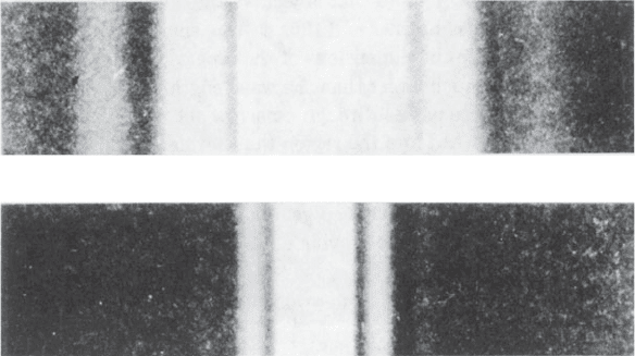
light when it passes through a single slit are given in Figure 3.1; in one,
(a)
(b)
Fig. 3.1 Diffraction patterns of single narrow slits.
The diffraction patterns of two single slits of different width, both illuminated with light
ofthesamesinglewavelength.
(a) The diffraction pattern of a narrow slit.
(b) The diffraction pattern of a slit 2.2 times wider than that used in (a). The diffraction
pattern is now narrower by a factor of 2.2.
Note that the wider slit gives the narrower diffraction pattern.
From Fundamentals of Optics by Francis A. Jenkins and Harvey E. White, 3rd edition
(1957) (Figure 16A). Copyright © 1957, McGraw-Hill Book Company. Used with permis-
sion of McGraw-Hill Book Company.