Glusker J.P., Trueblood K.N. Crystal Structure Analysis: A Primer
Подождите немного. Документ загружается.

78 The diffraction pattern obtained
B
iso
= 0.0 Å
2
B
iso
= 3.5 Å
2
6
0.2 0.4 0.6 0.8
f
e
–
(B
iso
sin
2
q / l
2
)
sin q / l
(c)
30
25
20
Br
–
Fe
++
Ca
++
CI
–
O
C
H
15
10
5
0
35
(a)
0.80.60.40.2 1.0 1.2
sin q / l
f
(b)
Direct beam
Direct beam
Small atom
Large atom
Small path difference
with respect to wavelength
at edges of atom
Bragg reflection
Very little intensity
reduction
Bragg reflection
Reduced intensity
Large path difference
with respect to wavelength
at edges of atom
PD
PD
2q
2q
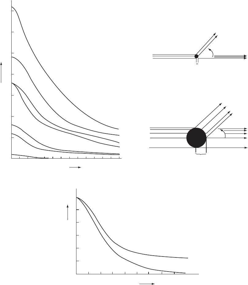
Fig. 5.4 Atomic-scattering-factor curves.
(a) Some atomic-scattering-factor curves for atoms, given as a function of sin Ë/Î so that they will be independent of wavelength.
(Remember that 2Ë is the deviation of the diffracted beam from the direct X-ray beam, wavelength Î.) The scattering factor
for an atom is the ratio of the amplitude of the wave scattered by the atom to that of the wave scattered by a single electron.
At sin Ë/Î = 0 the value of the scattering factor of a neutral atom is equal to its atomic number, since all electrons then scatter in
phase. Note that calcium (Ca
++
) and chloride (Cl
−
) are isoelectronic; that is, they have the same number of extranuclear electrons.

Scattering by an individual atom 79
factors as a function of sin Ë/Î; they are published and available in
International Tables. Some values are given in Appendix 5.
For most purposes in structure analysis it is adequate to assume that
atoms themselves are spherically symmetrical, but, with some of the
best data now available, small departures from spherical symmetry
(attributable to covalent bonding, lone pairs of electrons, and nonspher-
ical orbitals, for example) are detectable. However, in our discussions,
unless stated otherwise, we will assume spherical symmetry of atoms.
This means that the scattering by an assemblage of atoms—that is,
by the structure—can be very closely approximated by summing the
contributions to each scattered wave from each atom independently,
taking appropriate account of differences in the phase angles of each
wave. Some atomic scattering factors, plotted as a function of sin Ë/Î,
are shown in Figure 5.4a. Since the diffraction pattern is the sum of the
scattering from all unit cells, and this can be represented by the average
contents of a single one of these unit cells, vibrations or disorder may
be considered the equivalent of the smearing out of the electron density,
so that there is a greater fall-off in the intensity of the diffraction pattern
at a higher sin Ë/Î values (cf. the optical analogy in Figure 3.1: the wider
the slit, the narrower the diffraction pattern). This modification of the
fall-off by atomic vibration, motion or disorder, which results in a larger
apparent atomic size as shown in Figure 5.4b, increases the falloff in
scattering power as a function of scattering angle (Figure 5.4c). This fall-
off may be isotropic (equal in all directions) or anisotropic (greater in
certain directions in the unit cell than in others). Information obtained
from an analysis of such atomic motion or disorder is discussed in
Chapter 12. It leads, in nearly all crystal structures, to a model with
anisotropic displacement parameters representing an inexact register
of atomic positions from unit cell to unit cell. By contrast to X-ray scat-
tering, neutrons are scattered by atomic nuclei, rather than by electrons
around a nucleus, and hence, since the nucleus is so small (equivalent
to a “point atom”), the neutron scattering for a nonvibrating nucleus is
almost independent of scattering angle.
The positively charged calcium ion pulls electrons closer to the nucleus than does the chloride ion, which is negatively charged
and has a lower atomic number. The resulting “narrower atom” for Ca
++
will, for reasons shown in Figure 3.1, give a broader
diffraction pattern. This is shown at high values of sin Ë/Î by higher values of f for Ca
++
than for Cl
−
.
(b) When radiation is scattered by particles that are very small relative to the wavelength of the radiation, such as neutrons, the
scattered radiation has approximately the same intensity in all directions. When it is scattered by larger particles, the radiation
scattered from different regions of the particle will still be in phase in the forward direction, but at higher scattering angles there
is interference between radiation scattered from various parts of the particle. The intensity of radiation scattered at higher angles
is thus less than for that scattered in the forward direction. This effect is greater the larger the size of the particle relative to the
wavelength of the radiation used.
(c) The effects of isotropic vibration on the scattering by a carbon atom. Values are shown for a stationary carbon atom (B
iso
of
0.0Å
2
) and for one with a room temperature isotropic displacement factor (B
iso
of 3.5Å
2
) that corresponds to a root-mean-
square amplitude of vibration of 0.21 Å. Vibration and disorder result in an apparently relatively greater size for the atoms (since
we are considering an average of millions of unit cells), and consequently a decrease in scattering intensity with increasing
scattering angle. If B
iso
is large, no Bragg reflections may be detectable at high values of 2Ë; that is, a narrower diffraction pattern
is obtained from the “smeared-out” electron cloud of a vibrating atom (cf. Figure 3.1).
80 The diffraction pattern obtained
Scattering by a group of atoms (a structure)
The X radiation scattered by one unit cell of a structure in any direction
in which there is a diffraction maximum has a particular combination of
amplitude and relative phase, known as the structure factor and symbol-
ized by F or F (hkl ) (Sommerfeld, 1921). It is the ratio of the amplitude
of the radiation scattered in a particular direction by the contents of one
unit cell to that scattered by a single electron at the origin of the unit
cell under the same conditions. The intensity of the scattered radiation
is proportional to the square of the amplitude, |F (hkl )|
2
. In the manner
just discussed [see Eqn. (5.17)], the structure factor can be represented
either exponentially or as an ordinary complex number:
F (hkl)=|F(hkl)|e
i·(hkl)
= A(hkl)+iB(hkl) (5.18)
with |F| or |F (hkl)| representing the amplitude of the scattered wave,
and ·(hkl) its phase relative to the chosen origin of the unit cell.
*
As
*
The structure factor F may be repre-
sented as a vector, but it is not conven-
tionally written in bold face, so we, as is
common, will use F for the vector and |F |
for its amplitude.
before (Figure 5.1), · =tan
−1
(B/A) and c
r
= |F (hkl)| =(A
2
+ B
2
)
1/2
.The
quantities A and B, representing the components of the wave in its
vector representation (see Figure 5.2), can be calculated, if one knows
the structure, merely by summing the corresponding components of the
scattering from each atom separately. These components are [by Eqns.
(5.6) and (5.7)] the products of the individual atomic-scattering-factor
amplitudes, f
j
, and the cosines and sines of the phase angles, ·
j
,ofthe
waves scattered from the individual atoms:
A(hkl)=
j
f
j
cos ·
j
(5.19)
and
B(hkl)=
j
f
j
sin ·
j
(5.20)
But how do we calculate ·
j
for each atom?
If an atom lies at the origin of the unit cell and if other atoms lie one or
several unit-cell translations (a) from it, then this grating of atoms will
give a series of Bragg reflections h00 on diffraction. If there is another
atom between two of them, at a distance xa from the origin (where x is
less than 1), radiation scattered by this atom will interfere with the other
resultant Bragg reflection by an amount that depends on the value of x.
This can be generalized so that, for each h00 Bragg reflection, the phase
difference (interference) will depend on the value of hx as illustrated
in Figures 5.5 and 5.6. We show in Appendix 6 that the phase of the
wave scattered in the direction of a reciprocal lattice point (hkl)byan
atom situated at a position x, y, z in the unit cell (where x, y,andz are
expressed as fractions of the unit-cell lengths a, b,andc, respectively) is
just 2π(hx + ky+ lz) radians, relative to the phase of the wave scattered
in the same direction by an atom at the origin. This is important because
it defines the effect of the location of an atom in the unit cell. The “rela-
tive phase angle” for an atom at x,y, z, where these numbers are defined
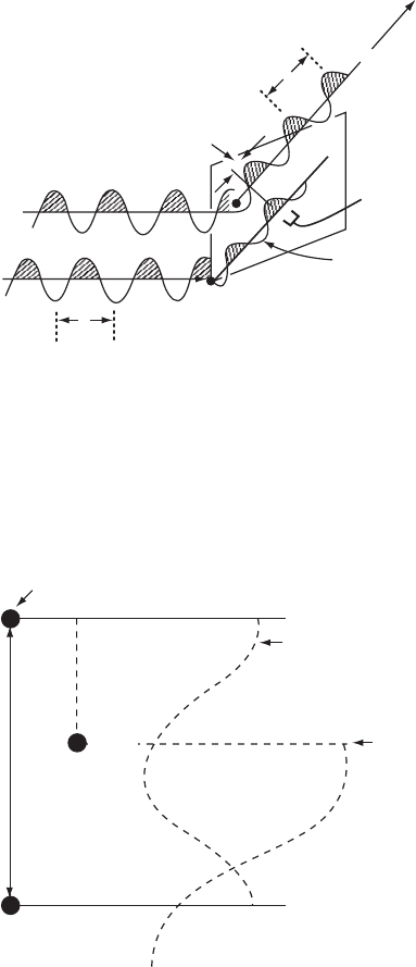
Scattering by a group of atoms (a structure) 81
=P
l
a
hkl
= Phase
relative to that
of wave at origin
l=q
Orgin
Phase difference
Wave scattered
from origin
hkl Bragg
reflection
Direct beamDirect beam
Fig. 5.5 The meaning of “relative phase.”
The relative phase angle ·(hkl) of a Bragg reflection hkl is the difference between the
phase of a wave scattered by an atom (shown as a black circle within the unit cell) and
the phase of a wave scattered in the same direction by an imaginary atom at the chosen
origin of the unit cell.
a
100 Bragg
reflection
scattered by atoms
at 0 and 1.0
Origin
Unit cell length
100
Bragg
reflection
scattered by
atom at x
(partially
out of phase)
x
Atom
Fig. 5.6 The relative phase angle on diffraction.
If a one-dimensional structure with a repeat distance a has an atom at 0.0 (and this is
repeated from unit cell to unit cell by other atoms at 1.0, 2.0, etc.) and an atom at x/a,the
phase difference between the atom at 0.0 and the atom at x/a is 2πhx radians. Suppose
that the atom at 0.0 is at the chosen origin of the system. Its phase angle for a cosine
function is 0
◦
. The phase angle of the atom at x is 2πhx radians. This is the difference of
its phase with that of the atom at the origin, and hence the radiation scattered by the atom
at x is considered to have a relative phase of 2πhx radians.
82 The diffraction pattern obtained
with respect to a chosen origin at 0, 0, 0, is 2π(hx + ky+ lz)radians.If
the location of the chosen origin is changed, the relative phase will also
be changed. Each structure factor is the sum of the scattering from all
atoms j in the unit cell. Thus Eqns. (5.19) and (5.20) (for all atoms j) can
be rewritten as
A(hkl)=
j
f
j
cos 2π(hx
j
+ ky
j
+ lz
j
) (5.21)
B(hkl)=
j
f
j
sin 2π( hx
j
+ ky
j
+ lz
j
) (5.22)
where the value of f
j
chosen is that corresponding to the value of
sin Ë/Î for the Bragg reflection in question, modified to take into account
any thermal vibration of the atom. A comparison with Eqns. (5.19) and
(5.20) shows that we now know the phase ·
j
. The magnitude of |F (hkl)|
depends only on the relative positions of the atoms in the unit cell,
except to the extent that f
j
is a function of the scattering angle. The size
and shape of the unit cell do not appear as such in the expressions for
A and B. In Figure 5.2, F is represented as a vector. Note that a shift in
the chosen origin of the unit cell will add a constant to the phase angle
of each atom [see Eqns. (5.21) and (5.22)]; that is, it will rotate the phase
diagrams in Figure 5.2 relative to the coordinate axes, but will leave the
length of |F (hkl)|, and hence the values of |F (hkl)|
2
and the intensity,
unchanged.
Effects of atomic vibration and displacements
on atomic scattering
Atomic vibrations in a crystal, that is, displacements from equilibrium
positions, have a frequency of the order of 10
13
per second. This is
much slower than the frequencies of X rays used to study crystals; these
are of the order of 10
18
per second. Therefore a vibrating atom will
appear stationary to X rays but displaced in a random manner within
the vibration amplitude. Atoms in other unit cells will also exhibit this
random deviation from their equilibrium positions, different for each
such atom in the various unit cells throughout the crystal. Because
minor static displacements of atoms appear similar to displacements
caused by atomic vibrations, it is usual to use the term “atomic dis-
placement parameter” rather than “atomic temperature factor” for the
correction factor. When 2Ë = 0, all electrons in the atom scatter in phase,
and the scattering power of an atom at this angle, expressed relative to
the scattering power of a free electron, is just equal to the number of
electrons present (the atomic number for neutral atoms).
However, an atom has size (relative to the wavelength of the X rays
used), with the result that X rays scattered from one part of an atom
interfere with those scattered from another part of the same atom at
all angles of scattering greater than 0
◦
. This causes the scattering to
Effects of atomic vibration and displacements on atomic scattering 83
fall off with increasing scattering angle or, more precisely, increasing
values of sin Ë/Î, as indicated in Figure 5.4b. The fall-off in intensity
with higher scattering angle (Figure 5.4c) increases as the vibrations of
atoms become greater, and these vibrations in turn increase in extent
with rising temperature. Atoms are in motion in the crystalline state,
however, even when the temperature is reduced to near absolute zero.
This vibration, coupled with displacements of some atoms, leads to
a significant reduction in intensity that can be approximated by an
exponential function that has a large effect at high 2Ë values (illustrated
in Figure 5.4c); this indicates, as noted by Peter J. W. Debye and Ivar
Waller, that atomic motion endows a larger “apparent size” to atoms
(Debye, 1914: Waller, 1923). Effectively, since atoms are displaced
different amounts from unit cell to unit cell at the given instant in time
that measurement occurs, atoms appear to have become smeared in the
average of all the unit cells in the crystal. If the displacement amplitude
is sufficiently high, essentially no diffracted intensity will be observed
beyond some limiting value of the scattering angle; that is, the “slit” is
effectively widened by the vibration and so the “envelope” is narrow
(Figure 3.5a). If the displacements are nearly isotropic—that is, do not
differ greatly in different directions—the exponential factor can be
written as exp(−2B
iso
sin
2
Ë/Î
2
), with B
iso
called the atomic displacement
factor.
**
B
iso
is equal to 8π
2
< u
2
>, where < u
2
> is the mean square
**
Many crystallographers omit the sub-
script “iso,” relying on the context to
avoid confusion with the quantity B
defined in F = A+iB.
amplitude of displacement of the atom from its equilibrium position.
The type of disorder found in a crystal may be static, with the atom
in one site in one unit cell and a different site in another unit cell.
Alternatively, it may be dynamic, which implies that the atom moves
from one site to another. The overall effect in both cases is a reduction
in the scattering factors of the atoms involved as sinË/Î increases
(see Willis and Pryor, 1975).
If the motion or disorder is anisotropic, it is necessary to replace B
iso
by six terms. This is usually necessary for all atoms except hydrogen
atoms; these have only weak scattering power. Atoms in crystals rarely
have isotropic environments. The six parameters define the orientations
of the principal axes of the ellipsoid that represents the anisotropic
displacements and the magnitudes of the displacements along these
axes. The results are often displayed in an ORTEP
†
diagram, in which
†
ORTEP = Oak Ridge Thermal Ellipsoid
Plot (Johnson, 1965).
the atomic displacement factors are drawn as ellipsoids (Johnson, 1965).
If the anisotropy is severe, the ellipsoid representing the displacement
probability and its direction may be abnormally extended in shape and
may be better represented as disorder in two positions.
Macromolecules, such as proteins, show interesting thermal and dis-
placement effects. While their structures are generally, but not always,
measured at a lower resolution than for small molecules, anisotropic
displacement parameters are rarely determined, but isotropic displace-
ments give information on the motion and flexibility of various portions
in the molecule. One domain of the molecule may appear to move
in a hingelike manner with respect to another part of the same mole-
cule. Also, side chains at the surface of the macromolecule may have
84 The diffraction pattern obtained
alternate atomic positions from unit cell to unit cell as they interact
with the various water molecules that fill nearly half of the crystal
volume.
Calculating a structure factor
With a method for expressing a structure factor by means of an equation
(Eqn. 5.18), and information on the components of this equation, it is
possible to obtain a calculated value for the structure factor. This can be
compared with the experimental value derived from I(hkl). The data
needed in order to calculate a structure factor include the values of x,
y,andz for each atom; h, k, l, and sin Ë/Î for the Bragg reflection under
consideration; and the scattering factor f
j
for each atom at that value of
sin Ë/Î, modified by atomic displacement factors. Then it is necessary to
calculate 2π(hx
j
+ ky
j
+ lz
j
) and its sine and cosine for each atom and
the Bragg reflection for which F (hkl) is being calculated. This gives all
the information necessary to sum the results for each atom and obtain
A(hkl)andB(hkl) according to Eqns. (5.21) and (5.22). These lead to
F (hkl), that is, (A
2
+ B
2
)
1/2
, and the relative phase angle ·(hkl ), that is,
tan
−1
(B/ A), for the Bragg reflection with indices h, k,andl when all the
atomic coordinates are known. This process has to be repeated for all of
the other Bragg reflections. It demonstrates how important computers
are to the X-ray crystallographer.
Information on the electron-density map will have to wait until
we know the phase of the structure factor (so that we can deter-
mine the atomic positions x, y,andz). All we have so far are the
experimentally measured structure amplitudes, |F (hkl)|, but we can
calculate F(hkl)=A(hkl)+iB(hkl) (including its relative phase angle
· =tan
−1
(B(hkl)/A(hkl)), see Eqns. 5.21 and 5.22) if we have x, y,and
z for a model in a unit cell of known dimensions and space group.
Summary
When X rays are diffracted by a crystal, the intensity of X-ray scattering
at any angle is the result of the combination of the waves scattered
from different atoms and the manner in which they modify this inten-
sity by various degrees of constructive and destructive interference.
A structure determination involves a matching of the observed inten-
sity pattern to that calculated from a postulated model, and it is thus
imperative to understand how this intensity pattern can be calculated
for any desired model. The combination of the scattered waves can be
represented in various ways:
(1) The waves may be drawn graphically and the displacements (ordi-
nates, vertical axis) at a given position (abscissae, horizontal axis)
summed.
Summary 85
(2) A wave may be represented algebraically as
x
j
= c
j
cos(ˆ + ·
j
) (for the jth wave)
and the displacements, x
j
, of several such waves summed to give
a resultant wave.
(3) The waves may be expressed as two-dimensional vectors in an
orthogonal coordinate system, amplitude c
j
, with the relative
phase angle ·
j
measured in a counterclockwise direction from the
horizontal axis. This is the equivalent of representing one com-
plete wavelength as 360
◦
, so that the periodicity of the wave is
expressed. The phase relative to some origin is given as a fraction
of a revolution. The vectors may then be summed by vectorial
addition of their components.
(4) The waves may be represented in complex notation
A
j
+iB
j
= c
j
e
i·
j
which is merely a convenient way of representing two orthogonal
vector components (at 0
◦
and 90
◦
) in one equation. By convention
A is the component at 0
◦
and B the component at 90
◦
.
X rays are scattered by electrons. The extent of scattering depends
on the atomic number of the atom and the angle of scattering, 2Ë,and
is represented by an atomic scattering factor f . For a group of atoms,
the amplitude (relative to the scattering by a single electron) and the
relative phase of the X rays scattered by one unit cell are represented by
the structure factor F (hkl)=A(hkl)+iB(hkl) for each Bragg reflection.
For a known structure with atoms j at positions x
j
, y
j
, z
j
, this may be
calculated from
A(hkl)=
j
f
j
cos 2π(hx
j
+ ky
j
+ lz
j
)
and
B(hkl)=
j
f
j
sin 2π( hx
j
+ ky
j
+ lz
j
)
where the summation is over all atoms in the unit cell. The relative
phase angle ·(hkl)istan
−1
(B/ A) and the structure factor amplitude
|F (hkl)|is {(A(hkl)
2
+ B(hkl)
2
}
1/2
. The value of F (hkl ) may be reduced as
a result of thermal vibration and atomic displacement so that if F
novib
is
the value for a structure containing stationary atoms, the experimental
values will correspond to F (hkl )=F
novib
exp(−B
iso
sin
2
Ë/Î
2
), where
B
iso
, the atomic displacement parameter, is a measure of the amount
of vibration and/or displacement (B
iso
=8π
2
< u
2
>, where < u
2
> is the
mean square amplitude of displacement). With precise experimental
data, it is possible to measure the anisotropy of vibration and displace-
ment.

The phase problem and
electron-density maps
6
In order to obtain an image of the material that has scattered X rays
and given a diffraction pattern, which is the aim of these studies, one
must perform a three-dimensional Fourier summation. The theorem of
Jean Baptiste Joseph Fourier, a French mathematician and physicist,
states that a continuous, periodic function can be represented by the
summation of cosine and sine terms (Fourier, 1822). Such a set of terms,
described as a Fourier series, can be used in diffraction analysis because
the electron density in a crystal is a periodic distribution of scattering
matter formed by the regular packing of approximately identical unit
cells. The Fourier series that is used provides an equation that describes
the electron density in the crystal under study. Each atom contains
electrons; the higher its atomic number the greater the number of elec-
trons in its nucleus, and therefore the higher its peak in an electron-
density map. We showed in Chapter 5 how a structure factor amplitude,
|F (hkl)|, the measurable quantity in the X-ray diffraction pattern, can
be determined if the arrangement of atoms in the crystal structure is
known (Sommerfeld, 1921). Now we will show how we can calculate
the electron density in a crystal structure if data on the structure factors,
including their relative phase angles, are available.
Calculating an electron-density map
The Fourier series is described as a “synthesis” when it involves struc-
ture amplitudes and relative phases and builds up a picture of the elec-
tron density in the crystal. By contrast, a “Fourier analysis” leads to the
components that make up this series. The term “relative” is used here
because the phase of a Bragg reflection is described relative to that of an
imaginary wave diffracted in the same direction at a chosen origin of
the unit cell (see Figure 6.1). The number of electrons per unit volume,
that is, the electron density at any point x, y, z, represented by Ò(xyz),
is given by the following expression (for an electron-density map,
86

Calculating an electron-density map 87
a Fourier synthesis):
Ò(xyz)=
1
V
c
all hkl
F (hkl) exp[−2πi(hx + ky+ lz)] (6.1)
Here V
c
is the volume of the unit cell, and F (hkl ) is the structure factor
for the Bragg reflection with indices h, k,andl. The triple summa-
tion is over all values of the indices h, k,andl. This summation, first
calculated in 1925, represents a mathematical analogy to the process
effected physically in the microscope (Duane, 1925; Havighurst, 1925;
Waser, 1968). As described in Chapter 4, the amplitude of F (hkl), that
is, |F(hkl)|, is easily derived [Eqn. (4.3)] from the intensity of the Bragg
reflection. The phase of that same Bragg reflection ·(hkl), however,
is not.
We will simplify the following equations by putting
ˆ =2π(hx + ky+ lz) (6.2)
We then abbreviate A(hkl)andB(hkl)toA and B, respectively,
*
and
*
Note that the exponential terms in the
expressions for F (the structure factor)
and Ò (the electron density) are opposite in
sign; F = ” f e
iˆ
and Ò =(1/ V)” F e
−iˆ
.This
is because these are Fourier transforms
of each other (Glasser, 1987a,b; Carslaw,
1930).
F (hkl)=|F(hkl)|e
iˆ
to F = A +iB. This leads to Eqn. (6.3) (from Eqns.
(5.16) to (5.18) for |F (hkl )| = F (hkl)e
−iˆ
. In this equation, e
−iˆ
= cos ˆ −
isinˆ and i
2
= −1:
F e
−iˆ
=(A+iB)(cos ˆ − isinˆ)=Acos ˆ + B sin ˆ − i(Asin ˆ − B cos ˆ)
(6.3)
Because the summation in Eqn. (6.1) is over all values of the indices
h, k,andl, it includes, in addition to every Bragg reflection hkl,the
corresponding one with all indices having the opposite signs, −h, −k,
−l (also denoted
h, k, l). The magnitude of each term (A(hkl ), B(hkl),
cos ˆ, and sin ˆ) is normally the same
**
for a Bragg reflection with
**
This implies that “Friedel’s Law”
|F(hkl)|
2
= |F (
¯
h
¯
k
¯
l)|
2
is obeyed (Friedel,
1913); deviations from this law are
considered in Chapter 10.
indices hkl as for that with indices −h, −k, −l.Thesign of the term
will change for these pairs of Bragg reflections if the term involves
sine functions [since sin(−x)=−sin x], but will remain unchanged if
it involves cosine functions [since cos(−x) = cos x]. Both A(hkl) [the
sum of cosines, by Eqn. (5.19)] and cos ˆ have the same sign for hkl as
for −h, −k, −l, whereas B(hkl ) [the sum of sines, by Eqn. (5.20)] and
sin ˆ have opposite signs for this pair of Bragg reflections. Therefore,
when Eqn. (6.3) is substituted in Eqn. (6.1) and the summation is made,
the i(Asin ˆ − B cos ˆ) terms cancel for each pair of Bragg reflections
hkl and
hkl and vanish completely. The remaining terms, A cos ˆ and
B sin ˆ, need be summed over only half of the Bragg reflections. All
those with any one index (for example, h) negative are omitted and a
factor of 2 is introduced to account for this. Therefore we may write, by
Eqns. (6.1) and (6.3),
Ò(xyz)=
1
V
c
F (000)
+2
∞
h ≥0, all k, l
excluding F (000)
(Acos ˆ + B sin ˆ)
(6.4)