Gladstone Geoffrey J., Black Evan H., Myint Sh. Oculoplastic Surgery Atlas
Подождите немного. Документ загружается.


teum. Often an endoscopic forehead lift is performed simultaneously,
so the entire arcus marginalis is exposed and released through a cen-
tral brow incision. However, if only the temporal lift is being per-
formed, it is still necessary to release the periosteal attachments
along the arcus marginalis medially to the area of the supraorbital
neurovascular bundle.
A fingertip inserted into the field is the best instrument for
strumming and identifying the remaining attachments. Most often I
find residual attachments at the inferolateral orbit and the zygomatic
arch; if these attachments are not released, the flap cannot elevate
and the surgery will have accomplished very little.
Once the flap is completely mobilized, the surgeon can grasp the
deep temporoparietal fascia with toothed forceps and elevate the en-
tire flap so the cheek rises. It is important to recognize that we can
always grasp the skin and elevate the flap; but only if the deep layer
lifts the flap without resistance is the flap adequately mobilized to
allow the surgery to be successful.
Once the flap is adequately mobilized, it is fixated in an elevated
position. This is done by placing a suture through the superficial tem-
poroparietal fascia (SMAS) 1 cm inferior to the cut skin edge and su-
turing the flap to the deep temporalis fascia in an elevated position.
The suture in the SMAS can dimple the skin, so it is best placed un-
derneath hair-bearing skin; moreover, an effort can be made to place
the suture bite in such a way that it minimizes skin dimpling. I do
not believe that a permanent suture offers any more success than a
long-lasting absorbable suture; hence I use an absorbing suture such
as 3-0 Vicryl to make the attachments. Two sutures can be placed. If
the assistant manually elevates the flap to relieve tension, it is eas-
ier for the surgeon to tighten the knot. Recognize, however, that it is
useless to try to lift the flap under extreme tension. We all know that
a surgical wound under tension does not heal. The flap must be ad-
equately released so fixation can be accomplished under normal sur-
gical tension and the surgery can be successful.
Sometimes a cable lift is performed simultaneously. When it is,
the cables are placed and fastened to the deep temporal fascia before
fixating the temporoparietal SMAS flap.
The short temporal incision can be closed with surgical staples. If
the incision was performed above the eyebrow, it is closed in layers
in standard fashion, with care taken to evert the wound to decrease
the chance of a depressed scar. The hair is washed with warm water
and baby shampoo, and no dressings are applied postoperatively.
Surgical Technique 61

REFERENCES
1. Sasaki GH, Cohen AT. Meloplication of the malar fat pads by percutaneous
cable-suture technique for midface rejuvenation: outcome study (392 cases, 6
years’ experience). Plast Reconstr Surg 2002;110:635–657.
2. Keller GS, Namazie A, Blackwell K, et al. Elevation of the malar fat pad with
a percutaneous technique. Arch Facial Plast Surg 2002;4:20–25.
62 Mid-face Lift

6
U
PPER BLEPHAROPLASTY
EVALUATION
U
pper blepharoplasty is a common procedure for aesthetic func-
tional treatment of excess eyelid skin, or dermatochalasis. In
fact, often a patient with this condition has a combination of
cosmetic and functional complaints. The initial evaluation should in-
clude the details of the patient’s particular concerns. A handheld mir-
ror is useful to help patients point out eyelid features that are both-
ersome to them. A complete medical and ophthalmic history, including
the use of topical and systemic medications and drug sensitivities, is
documented. Specifically ask the patient about aspirin and antico-
agulant use. Additional historical questions should focus on dry eye
symptoms, eyelid irritation or edema, and visual obscuration.
The complete ophthalmic examination is performed with particu-
lar attention to conditions that could adversely affect the outcome of
blepharoplasty, including dry eye, meibomian gland dysfunction, ker-
atitis, and corneal dystrophies. In addition to the biomicroscopic ex-
amination, one should measure lagophthalmos with gentle lid closure,
Bell’s phenomenon, and basic tear secretion using Schirmer’s filter
paper strips after applying a topical anesthetic.
There are specific measurements relevant to dermatochalasis.
These are the margin-reflex distance (MRD1), the margin-fold distance
(MFD), the margin-crease distance (MCD), and eyebrow position. The
MRD1 is the distance from the corneal light reflex (from a penlight on
which the patient is fixating) to the upper eyelid margin. This should
be normal (3.5–5.5 mm) in patients with isolated dermatochalasis. A
low MRD1 value indicates ptosis, and the patient should be worked up
63

accordingly. Certainly many patients have true ptosis with concomi-
tant dermatochalasis, but both problems must be addressed to achieve
an ideal outcome. The MFD is the distance from the upper eyelid mar-
gin to the lowest point on the fold of “hanging” upper eyelid skin. This
may be zero if the skin is resting on the eyelashes or even a negative
number if the skin hangs below the upper eyelid margin. The MCD is
the distance from the eyelid margin to the supratarsal eyelid crease.
This crease is often hidden under the fold of dermatochalasis and is
sometimes asymmetrical between the two eyelids. The normal MCD in
a Caucasian woman is about 10–12 mm and in a Caucasian man about
8–10 mm. The Asian lid crease is several millimeters lower.
In addition to the skin and lid crease evaluation, assessment of
the upper eyelid fat contours, lacrimal gland position, and brow po-
sition is essential to an ideal surgical outcome. The upper eyelid has
medial and central fat pads. The lacrimal gland may cause a visible
prominence laterally. These findings should be documented and ad-
dressed during the procedure. The brow position should be addressed
qualitatively. It is important to differentiate brow skin from eyelid
skin. If the patient plucks his or her eyebrows, the brow position ap-
pears misleadingly high, but close inspection reveals the change in
skin thickness and consistency as the transition from thin eyelid skin
to thicker brow skin occurs. If the primary cause of the patient’s com-
plaints is brow ptosis, this must be addressed with a brow elevation
procedure, not with blepharoplasty.
Visual field testing is generally not necessary prior to cosmetic
blepharoplasty. If functional dermatochalasis obscures the superior
visual field, it should be documented using automated or kinetic
perimetry with the lid in the natural position and then in a taped po-
sition. Preoperative photographs should be obtained including full-
face views and side or oblique views.
SURGICAL TECHNIQUE
The patient is taken to the operating room or procedure room, and
eyelid marking is performed with the patient awake, prior to local
anesthetic administration. A fine marking pen is used to define the
supratarsal lid crease from directly over the punctum medially to
directly over the lateral canthus laterally. The patient’s natural lid
crease is often appropriate, but it is sometimes necessary to correct
the lid crease position. If the lid creases are asymmetrical, the asym-
metry should be corrected. If the lid crease is significantly lower than
64 Upper Blepharoplasty
the normal MCD for that gender/ethnic group, it should be raised per
the preoperative surgical decision-making and discussion with the pa-
tient. The lid crease marking then continues laterally to address any
lateral “hooding” skin. Once lateral to the lateral canthus, the mark-
ing should angle slightly upward, preferably in a skin crease. The
length of the lateral skin extension depends on the amount of lateral
skin to be excised, but it generally extends 3–10 mm. The medial por-
tion of the excision must be demarcated with particular care owing to
potential problems with webbing or scarring. The line should slope
upward by about 30 degrees once it is medial to the punctum. The
pinch technique is then used to determine the width of skin to be ex-
cised from the eyelid. Nontoothed forceps are used to grasp the eye-
lid skin at the lid crease marking and pinch up the excess skin. A con-
servative approach should be used, and the brow must not be pulled
inferiorly with the forceps. The absolute limits of skin removal are
the previously marked lid crease, and the superior limit of the true
eyelid skin is about 7–10 mm below the lower eyebrow skin/hairs. It
must be noted, however, that the procedure should seldom, if ever, be
performed this aggressively. The superior marking is placed with the
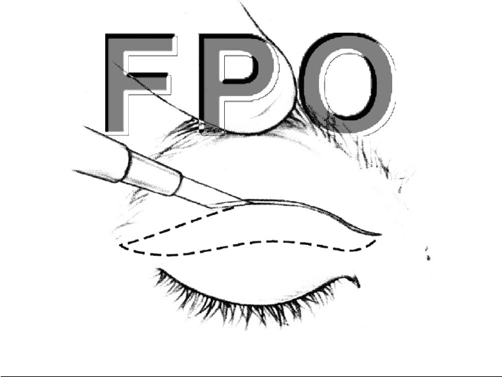
marking pen, creating a “double gull wing” (Fig. 6-1). A ruler or caliper
Surgical Technique 65
FIGURE 6-1. Marking the upper eyelid and incision with a No. 15 blade.

can be used to mark a symmetrical pattern on the contralateral eye-
lid, but the pinch technique should still be used to confirm that the
excision does not result in skin shortage. Remember, upper ble-
pharoplasty removes some skin and muscle, but that is not the only
goal of the procedure, so markings should be placed conservatively.
Local anesthetic is administered to the subcutaneous tissue. The
fat pads or lacrimal gland (or both) should be anesthetized if these
tissues are to reduced or repositioned. The eyelid skin is stretched,
and the markings are incised with a blade. The skin/orbicularis mus-
cle flap is then removed with Westcott scissors or cautery/laser, de-
pending on the surgeon’s preference (Fig. 6-2). The medial and cen-
tral fat pads may be removed by gently tenting the overlying septum
and opening it with the cautery, laser, or scissors. The fat pad is then
removed by the “clamp–cut–cautery” technique. The fat is gently
teased through the open septum with forceps or by applying gentle
pressure to the globe. The fat is clamped with a hemostat and excised
over the hemostat with scissors; and then the base is cauterized (Fig.
6-3). Forceps should be used to hold the stump as the hemostat is re-
leased until hemostasis is visually confirmed. This technique is con-
tinued until the appropriate amount of fat is removed from the re-
gion. Again, a conservative approach is recommended to avoid a
superior sulcus deformity. The septum should not be sutured.
66 Upper Blepharoplasty

Surgical Technique 67
FIGURE 6-2. A skin/muscle flap is removed with scissors.
FIGURE 6-3. Orbital fat is clamped with a hemostat and excised with scissors.

If the lacrimal gland is prominent, it should be posteriorly repo-
sitioned at this point in the procedure. The dissection is continued
through the lateral portion of the blepharoplasty incision by incising
the septum. In a small number of cases, significant orbital fat is pres-
ent between the septum and the lacrimal gland. This fat pad should
be carefully excised, exposing the orbital lobe of the lacrimal gland.
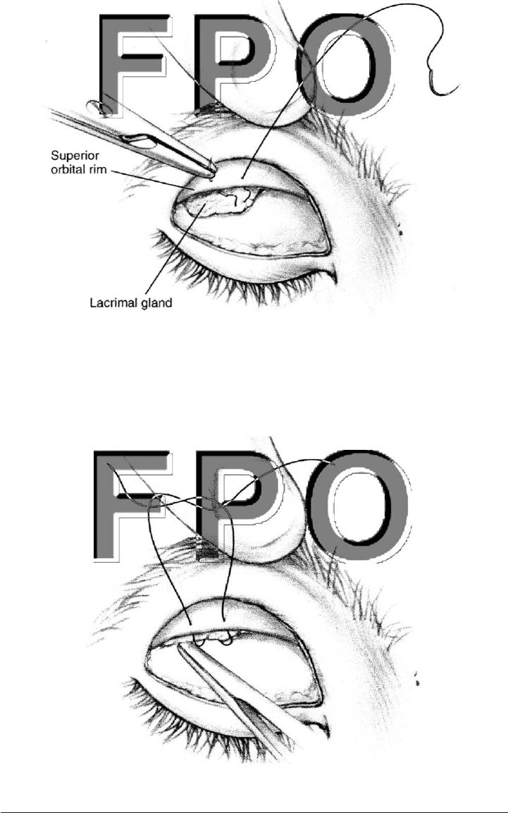
Dissection should extend to the superior orbital rim. A double-armed
5-0 polypropylene suture is passed in mattress fashion through the
inferior portion of the prolapsed gland (Fig. 6-4). Each arm of this su-
ture is then passed posteriorly to anteriorly through the periosteum
of the lacrimal gland fossa, just underneath the superior orbital rim
(Fig. 6-5). The suture is secured, pulling the lacrimal gland back into
the fossa.
Lid crease re-formation can be accomplished by several methods.
If the skin is closed with interrupted sutures, an intervening pur-
chase of the preaponeurotic tissue at the superior tarsal border is in-
cluded in several of the passes across the eyelid crease. If subcuticu-
lar lid closure is planned, it is simpler to place several interrupted,
buried 6-0 absorbable sutures (Vicryl) through the orbicularis at the
inferior incision including the preaponeurotic tissue at the superior
tarsal border. This maneuver locks the lid crease in place and facil-
itates subcuticular closure, which can then be accomplished in a run-
ning fashion with 6-0 polypropylene suture. The suture ends are left
untied, and the entire incision, including the suture ends, are cov-
ered with tincture of benzoin and suture strip tape (Steri-Strips).
68 Upper Blepharoplasty
Surgical Technique 69
FIGURE 6-4. Double-armed suture is passed through the prolapsed lacrimal
gland and through the periosteum.
FIGURE 6-5. Suture tightening pulls the lacrimal gland under the orbital rim.

70 Upper Blepharoplasty
POSTOPERATIVE CARE
Following surgery, the patient should be instructed to place cold or
ice packs on the eyelids for about 15 minutes each hour during the
day for 2 days. The patient should not engage in any lifting or bend-
ing activities and should sleep with his or her head elevated. The su-
tures are removed within 5–8 days by first peeling off the strip tape
and then pulling one end of the subcuticular suture until the entire
suture is free.