Fisher John P. e.a. (ed.) Tissue Engineering
Подождите немного. Документ загружается.


mikos: “9026_c032” — 2007/4/9 — 15:53 — page2—#2
32-2 Tissue Engineering
32.2 The Tooth and Its Supporting Structures
The crowns of teeth that are exposed in the oral cavity are covered by enamel. Under the enamel is
a thick layer of dentin and a soft central core, the pulp chamber (Figure 32.1). Enamel is the hardest
calcified structure as it is about 99.5% mineralized. It varies from 2 to 3 mm in thickness at the height
of cusps and narrows to a knife-edge thickness at the cementoenamel junction. Enamel is deposited
by ameloblasts, cells that are believed to undergo programmed cell death. Because enamel is acellular
and nonvital it cannot regenerate itself. Underlying enamel is dentin, a specialized mineralized matrix
that shares several biochemical characteristics with bone. In contrast to enamel, dentin is a vital tissue
that harbors odontoblastic processes and some nerve endings. The formation of dentin follows the same
principles that guide the formation of other hard connective tissues in the body, namely, cementum
and bone.
As described by Linde and Goldberg [1] and Butler and Ritchie [2], the composition of dentin matrix
and the process of dentinogenesis are highly complex. The organic phase of dentin is composed of
proteins, proteoglycans, lipids, various growth factors, and water. Among the proteins, collagen is the
most abundant and offers a fibrous matrix for the deposition of carbonate apatite crystals. The collagens
that are found in dentin are primarily type I collagen with trace amounts of type V collagen and some type
I collagen trimer. An important class of dentin matrix proteins is the noncollagenous proteins or NCPs [2].
The dentin-specific NCPs are dentin phosphoproteins (DPP) or phosphophoryns and dentin sialoprotein
(DSP). After type I collagen, DPP is the most abundant of dentin matrix proteins and represents almost
50% of the dentin extracellular matrix. DPP is a polyionic macromolecule that is rich in phosphoserine
and aspartic acid. Its high affinity for type I collagen as well as calcium makes it a strong candidate for
the initiation of dentin mineralization. DSP accounts for 5 to 8% of the dentin matrix and has a relatively
high sialic acid and carbohydrate content. Its role in dentin mineralization is unclear at the present time.
For several years it was believed that DSP and DPP were two independent proteins that were encoded
by individual genes. DPP and DSP are specific cleavage products of a larger precursor protein that was
translated from one large transcript [3]. This single gene encoding for DSP and DPP is named dentin
sialophosphoprotein or Dspp.
A second category of NCPs with Ca-binding properties are classified as mineralized tissue-specific
as they are found in all the calcified connective tissues, namely, dentin, bone, and cementum. These
FIGURE 32.1 Component parts of a tooth.

mikos: “9026_c032” — 2007/4/9 — 15:53 — page3—#3
The Bioengineering of Dental Tissues 32-3
include osteocalcin (OC) and bone sialoprotein (BSP). A serine-rich phosphoprotein called dentin matrix
protein 1, Dmp-1, whose expression was first described as being restricted to odontoblasts [4], was later
shown to be expressed by osteoblasts and cementoblasts [5]. Other NCPs include osteopontin (OP) and
osteonectin (secreted protein, acidic, cycteine-rich; SPARC). The fourth category of dentin NCPs are not
expressed in odontoblasts but are primarily synthesized in the liver and are released into the circulation.
An example of a serum borne protein is α2HS-glycoprotein. Diffusible growth factors that appear to be
sequestered within dentin matrix constitute the fifth group of dentin NCPs. This group includes the BMPs,
IGFs, and TGF-βs [6].
The central chamber of the tooth is occupied by a soft connective tissue called the dental pulp that is
comprised of a heterogeneous cell population of fibroblasts, undifferentiated mesenchymal cells, nerves,
blood vessels, and lymphatics. The regenerative capacity of dental pulp is well documented in the literature
and best illustrated by the formation of a layer of reparative dentin beneath a carious lesion or a cavity base.
As will be discussed later, somatic stem cells from the dental pulp are capable of regenerating several tissues
when transplanted in vivo. Cementum is another calcified tissue of mesodermal origin. The cementum
covering the apical third of the root is cellular (contains cementocytes), whereas that of the remaining
two-thirds is acellular. Since the fibers of the periodontal ligament are anchored within cementum, the
regeneration of this complex is important when bioengineering of whole tooth structures is considered.
32.3 Genetic Control of Tooth Development
32.3.1 Stages of Tooth Development
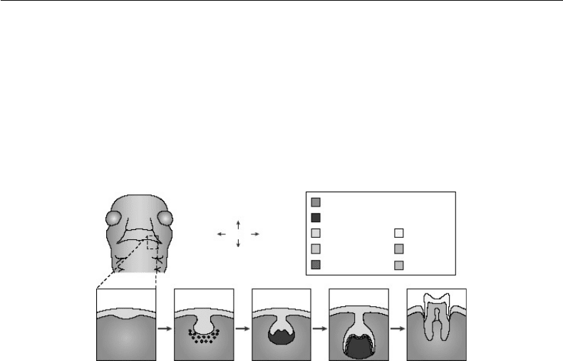
Teeth develop in distinct stages that are easily recognizable at the microscopic level. Hence, stages in odon-
togenesis are described in classic terms by the histologic appearance of the tooth organ. From early to late,
these stages are described as the lamina, bud, cap, and bell (early and late) stages of tooth development
[7,8]. Recent advances made in the understanding of the molecular control of tooth development have led
to the development of new terminology to describe tooth development as occurring in four phases: ini-
tiation, morphogenesis, cell or cyto-differentiation, and matrix apposition (Figure 32.2). The appearance
of the dental lamina marks the first visible sign of tooth initiation that is seen about five weeks of human
Oral
Distal
Proximal
Proximal
Aboral
Neual-crest-derived mesenchyme
Condensing dental mesenchyme
Oral epithelium Enamel
Ameloblasts Dentin
Obontoblasts Dental pulp
Thickening
E11.5
Bud
E13.5
Cap
E14.5
Bell
E18.5
Erupted tooth
P35
FIGURE 32.2 Stages of tooth development. A schematic frontal view of an embryo head at embryonic day (E)11.5
is shown with a dashed box to indicate the site where the lower (mandibular) molars will form. Below, the stages of
tooth development are laid out from the first signs of thickening at E11.5 to eruption of the tooth at around 5 weeks
after birth. The tooth germ is formed from the oral epithelium and neural-crest-derived mesenchyme. At the bell
stage of development, the ameloblasts and odontoblasts form in adjacent layers at the site of interaction between the
epithelium and mesenchyme. These layers produce the enamel and dentin of the fully formed tooth. (Reproduced
from Tucker, A. and Sharpe, P. Nat. Rev. Genet. 5: 499–508, 2004. With permission.)
mikos: “9026_c032” — 2007/4/9 — 15:53 — page4—#4
32-4 Tissue Engineering
development. The inductive influence of the dental lamina to dictate the fate of the underlying ectomesen-
chyme has been confirmed by several researchers [9]. The table below summarizes the molecules expressed
in the epithelium and mesenchyme at this inductive phase. The bud stage is characterized by the continual
growth of cells of the dental lamina and ectomesenchyme. The latter is condensed and termed the dental
papilla. At this stage, the inductive or tooth-forming potential is transferred from the dental epithelium to
the dental papilla. The transition from the bud to the cap stage is an important step in tooth development as
it marks the onset of crown formation. The tooth bud assumes the shape of a cap that is surrounded by the
dental papilla. The ectodermal compartment of the tooth organ is referred to as the dental or enamel organ.
The enamel organ and dental papilla become encapsulated by a sac called the dental follicle that separate
the tooth organ papilla from the other connective tissues of the jaws. A cluster of cells called the enamel
knot is an important organizing center within the dental organ and is important for the formation of cusps
[10,11]. The enamel knot expressesa unique set of signaling molecules that influence the shape of the crown
as well as the development of the dental papilla. Similar to the fate of signaling centers in other organizing
tissues like the developing limb bud, the enamel knot undergoes programmed cell death or apoptosis,
after cuspal patterning is completed at the onset of the early bell stage. As the dental organ assumes the
shape of a bell, several layers of cells continue to divide but at differential rates. A single layer of cuboidal
cells called the external or outer dental epithelium lines the periphery of the dental organ while cells that
border the dental papilla and are columnar in appearance form the internal or inner dental epithelium.
The latter gives rise to the ameloblasts, cells responsible for enamel formation. Cells located in the center
of the dental organ produce high levels of glycosaminoglycans that are able to sequester fluids as well as
growth factors that lead to its expansion. This network of star-shaped cells is named the stellate reticulum.
Interposed between the stellate reticulum and the internal dental epithelium is a narrow layer of flattened
cells termed the stratum intermedium that express high levels of alkaline phosphatase. The stratum inter-
medium is believed to influence the biomineralization of enamel. In the region of the apical end of the
tooth organ, the internal and external dental epithelial layers meet at a junction called the cervical loop.
At the early bell stage, each layer of the dental organ has assumed special functions and exchanges
molecular information that leads to cell differentiation at the late bell stage. The dental lamina that
connects the tooth organ to the oral epithelium gradually disintegrates at the late bell stage. At the
future cusp tips, cells of the internal dental epithelium stop dividing and assume a columnar shape. The
most peripheral cells of the dental papilla organize along the basement membrane and differentiate into
odontoblasts, the dentin-forming cells. At this time, the dental papilla is termed the dental pulp. After
the first layer of predentin matrix is deposited, cells of the internal dental epithelium differentiate into
ameloblasts or enamel-producing cells. As enamel is deposited over dentin matrix, ameloblasts retreat
to the external surface of the crown and are believed to undergo programmed cell death. In contrast,
odontoblasts line the inner surface of dentin and remain metabolically active throughout the life of a
tooth. Root formation then proceeds as epithelial cells proliferate apically and influence the differentiation
of odontoblasts from the dental papilla as well as cementoblasts from follicle mesenchyme. This leads to
the deposition of root dentin and cementum respectively. The dental follicle that gives rise to components
of the periodontium, namely the periodontal ligament fibroblasts, alveolar bone of the tooth socket and
the cementum also plays a role during tooth eruption which marks the end phase of odontogenesis.
32.3.2 Molecular Mechanisms That Determine Tooth Shape, Size, and
Structure
Similar to other organs like the limb bud, kidney, lung, and hair follicles, tooth development is regulated
by temporally and spatially restricted interactions between epithelial and mesenchymal compartments.
Molecular approaches used in expression analyses as well as functional in vivo and in vitro tooth recom-
binations and bead implantation assays have greatly increased our understanding about the molecular
control of tooth development. In addition, the use of genetic approaches involving transgenic mice with
targeted inactivation of various genes have provided a powerful means to delineate the in vivo functions
of individual molecules [12,13].
mikos: “9026_c032” — 2007/4/9 — 15:53 — page5—#5
The Bioengineering of Dental Tissues 32-5
In vivo and in vitro recombination studies have shown that during the formation of the epithelial bud
(E12), the inductive potential shifts to the dental mesenchyme that later influences the fate of the enamel
organ and its morphogenesis from the bud stage to the early bell stage (E16) [9,14–16]. Reciprocal inter-
actions between the morphologically distinct enamel organ and papilla mesenchyme at the late bell stage
(E18) then leads to the differentiation of dentin-forming odontoblasts and enamel-forming ameloblasts.
As morphogenesis advances, the matrices of dentin and enamel are deposited in an organized manner
and root formation begins. Interactions between the apical extension of the enamel organ (epithelial root
sheath) and papilla/follicle mesenchyme lead to the patterning of roots, the differentiation of cemento-
blasts and the formation of cementum. Hence, during crown and root development, morphogenesis, and
cytodifferentiation are controlled by epithelial-mesenchymal interactions.
As depicted in Table 32.1 [13], molecular changes in dental mesenchyme involve proteins in the bone
morphogenetic protein (BMP), fibroblast growth factor (FGF) and wingless-type MMTV integration
site family WNT families; sonic hedgehog (Shh) as well as transcriptional molecules like the Msx-1, -2
homeobox genes; lymphoid enhancer-binding factor 1 (Lef-1) and Pax-9, a member of the paired-box-
containing transcription factor gene family. The actions and interactions of these molecules are complex
and described eloquently in recent reviews [12,17].
The BMPs are among the best-characterized signals in tooth development. In addition to directly influ-
encing morphogenesis of the enamel organ (see discussion on enamel knot later), epithelial BMP-2 and -4
are able to induce expression of Msx1, Msx2, and Lef-1 in dental mesenchyme as shown in bead implant-
ation assays [18–20]. The shift in Bmp-4 expression from epithelium to mesenchyme occurs around
E12 and is coincident with the transfer of inductive potential from dental epithelium to mesenchyme [18].
In mesenchyme, Bmp-4 in turn, requires Msx-1 to induce its own expression [19]. The FGFs, in gen-
eral, are potent stimulators of cell proliferation and division both in dental mesenchyme and epithelium.
Fgf-2, -4,-8, and -9 expression are each restricted to dental epithelium and can stimulate Msx-1 but not
Msx-2 expression in underlying mesenchyme. Fgf-8 is expressed early in odontogenesis (E10.5 to E11.5),
in presumptive dental epithelium, and can induce the expression of Pax-9 in underlying mesenchyme.
Interestingly, BMP-4 prevents this induction and may share an antagonistic relationship with the FGFs
similar to what is observed in limb development [21]. Recent studies by Hardcastle et al., 1998, have
shown that Shh in beads cannot induce Pax9, Msx-1 or Bmp-4 expression in dental mesenchyme but is
able to stimulate other genes encoding the transmembrane protein patched (Ptc) and Gli1, a zinc finger
transcription factor [22–24]. Since neither FGF-8 nor BMP-4 can stimulate Ptc or Gli1, it can be assumed
at the present time that the Shh signaling pathway is independent of the BMP and FGF pathways during
tooth development [24]. Several Wnt genes are expressed during tooth development and may be required
for the formation of the tooth bud [12]. These genes are believed to play a role in activating the intracellular
pathway involving frizzled receptors, β-catenin and nuclear transport of Lef-1. Other signaling molecules
including the Notch genes, epidermal growth factor (EGF), hepatocyte growth factor (HGF) and, platelet
derived growth factor (PDGF) families may also influence tooth development, though the exact nature of
their involvement remains to be elucidated.
The enamel knot is a transient epithelial structure that appears at the onset of cusp formation. For
years, it was thought that the enamel knot controlled the folding of the dental epithelium and hence cuspal
morphogenesis. Recently, the morphological, cellular and molecular events leading to the formation and
disappearance of the enamel knot have been described, thus linking its role as an organizing center for
tooth morphogenesis [11,25,26]. Interestingly, cells of the enamel knot are the only cells within the enamel
organ that stop proliferating [10] and that undergo apoptosis [27]. Another intriguing finding linked p21,
a cyclin-dependent kinase inhibitor associated with terminal differentiation events, to apoptosis of the
enamel knot [11].
The enamel knot cells express several signaling molecule genes including Bmp-2, -4, -7; Fgf-4, -9;
Msx-2 and Shh [22,25,28–30]. Although the precise function of each morphogen is not known at the
present time, a model for the relationship of inductive signaling molecules involved has been proposed
by integrating morphological and molecular data [11]. Since the instructive signaling influence lies with
the dental mesenchyme prior to the development of the primary enamel knot, it is likely that this tissue

mikos: “9026_c032” — 2007/4/9 — 15:53 — page6—#6
32-6 Tissue Engineering
TABLE 32.1 Genes Expressed During Tooth Development in Mouse
a
Stage of Expressed in References Expressed in References
development epithelium mesenchyme
Up to epithelial thickening
(E10–E11) Fgf8,9 [21,60,63,64] Activin [70]
Bmp4 [21,35,60,75] Pax9 [21]
Shh [24,69] Barx1 [60]
Islet1 [65] Msx1 & Msx2 [19,83,84]
Pitx2 [66,67] Dlx1,2,3,5,6 [61,62]
Wnt 10b [76] Ptc [24]
Follistatin [70] Gli1,2,3 [24]
Lef1 [20] Lhx6,7 [63]
Eda, Edar [72]
Bud stage
(E12–E13) Eda, Edar [72] Runx2 [71]
Pitx2 [66,67] Bmp4 [35,75]
Msx1 [35]
Lef1 [20]
Fgf3,10 [73]
Dlx1,2 [62]
Lhx6,7 [63]
Cap stage
(E14–E15) Enamel knot Non-EK epithelium [74]
p21 [11] FgfR [72]
Shh [24] Eda
Edar [72]
Edaradd [68]
Bmp 2,4,7 [75]
Wnt10a [76]
Msx2 [11,29]
Fgf3,4 [10,73]
Bell stage
(E16 Onward) Amelogenin [77] Dspp [78]
Bmp5 [75]
a
This list indicates the expression pattern of several genes that are thought to be important in tooth
development in the mouse. A more comprehensive list of genes and their expression patterns can be found
at the Gene Expression in Tooth web site (http://bite-it.helsinki.fi/) [79]. Barx, BarH-like homeobox; Bmp,
bone morphogenetic protein; Dlx, distal-less homeobox; Dspp, dentin sialophosphoprotein; E, embryonic
stage; Eda, ectodysplasin-A; Edar, Eda receptor; Edaradd, EDAR (ectodysplasin-A receptor)-associated
death domain; EK, enamel knot; Fgf, fibroblast growth factor; FgfR, fibroblast growth factor receptor;
Gli, GLI-Kruppel family member; Lef, lymphoid enhancer binding factor; Lhx, LIM homeodomain genes;
Msx, homeobox, msh-like; p21 (CDKN1A), cyclin-dependent kinase inhibitor 1A; Pax, pairedbox gene;
Pitx, paired-related homeobox gene; Ptc, patched; Runx, runt homologue; Shh, sonic hedgehog; Wnt,
wingless-related protein.
Source: Reproduced from Tucker, A. and Sharpe, P. Nat. Rev. Genet. 5: 499–508, 2004. With permission.
influences enamel knot formation. In this regard, BMP-4 in condensing dental mesenchyme, functions
as a paracrine molecule that can upregulate Msx2 and p21 expression within the enamel knot [11,26].
It is hypothesized that p21 then prevents proliferation within the enamel knot allowing for the growth
stimulatory Fgf-4 to be expressed exclusively in this region [10]. FGF-4 in turn, may act singly or in concert
with Fgf-9 to influence patterning or to regulate expression of downstream genes like Msx1 in underlying
papilla mesenchyme [19,28]. Intriguingly, later in development, BMP-4 participates in the regulation of
apoptosis perhaps in an autocrine fashion by involving genes like Msx-2.
Mice genetically engineered with targeted mutations in transcription factor genes like Msx-1, Lef-1, and
Pax9 as well as activin-βA,amemberoftheTGF-β superfamily, have revealed important information.

mikos: “9026_c032” — 2007/4/9 — 15:53 — page7—#7
The Bioengineering of Dental Tissues 32-7
Knockouts of Bmp-2, -4, and Shh have proven less informative largely due to death that occurs in utero
prior to the onset of tooth development. In Msx-1, Lef-1, Pax9, and activin-βA mutant strains, tooth
development fails to advance beyond the bud stage. Thus, these molecules are important in directing
the fate of the dental mesenchyme and its ability to influence the progress of epithelial morphogenesis
to the cap stage [31–34]. Curiously, Msx2 molars develop fully but show abnormal cuspal patterning,
a poorly differentiated stellate reticulum and enamel matrix defects, suggesting that this homeobox gene
is involved in the patterning and differentiation of the enamel organ [17; Maas, personal communication].
As reviewed by Tucker and Sharpe [13], molecular information on tooth development can be used to alter
the shape and size of teeth. For example, when beads soaked with Bmp4 are placed on mesenchyme within
the presumptive incisor region, Msx1 expression is downregulated and expression of the transcription
factor Barx1 is upregulated. Since Barx1 is normally restricted to the molar region, its misexpression
within the incisor region results in the formation of a molar tooth organ instead of an incisor [35].
In addition to use of the mouse tooth organ model, the legacy of inheritable anomalies of human
dentition involving the failure of teeth to develop offers a powerful system for studying the genetic
pathways that control the development of human dentition. Familial tooth agenesis is the most common
dental anomaly that affects up to 25% of the population. It is transmitted either as an autosomal-dominant,
autosomal-recessive or X-linkedtrait, and presents in syndromic and nonsyndromic forms. Genes involved
in epithelial-mesenchymal interactions as shown by studies in the mouse are strong candidates for human
tooth agenesis. Until recently, mutations in two genes that encode for the key transcription factors PAX9
and MSX1 were associated with agenesis of molars and premolars [36]. Importantly, PAX9 and MSX1 have
each been excluded in other families with autosomal dominant forms of tooth agenesis. Recently, tooth
agenesis has been linked to a mutation in AXIN2, a molecule known to regulate cell homeostasis [37].
Several members of this four-generation family are affected by or are at risk for colon cancer suggesting a
broader role for this molecule in cell proliferation.
Taken together, the data from mouse and human studies have provided valuable insights into the
molecular and genetic control of tooth development. As illustrated later, such basic information provided
the rationale for tooth bioengineering initiatives for the regeneration of dentin matrix and whole tooth
forms.
32.4 Tooth Regenerative Strategies
Over the years, the research on the use of stem cells for clinical therapies has been growing, especially
after researchers have found that hematopoietic stem cells, a well-characterized population of postnatal
stem cells, have been successfully utilized in clinics to treat hematopoietic diseases [38] autoimmune
diseases [39] and solid tumors [40]. Stem cells are defined as cells that have clonogenic and self-renewing
capabilities and that differentiate into multiple-cell lineages. In general, there are two kinds of stem cells:
embryonic and postnatal stem cells. Embryonic stem cells are derived from mammalian embryos in the
blastocyst stage and have the ability to generate any terminally differentiatedcell in the body; postnatal stem
cellsare part of tissue-specific cells of the postnatal organism intowhich they are committed to differentiate.
Stem cell-based tissue regeneration has great clinical potential to regain physiological functions that have
been damaged by various diseases.
32.4.1 Human Dental Pulp Stem Cells
Isolation and identification of stem cells are the first step in studying the potential of stem cell-mediated
therapy. Postnatal stem cells have been isolated from a variety of tissues including but not limited to
skin, liver, brain, bone marrow, and peripheral blood. Recently, dental pulp stem cells (DPSCs) have
been successfully isolated from adult dental pulp in extracted human teeth [41]. Similar to the other
mesenchymal stem cells, DPSCs are able to generate clonogenic cell colonies in vitro. The majority of the
individual colonies (67%) failed to proliferate beyond 20 population doublings in the culture, suggesting

mikos: “9026_c032” — 2007/4/9 — 15:53 — page8—#8
32-8 Tissue Engineering
that only a small portion of cells maintain high proliferation potential in vitro [42]. Mixed multi-colony
DPSCs show a higher proliferation rate than bone marrow stromal stem cells (BMSSCs) in culture. cDNA
microarray analysis demonstrated that highly expressed cyclin-dependent kinase 6 (cdk6) and IGF-2 in
DPSCs might be, at least partially, responsible for the promoted progression of cells through G1 to the
start of DNA synthesis [43–45], leading to an elevated replicative proliferation. Most postnatal stem
cells reside in a specific niche microenvironment to maintain their stemness. To elucidate the DPSCs’
niche environment, DPSCs were first found to express various markers associated with endothelial and/or
smooth muscle cells such as STRO-1, 3G5, VCAM-1, MUC-18 and α-smooth muscle actin [41,46].
Then, immunohistochemical staining and magnetic beads sorting were applied to confirm that DPSCs,
similar to BMSSCs, reside in a perivascular niche microenvironment [46]. Taken together with their
clonogenic nature, higher proliferation rate, and specific niche microenvironment, DPSCs satisfy three
criteria characteristic of human postnatal somatic stem cells.
32.4.2 Dentin Tissue Regeneration
One of the most important characteristics of DPSCs is their capability to form a dentin/pulp-like
complex upon in vivo transplantation in conjunction with hydroxyapatite/tricalcium phosphate as a
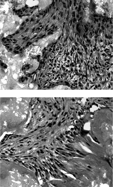
carrier (Figure 32.3). Backscatter EM analysis demonstrated that the dentin-like material formed in the
(a)
(b)
FIGURE 32.3 Hematoxylin and eosin staining of representative DPSC transplants. (a) After one week posttrans-
plantation, DPSC transplants contain connective tissue (CT) around HA/TCP carrier (HA), without any sign of dentin
formation. (b) After six week posttransplantation, DPSCs differentiate into odontoblasts (arrows) that are responsible
for the dentin formation on the surface of HA/TCP (HA). Original magnification: 40×.
mikos: “9026_c032” — 2007/4/9 — 15:53 — page9—#9
The Bioengineering of Dental Tissues 32-9
transplants had a globular appearance consistent with the structure of dentin in situ [42]. DPSC-mediated
odontogenesis is differentiable from BMSSC-mediated osteogenesis by regenerating different organ-like
structures and involving different regulating molecules [47]. This implies that critical factor(s) may regu-
late mineralized matrix forming stem cells to generate defined mineralized tissue along with associated soft
tissues. The property of multipotential differentiation of DPSCs has been demonstrated by the findings
that under the proper culture conditions, DPSCs are capable of differentiating into osteo/odontogenic
cells, adipocytes, and neural cells [42]. However, the assets of multipotential differentiation of DPSCs at
functional levels remain to be confirmed.
The findings on DPSCs may provide a potential for utilizing them for dentin and pulp tissues regen-
eration. Human teeth do not undergo the type of remodeling that is seen in other mineralized tissues
such as bone, which remodels to maintain organ integrity. Once a tooth has erupted, dentinal damage
caused by mechanical trauma, exposure to chemicals or by infectious processes, induces the forma-
tion of reparative dentin that is even though structurally poorly organized, but serves as a protective
barrier to the dental pulp with limited capacity [48–52]. It was reported that bone morphogenetic
protein-7 is capable of stimulating tertiary dentin formation when applied to freshly cut dentin both
in vitro and in vivo [53,54]. This probably occurs through an osteo/odontogenic induction property of
BMP-7, since BMP-7 transfected human fibroblasts were able to express osteogenic characteristic and
form bone tissue in vivo [55]. DPSCs were also capable of forming reparative dentin structure on the
surfaces of regular human dentin [47]. However, it seems that DPSCs exhibit a decreased and altered
in vivo odontogenic capacity when loaded on the surface of human dentin. Although the reason is not
known, it may be associated with the microenvironment that accommodates in vivo differentiation of
DPSCs [47]. Recently, it was demonstrated that autogenous transplantation of BMP2-treated DPSCs
was able to stimulate reparative dentin formation on the amputated pulp [56]. This finding suggests
that combination therapy using stem cells and growth factors may improve stem cell-mediated dentin
regeneration.
32.4.3 Tooth Regeneration
Recently, whole tooth regeneration in vivo has become a hot topic in dental research. Tooth development
involves a mutual signaling interaction between epithelial and mesenchymal cells of neural ectodermal
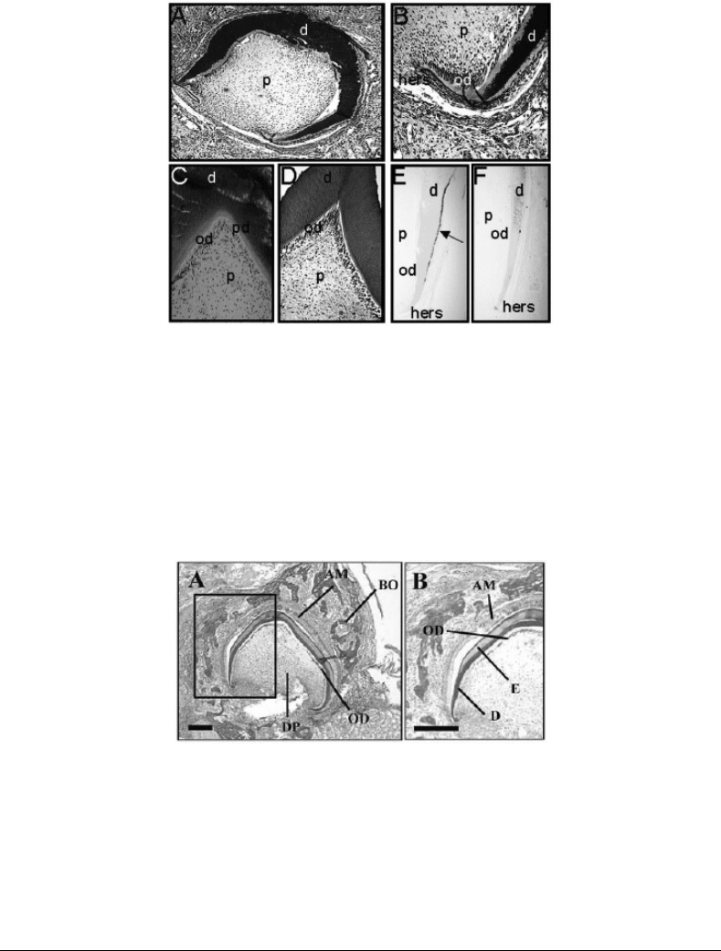
origin. It was demonstrated that tooth crown structures including dentin, odontoblasts, pulp chamber,
putative Hertwig’s root sheath epithelia, putative cementoblasts, and enamel organ could be regenerated
using dissociated cells from pig tooth bud tissues (Figure 32.4) [57]. Further, the same research group
identified that cultured cells from rat tooth bud were also able to regenerate tooth structure when loaded
on the PGA or PLGA scaffolds [58]. These studies demonstrate for the first time that mammalian tooth
structure can be regenerated in a system consisting of tooth bud progenitors and the proper scaffold.
Moreover, Sharpe’s group conducted a promising study to demonstrate that mice embryonic oral epi-
thelium along with nondental stem cells can induce an odontogenic response, showing the expression of
odontogenic mesenchymal cell associated genes such as Msx1, Lhx7, and Pax9 [59]. After being trans-
planted into adult renal capsules, the recombination of embryonic oral epithelium with nondental stem
cells (embryonic, neural, and bone marrow stem cells) gave rise to both tooth structure and bone tissue
(Figure 32.5) [59]. Also, transplanted embryonic tooth primordial were able to maintain their tooth
development potential within an adult environment [59]. This study clearly indicates that the inductive
function of embryonic oral epithelium may be an important driving force for future prospects of achieving
entire tooth regeneration in vivo.
Human DPSCs have been successfully isolated and characterized, which open the door for using
these cells for potential tooth structure regeneration. In addition, stem cell-mediated whole tooth
structure regeneration implies a great potential for regenerating functional entire tooth in vivo.
However, substantial experimentation is still required for translating these technologies into clinical
applications.

mikos: “9026_c032” — 2007/4/9 — 15:53 — page 10 — #10
32-10 Tissue Engineering
FIGURE 32.4 Histology and immunohistochemistry of a 20-week implant. (a) Von Kossa stain for calcified min-
eralization in bioengineered tooth crown (50× magnification). Dark brown stain is positive for mineralized tissues.
(b) A high-magnification (400×) photomicrograph of the Hertwig’s epithelial root sheath is shown, stained by the Von
Kossa method to detect calcified mineralization. (c) High-magnification (200×) photomicrograph of cuspal region in
bioengineered tooth crown. The tissue was stained by the Von Kossa method. (d) Hematoxylin and eosin (H&E) stain
of a positive control porcine third molar cuspal region demonstrates morphology similar to that of the bioengineered
tooth structure (200×). (e) BSP immunostain of 20-week bioengineered tooth crown (100×). Positive BSP expression
is indicated by the arrow. (f) Negative preimmune control immunostain for BSP in bioengineered tooth crown (100×).
Abbreviations: d, dentin; od, odontoblasts; p, pulp; pd, predentin; hers, Hertwig’s epithelial root sheath.
FIGURE 32.5 Recombinant explant between bone-marrow-derived cells and oral epithelium following 12 days of
development in a renal capsule. All the tissues visible are donor-derived, since the host kidney makes no cellular
contribution to the tissue. Where epithelium in the recombinations was from GFP mice, in situ hybridization of
sections of these tissues confirmed that all mesenchyme-derived cells were of wildtype origin (not shown). BO, bone;
Am, ameloblasts; DP, dental pulp; OD, odontoblasts; E, enamel; D, dentin. Scale bar: 80 µm. (Reproduced from
Ohazama, et al. J. Dent. Res. 83: 518–522. With permission.)
32.5 Conclusions
The last decade has witnessed an explosion of scientific and technological advances that will undoubtedly
propel the field of tooth bioengineering forward. This chapter was limited in scope in as much as only
a few tooth regenerative strategies were discussed. Therefore, readers should be mindful of several other
dimensions of research that exist. As presented in the current literature, there is much interest in under-
standing the structural, biomechanical, and bioregulatory features of dentin and bone matrices as well as
the complex process of enamel biomineralization and remineralization. Such basic knowledge is essential
for the development of tooth-specific biological substitutes that will best restore, maintain, or improve
mikos: “9026_c032” — 2007/4/9 — 15:53 — page 11 — #11
The Bioengineering of Dental Tissues 32-11
the functions of normal dentition. The legacy of inheritable anomalies that involve tooth patterning and
extracellular matrices will continue to provide a powerful means of identifying new molecular pathways
that influence normal and abnormal development. Clearly, advances in the field of tooth bioengineering
will depend on the clever integration of basic science knowledge from animal and human developmental
and genetic studies with emerging technologies in the field of stem cell biology, autologous cell therapy,
gene therapy, materials sciences, and nanotechnology. Although the clinical applications for the use of
bioengineered tooth forms and matrices remain limitless, several challenges must be surmounted prior to
successful therapeutic interventions. As important as the timely diagnosis, accurate prognosis and proper
treatment of diseases affecting dentition will be the preparation of host sites within the oral cavity to receive
bioengineered materials. In every respect, the field of tooth bioengineering encompasses broad strategies
and multidisciplinary approaches directed at restoring one of the most complex organs in vertebrates.
Acknowledgments
The authors acknowledge the support of the National Institute of Dental and Craniofacial Research
(NIDCR), National Institutes of Health (NIH). The research program of RDS has been funded through
NIH grants DE10517; DE07252; DE12269; DE11663 and DE13368. STS is supported by the Division of
Intramural Research at the NIDCR.
References
[1] Linde, A. and Goldberg, M. Dentinogenesis. Crit. Rev. Oral. Biol. Med. 4: 679–728, 1993.
[2] Butler, W.T. and Ritchie, H. The nature and functional significance of dentin extracellular matrix
proteins. Int. J. Dev. Biol. 39: 169–179, 1995.
[3] MacDougall, M. et al. Dentin phosphoprotein and dentin sialoprotein are cleavage products
expressed from a single transcript coded by a gene on human chromosome 4. Dentin phosphopro-
tein DNA sequence determination. J. Biol. Chem. 272: 835–842, 1997.
[4] George, A. et al. Characterization of a novel dentin matrix acidic phosphoprotein. Implications for
induction of biomineralization. J. Biol. Chem. 268: 12624–12630, 1993.
[5] D’Souza, R.N. et al. Gene expression patterns of murine dentin matrix protein 1 (Dmp1) and dentin
sialophosphoprotein (DSPP) suggest distinct developmental functions in vivo. J. Bone Miner. Res.
12: 2040–2049, 1997.
[6] Smith, A.J. and Lesot, H. Induction and regulation of crown dentinogenesis: embryonic events as a
template for dental tissue repair? Crit. Rev. Oral Biol. Med. 12: 425–437, 2001.
[7] Ten Cate, A.R. Oral Histology: Development, Structure, and Function, 5th ed. St. Louis, MO, Mosby,
Inc. 1998.
[8] Avery, J.K. Oral Development and Histology. Pine, J.W. (ed.) Baltimore, MD, Waverly Press, 1987.
[9] Mina, M. and Kollar, E.J. The induction of odontogenesis in non-dental mesenchyme combined
with early murine mandibular arch epithelium. Arch. Oral Biol. 32: 123–127, 1987.
[10] Jernvall, J. et al. Evidence for the role of the enamel knot as a control center in mammalian tooth
cusp formation: non-dividing cells express growth stimulating Fgf-4 gene. Int. J. Dev. Biol. 38:
463–469, 1994.
[11] Jernvall, J. et al. The life history of an embryonic signaling center: BMP-4 induces p21 and is
associated with apoptosis in the mouse tooth enamel knot. Development 125: 161–169, 1998.
[12] Thesleff, I. and Sharpe, P. Signalling networks regulating dental development. Mech. Dev. 67:
111–123, 1997.
[13] Tucker, A. and Sharpe, P. The cutting-edge of mammalian development; how the embryo makes
teeth. Nat. Rev. Genet. 5: 499–508, 2004.
[14] Lumsden, A.G.S. Spatial organization of the epithelium and the role of neural crest cells in the
initiation of the mammalian tooth. Development 103: 155–169, 1988.
