Fisher John P. e.a. (ed.) Tissue Engineering
Подождите немного. Документ загружается.


mikos: “9026_c021” — 2007/4/9 — 15:52 — page 12 — #12
21-12 Tissue Engineering
(a)
(b)
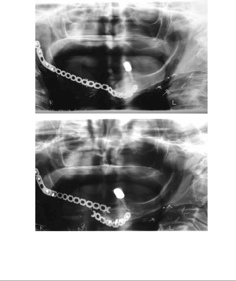
FIGURE 21.5 Panoramic radiographs of a patient after right mandible reconstruction with only a titanium recon-
struction plate showing appearance after surgery (a) and appearance several months later after fracture of the plate
due to metal fatigue (b).
21.5 Conclusion
Tissue engineering may offer a new dimension of therapeutic care for patients. The design and devel-
opment of the tissue engineered therapeutics must be guided by basic, fundamental biological pathways
targeting specific clinical applications. Targeted clinical performance standards for the tissue engineered
therapeutic must be achieved in the design, validated by the appropriate standardized characterization
protocols (e.g., ASTM, ISO 10993), preclinical and clinical phase I–III studies. To fulfill stringent clinical
requirements for a tissue engineered bone therapeutic, specific clinical targets must be identified. Is the
target a nonload bearing or load bearing bone? Is the target a fracture in the tibia of a healthy, young male?
Or is the clinical target a distal radius fracture in a postmenopausal osteoporotic?
In this chapter, we have briefly reviewed the pathways of bone formation and some of the key signaling
molecules cuing discrete outcome events. Tissue engineered products must integrate into their design
fundamental biological elements consistent with bone formation pathways. Many of the key temporal and

mikos: “9026_c021” — 2007/4/9 — 15:52 — page 13 — #13
Tissue Engineering Applications — Bone 21-13
(a)
(b)
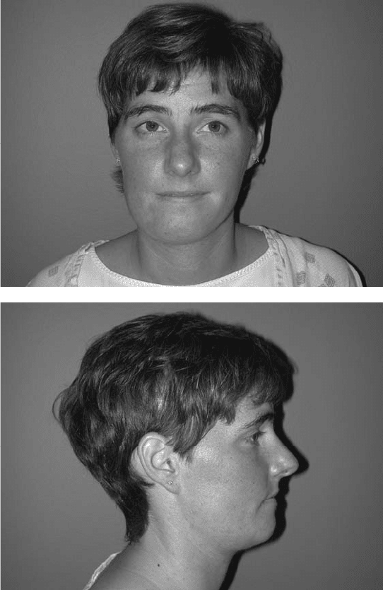
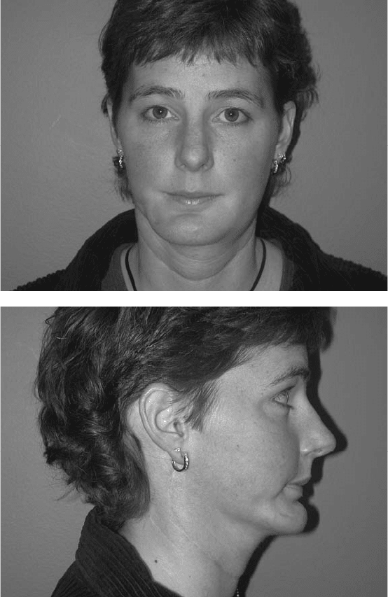
FIGURE 21.6 Appearance of a young woman treated with surgery for a tumor involving the right mandible. The
entire mandible on the right was removed and replaced using a bone flap harvested from the fibula. Appearance before
surgery (a),(b) and 1 year after surgery (c),(d). Although surgery resulted in a durable reconstruction, note the changes
in appearance due to the difference in the shape of the fibula and native mandible.
spatial aspects of bone formation (developmental, homeostatic, healing) remain a mystery. Unless these
key aspects are elucidated, rational, effective tissue engineered therapeutics will not develop.
A broad understanding by tissue engineers of the complex anatomical and physiological issues chal-
lenging surgeons and patients will provide tissue engineers with an important foundational head start
for robust design options that may include uniquely engineered compositions of cells, genes, and biomi-
metic extracellular matrices (e.g., bioactive matrices). The biomimetic extracellular matrix was especially
emphasized in this chapter to stress the importance of spatially and temporally directing the process of
tissue regeneration. Matrix design is the pivotal tissue engineering challenge.
We provided some clinical examples where bone tissue engineering will benefit craniofacial recon-
struction. The craniofacial surgeon faces different clinical challenges than the orthopedic surgeon. Tissue

mikos: “9026_c021” — 2007/4/9 — 15:52 — page 14 — #14
21-14 Tissue Engineering
(c)
(d)
FIGURE 21.6 Continued.
engineering must emphasize a regional-anatomic and physiological approach to design and development.
A single platform treatment for a tibial diaphyseal fracture in the healthy adult male may be inadequate
for the patient with an avulsive bone wound of the oral–antral complex.
There are significant opportunities for the tissue engineer to improve the quality of patient care. The
bedrock underscoring tissue engineering is basic biology, clinical performance standards, and patient
application focus.
References
[1] Boskey A.L. 2003. Biomineralization: an overview. Connect Tissue Res. 44: 5.
[2] Barckhaus R.H. and Hohling H.J. 1978. Electron microprobe analysis of freeze dried and unstained
mineralized epiphyseal cartilage. Cell Tissue Res. 18693: 541–549.
mikos: “9026_c021” — 2007/4/9 — 15:52 — page 15 — #15
Tissue Engineering Applications — Bone 21-15
[3] Marks S.C. and Odgreen P.R. 2002. Structure and development of the skeleton. In J.P. Bilezikian,
L.G. Raisz, and G.A. Rodan (Eds.), Principles of Bone Biology, Vol. 1, pp. 3–14, New York, Academic
Press.
[4] Gay C.V., Donahue H.J., Siedlecki C.A., and Vogler E. 2004. Cellular elements of the skeleton:
osteoblasts, osteocytes, osteoclasts, bone marrow stromal cells. In J.O. Hollinger, T.A. Einhorn,
and B.A. Doll (Eds.), Bone Tissue Engineering, Chapter 3, Boca Raton, FL, CRC Press.
[5] Olsson S.E. and Ekman S. 2002. Morphology and physiology of the growth cartilage under normal
and pathologic conditions. In G.E. Fackelman (Ed.), Bone in Clinical Orthopedics, p. 117, Stuttgart,
Germany, AO Publishing.
[6] Scott, C.K. and Hightower J.A. 1991. The matrix of endochondral bone differs from the matrix of
intramembranous bone. Calcif. Tissue Int. 49: 349–354.
[7] Sandberg M.M. 1991. Matrix in cartilage and bone development: current views on the function
and regulation of the major organic components. Ann. Med. 23: 207–217.
[8] Tuan R.S. 1994. Developmental skeletogenesis. In C.T. Brighton, G. Friedlaender, and J.M. Lane
(Eds.), Bone Formation and Repair, pp. 13, Rosemont, IL, AAOS.
[9] Doll, B.A. 2004. Developmental biology of the skeletal system. In J.O. Hollinger, T.A. Einhorn, and
B.A. Doll (Eds.), Bone Tissue Engineering, Chapter 1, Boca Raton, FL, CRC Press.
[10] Wolff J. 1892. The Law of Bone Remodelling. Translatedby Maquet P. and FurlongR. 1986. NewYork,
NY, Springer-Verlag.
[11] Mikuni-Takagaki Y. 1999. Mechanical responses and signal transduction pathways in stretched
osteocytes. J. Bone Miner. Metab. 17: 57–60.
[12] Dehority W., Halloran B.P., Bikle D.D., Curren T., Kostenuik P.J., Wronski T.J., Shen Y., Rabkin B.,
Bouraoui A., and Morey-Holton E. 1999. Bone and hormonal changes induced by skeletal
unloading in the mature male rat. Am. J. Physiol. 276: E62–E69.
[13] Montufar-Solis D., Duke P.J., and Morey-Holton E. 2001. The Spacelab 3 stimulation: basis for a
model of growth plate response in microgravity in the rat. J. Gravit. Physiol. 8: 67–76.
[14] Cullinane D.M. and Salisbury K.T. 2004. Biomechanics. In J.O. Hollinger, T.A. Einhorn, and
B.A. Doll (Eds.), Bone Tissue Engineering, Chapter 10, Boca Raton, FL, CRC Press.
[15] Ferretti J.L., Capozza R.F., and Zanchetta J.R. 1996. Mechanical validation of a tomographic QCT
index for noninvasive estimation of rat femur bending strength. Bone 18: 97–102.
[16] Beck T.J., Mourtada F.A., Ruff C.B., Scott W.W., and Kao G. 1998. Experimental testing of a
DEXA-derived curved beam model of the proximal femur. J. Orthop. Res. 16: 394–398.
[17] Toyras J., Nieminen M.T., Kroger H., and Jurvelin J.S. 2002. Bone mineral density, ultrasound
velocity, and broadband attention predict mechanical properties of trabecular bone differently.
Bone 31: 503–507.
[18] Stigbrand T. 1984. Present Status and Future Trends of Human Alkaline Phosphatases, pp. 3–14,
New York, NY, Alan R. Liss.
[19] Cole D.E. and Cohen M.M. 1990. Mutations affecting bone forming cells. In B.K. Hall (Ed.), Bone:
The Osteoblast and the Osteocyte, pp. 442–452, New Jersey, The Telford Press.
[20] Risteli L. and Risteli J. 1993. Biochemical markers of bone metabolism. Ann. Med. 25:
385–393.
[21] Butler W.T. 1989. The nature and significance of osteopontin. Connect Tissue Res. 23:
123–136.
[22] Hoang Q.Q., Sicheri F., Howard A.J., and YANG D.S.C. 2003. Bone recognition mechanism of
porcine osteocalcin from crystal structure. Nature 425: 977–980.
[23] Hunter G.K. and Goldberg H.A. 1993. Nucleation of hydroxyapatite by bone sialoprotein. Proc.
Natl Acad. Sci. USA 90: 8562–8565.
[24] Barnes G.L., Kostenuik P.J., Gerstenfeld L.C., and Einhorn T.A. 1999. Growth factor regulation of
fracture repair. J. Bone Miner. Res. 14: 1805–1815.
[25] Learnmoth I.D. 2004. The management of periprosthetic fractures around the femoral stem.
J. Bone Joint Surg. Br. 86: 13–19.
mikos: “9026_c021” — 2007/4/9 — 15:52 — page 16 — #16
21-16 Tissue Engineering
[26] Sfeir C., Jadlowiec J.J., Koch H., and Campbell P.G. 2004. Signaling molecules for tissue
engineering. In J.O. Hollinger, T.A. Einhorn, and B.A. Doll (Eds.), Bone Tissue Engineering,
Chapter 5, Boca Raton, FL, CRC Press.
[27] Mohan S. and Baylink D.J. 2002. IGF-binding proteins are multifunctional and act via IGF-
dependent and -independent mechanisms. J. Endocrinol. 175: 19–31.
[28] Nunes I., Gleizes P.E., Metz C.N., and Rifkin D.B. 1997. Latent transforming growth factor-beta
binding protein domains involved in activation and transglutaminase-dependent cross-linking of
latent transforming growth factor-beta. J. Cell Biol. 136: 1151–1163.
[29] Balemans W. and HuI W.V. 2002. Extracellular regulation of BMP signaling in vertebrates: a
cocktail of modulators. Dev. Biol. 250: 231–250.
[30] Baxter, R.C. 2000. Insulin-like growth factor (IGF)-binding proteins: interactions with IGFs and
intrinsic bioactivities. Am. J. Physiol. Endocrinol. Metab. 278: E967–E976.
[31] Saltzman W.M. and Olbreicht W.L. 2002. Building drug delivery into tissue engineering. Nat. Rev.:
Drug Dis. 1: 177–186.
[32] Richardson T.P., Peters M.C., Ennett A.B., and Mooney D.J. 2001. Polymeric system for dual
growth factor delivery. Nat. Biotechnol. 19: 1029–1034.
[33] Nixon A.J., Brower-Toland B.D., Bent S.J., Saxer R.A., Wilke M.J., Robbins P.D., and Evans C.H.
2000. Insulin-like growth factor-I gene therapy applications for cartilage repair. Clin. Orthop. 379:
S201–1113.
[34] Zhang M., Xuan S., Bouxsein M.L., von Stechow D., Akeno N., and Fougere M.C. et al. 2002.
Osteoblast-specific knockout of the IGF receptor gene reveals an essential role of IGF signaling in
bone matrix mineralization. J. Biol. Chem. 277: 44005–44012.
[35] McCarthy T.L., Centrella M., and Canalis E. 1989. Regulatory effects of insulin-like growth factors
I and II on bone collagen synthesis in rat calvarial cultures. Endocrinology 124: 301–309.
[36] Ogata N., Chikazu D., Kubota N., Terauchi Y., and Tobe K. et al. 2000. Insulin receptor substrate-1
in osteoblast is indispensable for maintaining bone turnover. J. Clin. Invest. 105: 935–943.
[37] Zhang M., Faugere M.C., Malluche H., Rosen C.J., Chernausek S.D., and Clemens T.L. 2003.
Paracrine overexpression of IGFBP-4 in osteoblasts of transgenic mice decreases bone turnover
and causes global growth retardation. J. Bone Miner. Res. 18: 836–843.
[38] Raile K., Hoflich A., Hessler U., Yang Y., Pfuender M., Blum W.F., Kolb H., Schwartz H.B.,
and Kiess W. 1994. Human osteosarcoma (U-2 OS) cells express both insulin-like growth factor-I
(IFG-I) receptors and insulin-like growth factor-II/mannose-6-phosphate (IGF-II/M6P) receptors
and synthesize IGF-II: autocrine growth stimulation by IGF-II via the IGF-I receptor. J. Cell Physiol.
159: 531–541.
[39] Rosenfeld R.G. and Roberts C.T. 1999. The IGF System: Molecular Biology, Physiology and Clinical
Applications, p. 19, NJ, Homana Press.
[40] Chan S.J., Magamatsu S., Cao Q.-P., and Steiner D.F. 1992. Structure and evolution of insulin and
insulin-like growth factors in chordates. Prog. Brain Res. 1992: 15–24.
[41] Mohan S. and Baylink D.J. 1996. The I.G.Fs as Potential therapy for metabolic bone diseases.
In J.P. Bilezikian, L.G. Raisz, and G.A. Rodan (Eds.), Principles of Bone Biology, pp. 1100–1107,
New York, Academic Press.
[42] Gray A., Tam A., Dull W., Hayflick T.J., Pintar J., Cavenee W.K., Koufos A., and Ulrich A.
1987. Tissue-specific and developmentally-regulated transcription of the IGF 2 gene. DNA 6:
283–295.
[43] Trippel, S.B. 1994. Biologic regulation of bone growth. In C.T. Brighton, G. Friedlaender, and
J.M. Lane (Eds.), Bone Formation and Repair, p. 43, Rosemont, IL, AAOS.
[44] Underwood L.E. and Van Wyk J.J. 1992. Normal and aberrant growth. In J.D. Wilson and
D.W. Foster (Eds.), Williams Textbook of Endocrinology, pp. 1079–1138, Philadelphia, PA,
WB Saunders.
[45] Schoenle E., Zapf J., Hauri C. et al. 1985. Comparison of in vivo effects of insulin-like growth factors
I and II and of growth hormone in hypophysectomized rats. Acta Endocrinol. 108: 167–174.
mikos: “9026_c021” — 2007/4/9 — 15:52 — page 17 — #17
Tissue Engineering Applications — Bone 21-17
[46] DeChiara T.M., Efstratiadis A., and Robertson E.J. 1990. A growth factor deficiency phenotype in
heterozygous mice carrying an insulin-like growth factor II gene disrupted by targeting. Nature.
345: 78–80.
[47] Thaler S.R., Dart A., and Tesluk H. 1993. The effect of insulin-like growth factor-I on critical sized
calvarial defects in Sprague-Dawley rats. Ann. Plast. Surg. 31: 429–433.
[48] Tanaka H., Quarto R., Williams S., Barnes J., and Liang C.T. 1994. In vivo and in vitro effects of
insulin-like growth factor-I (IGF-I) on femoral mRNA expression in old rats. Bone 15: 647–653.
[49] Jadlowiec J.J., Celil A.B., and Hollinger J.O. 2003. Bone tissue engineering: recent advances and
promising therapeutic agents. Exp. Opin. Biol. Ther. 3: 409–423.
[50] Kurtz C.A., Loebig T.G., Anderson D.D., Demeo P.J., and Campbell P.G. 1999. Insulin-like growth
factor I accelerates functional recovery from Achilles tendon injury in a rat model. Am. J. Sports
Med. 27: 363–369.
[51] Kandziora F., Schmidmaier G., Schollmeier G. et al. 2002. IGF-I and TGF-beta application by a
poly-(d,l-lactide)-coatedcage promotes intervertebral bone matrix formation in the sheep cervical
spine. Spine 27: 1710–1723.
[52] Illi O.E. and Feldmann C.P. 1998. Stimulation of fracture healing by local application of humoral
factors integrated in biodegradable implants. Eur. J. Pediatr. Surg. 8: 251–255.
[53] Trippel S.B., Whelan M.C., Klagsbrun M. et al. 1992. Interaction of basic fibroblast growth factor
with bovine growth plate chondrocytes. J. Orthop. Res. 10: 638–646.
[54] Canalis E., McCarthy T.L., and Centrella M. 1989. Effects of platelet-derived growth factor on
bone formation in vitro. J. Cell Physiol. 140: 530–537.
[55] Jingushi S., Heydemann A., Kana S.R., Macey L.R., and Bolander M.E. 1990. Acidic fibro-
blast growth factor (aFGF) injection stimulates cartilage enlargement and inhibits cartilage gene
expression in rat fracture healing. J. Orthop. Res. 8: 364–371.
[56] Dunstan C.R., Boyce R., Boyce B.F. et al. 1999. Systemic administration of acidic fibroblast growth
factor (FGF-1) prevents bone loss and increases new bone formation in ovariectomized rats. J. Bone
Miner. Res. 14: 953–959.
[57] Pandit A.S., Feldman D.S., Caulfield J. et al. 1998. Stimulation of angiogenesis by FGF-1 delivered
through a modified fibrin scaffold. Growth factors 15: 113–123.
[58] Kimoto T., Hosokawa R., Kubo T., Maeda M., Sano A., and Akagawa Y. 1998. Continuous
administration of basic fibroblast growth factor (FGF-2) accelerates bone induction on rat calvaria
— an application of a new drug delivery system. J. Dent. Res. 77: 1965–1969.
[59] Inui K., Maeda M., Sano A. et al. 1998. Localapplication of basic fibroblast growth factor minipellet
induces the healing of segmental bony defects in rabbits. Calcif. Tissue Int. 63: 490–495.
[60] Hosokawa R., Kikuzaki K., Kimoto T. et al. 2000. Controlled local application of basic fibroblast
growth factor (FGF-2) accelerates the healing of GBR. An experimental study in beagle dogs. Clin.
Oral Implants Res. 11: 345–353.
[61] Goodman S.B., Song Y., Yoo J.Y., Fox N., Trindale M.C., Kajiyama G., Ma T., Regula D., Brown J.,
and Smith R.l. 2003. Local infusion of FGF-2 enhances bone ingrowth in rabbit chambers in the
presence of polyethylene particles. J. Biomed. Res. A: 454–461.
[62] Bates D.O., Lodwick D., and Williams B. 1999. Vascular endothelial growth factor and
microvascular permeability. Microcirculation 6: 83–96.
[63] Stone D., Phaneuf M., Sivamurthy N. et al. 2002. A biologically active VEGF construct in vitro:
implications for bioengineering-improved prosthetic vascular grafts. J. Biomed. Mater. Res. 59:
160–165.
[64] Bauters C., Asahara T., Zheng L.P. et al. 1995. Recovery of disturbed endothelium-dependent flow
in the collateral-perfused rabbit ischemic hindlimb after administration of vascular endothelial
growth factor. Circulation 91: 2802–2809.
[65] Takeshita S., Pu L.Q., Stein L.A. et al. 1994. Intramuscular administration of vascular endothelial
growth factor induces dose-dependent collateral artery augmentation in a rabbit model of chronic
limb ischemia. Circulation 90: II228–II234.
mikos: “9026_c021” — 2007/4/9 — 15:52 — page 18 — #18
21-18 Tissue Engineering
[66] Takeshita S., Tsurumi Y., Couffinahl T. et al. 1996. Gene transfer of naked DNA encoding for
three isoforms of vascular endothelial growth factor stimulates collateral development in vitro.
Lab. Invest. 75: 487–501.
[67] Pepper M.S., Ferrara N., Orci L. et al. 1992. Potent synergism between vascular endothelial growth
factor and basic fibroblast growth factor in the induction of angiogenesis in vitro. Biochem. Biophys.
Res. Commun. 189: 824–831.
[68] Murphy W.L., Simmons C.A., Kaigler D., and Mooney D.J. 2004. Bone regeneration via a mineral
substrate and induced angiogenesis. J. Dent. Res. 83: 204–210.
[69] Massague J. and Wotton D. 2000. Transcriptional control by the TGF-beta/Smad signaling system.
EMBO J. 19: 1745–1754.
[70] Bourque W.T., Gross M., and Hall B.K. 1993. Expression of four growth factors during fracture
repair. Int. J. Dev. Biol. 37: 573–579.
[71] Robey P.G., Young M.F., Flanders K.C. et al. 1987. Osteoblasts synthesize and respond to
transforming growth factor type beta (TGF-beta) in vitro. J. Cell Biol. 105: 457–463.
[72] Abe N., Lee Y.P., Sato M. et al. 2002. Enhancement of bone repair with a helper-dependent
adenoviral transfer of bone morphogenetic protein-2. Biochem. Biophys. Res. Commun. 297:
523–527.
[73] Hassel S., Schmitt S., Hartung A. et al. 2003. Initiation of Smad-dependent and Smad independent
signaling via distinct BMP-receptor complexes. J. Bone Joint Surg. Am. 85: 44–51.
[74] Sciadini M.F. and Johnson K.D. 2000. Evaluation of recombinant human bone morphogenetic
protein-2 as a bone graft substitute in a canine segmental defect model. J. Orthop. Res. 18:
289–302.
[75] Salkeld S.L., Parton L.P., Barrack R.L., and Cook S.D. The effect of osteogenic protein-1 on the
healing of segmental bone defects treated with autograft or allograft bone. JBJS Am. 2001: 83–1:
803–816.
[76] Grauer J.N., Patel T.C., Erulkar J.S., Troiano N.W., Panjabi M.M., and Friedlaender G.E.
2001. Young Investigator research award winner: evaluation of OP-1 as a graft substitute for
intertransverse process lumbar fusion. Spine 26: 127–133.
[77] Magin M.N. and Delling G. 2001. Improved lumbar vertebral interbody fusion using rhOP-1:
a comparison of autogenous bone graft, bovine hydroxyapatite and BMP-7 in sheep. Spine 26:
469–478.
[78] Gerhart T.N., Kirker-Head C.A., Kriz M.J. et al. 1993. Healing segmental defects in sheep using
recombinant human bone morphogenetic protein. Clin. Orthop. 293: 317–326.
[79] Bostrom M., Lane J.M., Tomin E. et al. 1996. Use of bone morphogenetic protein-2 in the rabbit
ulnar nonunion model. Clin. Orthop. 327: 272–282.
[80] Yasko A.W., Lane J.M., Fellinger E.J., Rosen V., Wozney J.M., and Wang E.A. 1992. The healing of
segmental defects, induced by human bone morphogenetic protein (rhBMP-2). A radiographic,
histological, and biomechanical study in rats. J. Bone Joint Surg. Am. 74: 659–670.
[81] Geesink R.G., Hoefnagels N.H., and Bulstra S.K. 1999. Osteogenic activity of OP-1 bone
morphogenetic protein (BMP-7) in a human fibular defect. J. Bone Joint Surg. Br. 81:
710–718.
[82] Boden S.D., Zdeblick T.A., Sandhu H.S., and Heim S.E. 2000. The use of rhBMP-2 in interbody
fusion cages: definitive evidence of osteoinduction in human—apreliminary report. Spine 25:
376–381.
[83] Turgeman G., Pittman D.D., Muller R. et al. 2001. Engineered human mesenchymal stem cells: a
novel platform for skeletal cell mediated gene therapy. J. Gene Med. 3: 240–251.
[84] Issack P.S. and DiCesare P.E. 2003. Recent advances toward the clinical application of bone
morphogenetic proteins in bone and cartilage repair. Am.J.Orthop.32: 429–436.
[85] Westermark B. and Heldin C.H. 1993. Platelet-derived growth factor. Acta Oncol. 32: 101–105.
[86] Pierce G.F. and Musote T.A. 1995. Pharmacologic enhancement of wound healing. Annu. Rev.
Med. 46: 467–481.
mikos: “9026_c021” — 2007/4/9 — 15:52 — page 19 — #19
Tissue Engineering Applications — Bone 21-19
[87] Centrella M., McCarthy T., Kusik W., and Canalis E. 1992. Isoform specific regulation of platelet
derived growth factor activity and binding in osteoblast-enriched cultures from fetal rat bone.
J. Clin. Invest. 89: 1076–1084.
[88] Rodan S.B. and Rodan G.A. 1992. Fibroblast growth factor and platelet derived growth factor. In
Gowan M. (Ed.), Cytokines and Bone Metabolism, pp. 116–140, Boca Raton, FL, CRC Press.
[89] Andrew J.J., Hoyland J., Freemont A., and Marsh D. 1995. Platelet derived growth factor expression
in normally healing human fractures. Bone 16: 455–460.
[90] Gruber R., Varga F., Fishcer M., and Watzek G. 2002. Platelets stimulate proliferation of bone
cells: involvement of platelet derived growth factors, microparticles and membranes. Clin. Oral
Implant. Res. 3: 529–535.
[91] Lynch, S.E., Colvin R.B., and Antoinades H.N. 1989. Growth factors in wound healing: single and
synergistic effects on partial thickness porcine skin wounds. J. Clin. Invest. 84: 640–646.
[92] Pierce G.F., Mustoe T.A., Altrock B.W., Deuel T.F., and Thomason A. 1991. Role of platelet derived
growth factor in wound healing. J. Cell Biochem. 10: 131–138.
[93] Canalis E. and Rydziel S. 1996. Platelet-derived growth factor and the skeleton. In J.P. Bilezikian,
L.G. Raisz, and G.A. Rodan (Eds.), Principles of Bone Biology, p. 621, New York, Academic
Press.
[94] Mitlak B., Finkelman R., Hill E., Li J., Martin B., Smith T., D’Andrea M., Antoniades H., and
Lynch S. 1996. The effect of systemically administered PDGF-BB on the rodent skeleton. J. Bone
Miner. Res. 11: 238–247.
[95] Howes R., Bowness J., Grotendorst G., Martin G., and Reddi A. 1988. Platelet derived growth
factor enhances demineralized bone matrix-induced cartilage and bone formation. Calcif. Tissue
Int. 42: 34–38.
[96] Nash T., Howlett C., Martin C., Steele J., Johnson K., and Hicklin D. 1994. Effect of platelet derived
growth factor on tibial osteotomies in rabbit. Bone 15: 203–208.
[97] Guler H.P., Zapf J., and Froesch E.R. 1987. Short-term metabolic effects of recombinant human
insulin-like growth factor I in healthy adults. N.Engl.J.Med.317: 137–140.
[98] Boden S.D., Zdeblick T.A., Sandhu H.S., and Heim S.E. 2002. Use of recombinant human bone
morphogenetic protein-2 to achieve postrolateral lumbar spine fusion in humans: a prospective,
randomized clinical pilot trial. Spine 27: 2661–2673.
[99] Boyne P.J., Marx R.E., Nevins M. et al. 1997. A feasibility study evaluating rhBMP-2/absorbable
collagen sponge for maxillary sinus floor augmentation. Int. J. Periodont. Restorat. Dent. 17: 11–25.
[100] Abe E., Yamamoto M., Taguchi Y. et al. 2000. Essential requirement of BMPs-2/4 for both osteoblast
and osteoclast formation in murine bone marrow cultures from adult mice: antagonism by noggin.
J. Bone Miner. Res. 15: 663–673.
[101] Aspenberg P., Jeppson C., and Economides A.N. 2001. The bone morphogenetic proteins
antagonist Noggin inhibits membranous ossification. J. Bone Miner. Res. 3: 15.
[102] Poynton A.R. and Lane J.M. 2002. Safety profile for the clinical use of bone morphogenetic proteins
in the spine. Spine 27: S40–S48.
[103] Morizono K., De Ugarte D.A., Zhu M. et al. 2003. Multilineage cells from adipose tissue as gene
delivery vehicles. Hum. Gene Ther. 14: 59–66.
[104] Jankowski R.J., Deasy B.M., and Huard J. 2002. Muscle-derived stem cells. Gene Ther. 9:
642–647.
[105] Lee J.Y., Peng H., Usas A. et al. 2002. Enhancement of bone healing based on ex vivo gene therapy
using human muscle-derived cells expressing bone morphogenetic protein-2. Hum. Gene Ther.
13: 1201–1211.
[106] Bruder S.P., Kurth A.A., Shea M., Hayes W.C., Jaiswal N., and Kadiyala S. 1998. Bone regeneration
by implantation of purified, culture-expanded human mesenchymal stem cells. J. Orthop. Res. 16:
155–162.
[107] Shang Q., Wang Z., Liu W., Shi Y., Cui L., and Cao Y. 2001. Tissue engineered bone repair of sheep
cranial defects with autologous bone marrow stromal cells. J. Craniofac. Surg. 12: 586–593.
mikos: “9026_c021” — 2007/4/9 — 15:52 — page 20 — #20
21-20 Tissue Engineering
[108] Hannallah D., Peterson B., Lieberman J.R., Fu F.H., and Huard J. 2003. Gene therapy in orthopedic
surgery. Inst. Course Lect. 52: 753–768.
[109] Gamradt S.C. and Lieberman J.R. 2004. Genetic modification of stem cells to enhance bone repair.
Ann. Biomed. Eng. 32: 136–147.
[110] Alden T.D., Pittman D.D., Beres E.J. et al. 1999. Percutaneous spinal fusion using bone
morphogenetic protein-2 gene therapy. J. Neurosurg. 90: 109–114.
[111] Alden T.D., Pittman D.D., Hankins G.R., Beres E.J. et al. 1999. In vivo endochondral bone
formation using a bone morphogenetic protein-2 adenoviral vector. Hum. Gene Ther. 10:
2245–2253.
[112] Kakar S. and Einhorn T.A. 2004. Tissue engineering of bone. In J.O. Hollinger, T.A. Einhorn, and
B.A. Doll (Eds.), Bone Tissue Engineering, Chapter 11, Boca Raton, FL, CRC Press.
[113] Hutmacher D.W. 2000. Scaffolds in tissue engineering bone and cartilage. Biomaterials 21:
2529–2543.
[114] Gerhart T.N., Kirker-Head C.A., Kriz M.J. et al. 1993. Healing segmental femoral defects in sheep
using recombinant human bone morphogenetic protein. Clin. Orthop. 293: 317–326.
[115] Li R.H. and Wozney J.M. 2001. Delivering on the promise of bone morphogenetic proteins. Trends
Biotechnol. 19: 255–265.
[116] Radomsky M.L., Aufdemorte T.B., Swain L.D. et al. 1999. Novel formulation of fibroblast growth
factor-2 in a hyaluronan gel accelerates fracture healing in nonhuman primates. J. Orthop. Res. 17:
607–614.
[117] Sellers R.S., Zhang R., Glasson S.S. et al. 2000. Repair of articular cartilage defects one year after
treatment with recombinant human bone morphogenetic protein-2 (rhBMP-2). J. Bone Joint Surg.
Am. 82: 151–160.
[118] Boden S.D. 1999. Bioactive factors for bone tissue engineering. Clin. Orthop. 367: S84–S94.
[119] Wang S. 2000. Study on the mechanism of bone formation of bioactive materials BMP/Beta-TCP
restoring bone defects by using quantitative analysis methods. Society for Biomaterials, 6th World
Biomaterials Conference, Hawaii, USA.
[120] Jin Q.M., Takita H., Kohgo T. et al. 2000. Effects of geometry of hydroxyapatite as a cell substratum
in BMP-induced ectopic bone formation. J. Biomed. Mater. Res. 51: 491–499.
[121] Ijiri S., Nakamura T., Fujisawa Y. et al. 1997. Ectopic bone induction in porous apatite-
woolastonite-containing glass ceramic combined with bone morphogenetic protein. J. Biomed.
Mater. Res. 35: 421–432.
[122] Winn S.R., Uludag H., and Hollinger J.O. 1999. Carrier systems for bone morphogenetic proteins.
Clin. Orthop. 3679: S95–S106.
[123] Whang K., Goldstick T.K., and Healy K.E. 2000. A biodegradable polymer scaffold for delivery of
osteotropic factors. Biomaterials 21: 2545–2551.
[124] Lee S.C., Shea M., Battle M.A. et al. 1994. Healing of large segmental defects in rat femurs is aided
by rhBMP-2 in PLGA matrix. J. Biomed. Mater. Res. 28: 1149–1156.
[125] Hollinger J.O. and Leong K. 1996. Poly(alpha-hydroxy acids): carriers for bone morphogenetic
proteins. Biomaterials 17: 187–194.
[126] Lucas P.A., Laurencin C., Syftestad G.T. et al. 1990. Ectopic induction of cartilage and bone by
water soluble proteins from bovine bone using a polyanhydride delivery vehicle. J. Biomed. Mater.
Res. 24: 901–911.
[127] Clokie C.M. and Urist M.R. 2000. Bone morphogenetic protein excipients: comparative
observations on poloxamer. Plast. Reconstr. Surg. 105: 628–637.
[128] Saito N., Okada T., Toba S. et al. 1999. New synthetic absorbable polymers as BMP carriers: plastic
properties of poly-D,L-lactic acid-polyethylene glycol block copolymers. J. Biomed. Mater. Res. 47:
104–110.
[129] Kenley R., Marden L., Turek T. et al. 1994. Osseous regeneration in the rat calvarium using novel
delivery systems for recombinant human bone morphogenetic protein-2 (rhBMP-2). J. Biomed.
Mater. Res. 28: 1139–1147.
mikos: “9026_c021” — 2007/4/9 — 15:52 — page 21 — #21
Tissue Engineering Applications — Bone 21-21
[130] Gao T., Lindholm T.S., Marttinen A. et al. 1996. Composite of bone morphogenetic protein (BMP)
and Type I. V. collagen, coral-derived coral hydroxyapatite, and tricalcium phosphate ceramics.
Int. Orthop. 20: 321–325.
[131] Higuchi T., Kinoshita A., Takahashi K. et al. 1999. Bone regeneration by recombinant human
bone morphogenetic protein-2 in rat mandibular defects. An experimental model of defect filling.
J. Periodontol. 70: 1026–1031.
[132] Ripamonti U. 1996. Osteoinduction in porous hydroxyapatite implanted in heterotropic sites of
different animal models. Biomaterials 17: 31.
[133] Galgut P.N. 1994. A technique for treatment of extensive periodontal defects: a case study. J. Oral
Rehabil. 21: 27–32
[134] Askar I., Gultan S.M., Erden E., and Yormuk E. 2003. Effects of polyglycolic acid bioabsorbable
membrane and oxidized cellulose on the osteogenesis in bone defects: an experimental study. Acta
Chir. Plast. 45: 131–138.
[135] Tsuchiya K., Mori T., Chen G., Ushida T., Tateishi T., Matsuno T., Sakamoto M., and Umezawa A.
2004. Custom shaping system for bone regeneration by seeding marrow stromal cells onto a
web-like biodegradable hybrid sheet. Cell Tissue Res. 316: 141–153.
[136] Sharma B. and Elisseeff J.H. 2004. Engineering structurally organized cartilage and bone tissues.
Ann. Biomed. Eng. 32: 148–159.
[137] Aurell C.J. and Flodin P. 2002. New Linear Block Polymer. July 11, 2002.
[138] Aurell C.J. and Flodin P. 2003. A Method for Preparing an Open Porous Polymer Material and an
Open Porous Polymer Material. EP1353982.
[139] Zhang J.Y., Beckman E.J., Hu J., Yuang G.G., Agarwal S., and Hollinger J.O. 2002. Synthesis,
biodegradability, and biocompatibility of lysine diisocyanate-glucose polymers. Tissue Eng. 8:
771–785.
[140] Zhang J.Y., Beckman E.J., Piesco N.J., and Agarwal S. 2002. A new peptide-based urethane poly-
mer: synthesis, biodegradation, and potential to support cell growth in vitro. Biomaterials 21:
1247–1258.
[141] Zhang J.Y., Doll B.A., Beckman E.J., and Hollinger J.O. 2003. Three-dimensional biocompatible
ascorbic acid-containing scaffold for bone tissue engineering. Tissue Eng. 9: 1143–1157.
[142] Doll B.A., Beckman E.J., and Hollinger J.O. 2003. A biodegradable polyurethane–ascorbic acid
scaffold for bone tissue engineering. J. Biomed. Mater. Res. 67A: 389–400.
[143] De Groot J.H., Kuijper H.W., and Pennings A.J. 1997. A novel method for fabrication of
biodegradable scaffolds with high compression moduli. J. Mater. Sci.: Mater. Med. 8: 707–712.
[144] De Groot J.H., de Vrijer R., Pennings A.J., Klompmaker J., Veth R. P.H., and Jansen H.W.B. 1996.
Use of porous polyurethanes for meniscal reconstruction and meniscal prosthesis. Biomaterials
17: 163–174.
[145] Szycher M. Biostability of polyurethane elastomers: a critical review. 1988. J. Biomater. Appl. 3:
297–402.
[146] Spaans C.J., De Groot J.H., Van der Molen L.M., and Pennings A.J. 2001. New biodegrad-
able polyurethane-ureas, polyurethane and polyurethane-amide for in-vivo tissue engineering:
structure–properties relationships. Polym. Mater. Sci. Eng. 85: 61–62.
[147] Spaans C.J., De Groot J.H., Belgraver V.W., and Pennings A.J. 1988. A new biomedical polyurethane
with a high modulus based on 1,4-butanediisocyanate and e-caprolactone. J. Mater. Sci.: Mater.
Med. 9: 675–678.
[148] Skarja G.A. and Woodhouse K.A. 2000. Structure–property relationships of degradable poly-
urethane elastomers containing an amino acid-based chain extender. J.Appl.Polym.Sci.75:
1522–1534.
[149] Oertel G. Polyurethane Handbook. 1994. 2nd ed., Berlin, Germany, Hanser Gardner Publications.
[150] Spaans C.J., Belgraver V.W., Rienstra O., De Groot J.H., Veth R.P.H., and Pennings A.J. 2000.
Solvent-free fabrication of micro-porous polyurethane-amide and polyurethane-urea scaffolds
for repair and replacement of the knee-joint meniscus. Biomaterials 21: 2453–2460.
