Fisher John P. e.a. (ed.) Tissue Engineering
Подождите немного. Документ загружается.

mikos: “9026_c021” — 2007/4/9 — 15:52 — page2—#2
21-2 Tissue Engineering
Woven and lamellar bone are the two types of bone observed at the microscopic level. Woven bone
is a disoriented arrangement of collagen fibers [9] and is the first bone formed in the embryo. After
birth, woven bone is gradually replaced by lamellar bone except in a few places (e.g., tooth sockets).
Lamellar bone is highly organized and has collagen bundles oriented in the same direction [9]. Structural
organization of woven and lamellar bone is in two categories:
1. Trabecular bone (spongy, cancellous)
2. Cortical bone (compact)
The inherent architecture of bone is influenced by the mechanical stresses. The structure–function
relationship of bone was described in 1892 by Wolff’s law [10], which states that mechanical stress
is responsible for the architecture of the bone. Bone undergoes adaptive changes in response to the
demands of mechanical stress from the environment [5]. It has been shown that bone formation is
upregulated in response to increased load application [11,12] and that bone tissue is removed from
the skeleton in response to reduced loading as in microgravity [13,14]. The mechanical signals to the
osteoprogenitor cells of the bone are also important in determining the formation of the tissue that forms
during development and healing [14]. Several noninvasive techniques are currently used to evaluate
bone mechanical properties in the clinic such as quantitative computer tomography (QCT), magnetic
resonance imaging (MRI), ultrasound, and dual-energy x-ray absorptiometry (DEXA) [15–17]. In a
laboratory setting several bone marker genes are used to evaluate differentiation into the osteoblast
phenotype and formation of bone. Among these marker genes are: alkaline phosphatase (alp), type I
collagen (type I col), osteopontin (opn), osteocalcin (ocn) and bone sialoprotein (bsp). Alp and type I col
are induced earlier in differentiation, whereas ocn and bsp are considered to be late markers. Techniques
such as conventional and quantitative real-time PCR and northern blot allow the detection of these genes
at the RNA level. For genome wide analysis microarray technology can be used for the analysis of a large
sample population.
Alkaline phosphatase (Alp) is an enzyme that catalyzes the hydrolysis of phosphate esters at an alkaline
pH. It has several different isoforms: tissue nonspecific, placental, and intestinal [18]. Three isoforms
exist for the tissue nonspecific isoenzyme including bone, liver, and kidney. The skeletal isoform is a
glycoprotein on the cell membrane of osteoblasts [19]. Alp is important in bone matrix mineralization
and its activity is recognized as an indicator of osteoblast function.
Type I collagen (type I col) is the major organic component of bone matrix. Collagen has a basic
structure of repeating primary amino acid sequence of -gly-X-Y. Osteoblasts synthesize type I collagen
molecules to form fibrils, which give the characteristic cross-banding pattern. Collagen secretion by
osteoblasts promotes their differentiation into a more mature phenotype [20].
Osteopontin (Opn) is a noncollagenous, acidic, sialic acid-rich phosphorylated glycoprotein; it binds
to hydroxyapatite and is abundant in the mineral matrix of bones [21].
Osteocalcin (Ocn) is the most abundant noncollagenous protein in bone, comprising approximately
2% of total protein in the human body. It is important in bone metabolism and its recently revealed
structure indicates a negatively charged protein surface that places calcium ions in positions complement-
ary to those in hydroxyapatite. Ocn could potentially modulate the crystal morphology and growth of
hydroxyapatite [22].
Bone sialoprotein (Bsp) is a noncollagenous protein that promotes RGD (ARG-GLY-ASP) dependent
cell attachment via integrins. Bsp is thought to be involved in the nucleation of hydroxyapatite for
mineralization of bone [23].
The restoration of skeletal tissue to its normal state and function, also known as fracture repair, is
dependent on the careful arrangement of the action of several factors. A number of growth factors,
cytokines, and their receptors are present around the fracture site to start the repair process. Whereas
many of these components are expressed in the skeletal tissue at all times, several other molecules are
released from the inflammatory cells at the site of injury. Fracture repair has been discussed in several
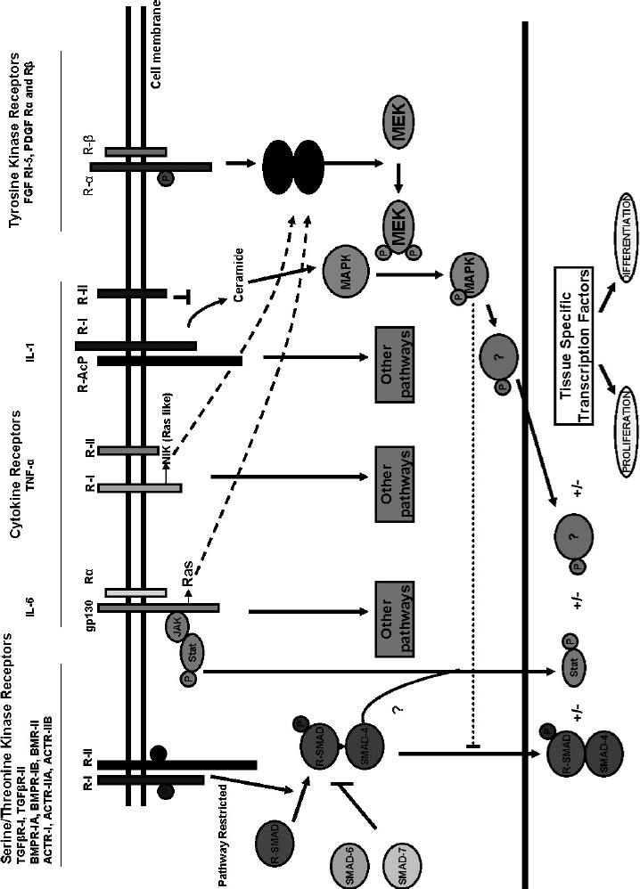
reviews and some of the factors and signaling pathways involved in this process are illustrated in Figure 21.1
[24,25]. In Section 21.2 we will overview some of the signaling molecules in bone formation and repair.

mikos: “9026_c021” — 2007/4/9 — 15:52 — page3—#3
Tissue Engineering Applications — Bone 21-3
FIGURE 21.1
Major growth factors and cytokines involved in fracture repair
. (Reproduced from
J. Bone Miner. Res.
1999, 14:1808 with permission of the American Society for
Bone and Mineral Research.)

mikos: “9026_c021” — 2007/4/9 — 15:52 — page4—#4
21-4 Tissue Engineering
BMPs Growth factors
ECM
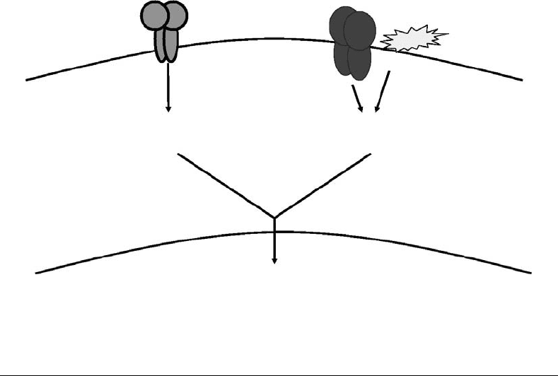
MAPK signaling/other pathwaysSmad dependent or independent
signaling
Bone-specific gene expression → mineralization → bone
FIGURE 21.2 Growth factors, ECM proteins, BMPs exert their effects on cells to promote osteoblastic differentiation
and bone formation.
21.2 Signaling Molecules for Bone
21.2.1 Growth Factors
Bone formation and development is the arrangement of the actions of a wide variety of signaling molecules
(Figure 21.2). These signaling molecules include growth factors, hormones, vitamins, and cytokines.
Hormones may be several categories including amino acid derivatives, peptides, steroids and fatty
acid derivatives. Growth factors are peptide hormones, which induce cellular proliferation. Cytokines,
which are also peptide-based can affect inflammatory and immune responses of the metabolism. The
orchestration of these factors regulates mitogenesis, cell shape, movement, differentiation, and apoptosis.
Growth factor effects are concentration-dependent and are exerted through their receptors on the cell
surface. A secreted growth factor may bind to matrix molecules, carrier molecules, or binding proteins to
regulate its activity and stabilization [26]. Growth factors can associate with specific binding proteins that
limit access to their receptors to control the bioavailability of the growth factor (e.g., IGF-I, IGF-II, TGFβ,
BMPs) [27–29]. The conversion of a growth factor to a bioactive state requires an activation event. Using
IGF-I as an example, greater than 99% are bound by IGFBPs (IGF binding proteins) in fluid and solid phase
[26,30] and requires protease activation to release IGF-I. This mechanism of sequestration allows temporal
and spatial regulation. For tissue engineering applications it is crucial to control the concentration and
physical placement and sequestration of a growth factor. Several methods are available for controlling the
physical placement of a growth factor including, but not limited to microfluidics, microencapsulation,
entrapment and release from polymeric systems, and nonspecific adsorption to matrices [26,31]. Growth
factors can also be immobilized to engineered matrices to localize delivery [32], but spatial patterning
remains a challenge.
The design of a tissue engineering application requires an appreciation of the mechanism by which a
factor elicits its signal and the downstream effect originating from this signal. In this section we will briefly
overview some of the factors involved in bone formation and regulation.
21.2.1.1 Insulin-Like Growth Factors
The putative functions of insulin-like growth factors (IGF-I and IGF-II) include embryonic and natal
growth, bone matrix mineralization, cartilage development and homeostasis [33,34]. IGFs can stimulate
mikos: “9026_c021” — 2007/4/9 — 15:52 — page5—#5
Tissue Engineering Applications — Bone 21-5
collagen production and prevent collagen degradation by reducing collagenase synthesis [35]. IGF-I is
known to activate osteocalcin expression [36] inside the cell. It has been reported to be important for
maintaining bone mass and promoting longitudinal bone growth [37].
The IGFs transduce their signals via two different receptors known as IGF-I and IGF-II receptors [38].
IGF-II mediates its signal through the type II IGF receptor although it can tissue-specifically activate the
type I IGF receptor. IGF-I is a single chain peptide with a structure similar to proinsulin, but consisting
of four domains (insulin contains three domains) [39]. During posttranslational modification of the
molecule, one of the peptide domains is cleaved from the rest of the domains and the rest are joined to one
another by forming disulfide bonds [39,40]. Growth hormone induces IGF-I synthesis [41]. The kidney,
muscle, and bone also contribute to the circulating IGF-I levels [41].
IGF-II has 60% homology to IGF-I and acts independent of growth hormone [41,42]. It appears
to be more important during fetal growth than in postnatal growth [43,44]. In a study where it was
systemically administered to rats, IGF-II was found to be less potent than IGF-I in stimulating skeletal
growth [45]. A study on chimeric mice carrying one inactivated IGF-II allele indicated that these mice were
smaller than their wild type littermates [46] indicating the significance of IGF-II on the skeleton during
early stages of growth. More studies have been conducted on IGF-I than IGF-II for tissue engineering
applications. Studies in rats suggest that IGF-I increases intramembranous ossification [47], improves the
effects of age-related osteopenia [48,49], and accelerates functional recovery from Achilles tendon injury
[50]. IGF-I has also been used for spinal fusion application in sheep, giving a successful outcome when
delivered via poly-(D, L-lactide) (PDLLA)-coated titanium [51]. In another study, IGF-I was delivered
via polyacetate (PLLA) microspheres to metacarpal defects in calves of pigs and showed enhancement of
bone formation [52].
21.2.1.2 Fibroblast Growth Factors
Fibroblast growth factors FGF-1 and -2, also known as acidic and basic FGF, respectively, belong to
a family of growth factors with heparin binding domains. FGFs regulate mitogenesis, differentiation,
protease production, receptor modulation, and cell maintenance [53]. FGF-2 (also called basic FGF or
bFGF) is produced by the osteoblasts and stored in skeletal tissues [54]. FGF-1 has been associated with
chondrocyte proliferation [55].
The FGF-1 and FGF-2 systemically and locally administered to ovariectomized rats increased new bone
formation and bone density [56,57]. Systemic delivery of FGF-1 appeared to be effective in restoring
the microarchitecture of bone and preventing bone loss associated with estrogen withdrawal [56]. In a
rabbit ulcer model, FGF-1 delivery within a modified fibrin matrix stimulated angiogenic and fibroblastic
responses in addition to an increased epithelialization rate [57]. Several studies indicated that scaffold
mediated delivery rather than direct injection of FGF-2 was more effective in improving bone healing in
rats [58], rabbits [59], and dogs [49,60]. Local infusion of recombinant FGF-2 increased bone ingrowth
in a rabbit tibia model in the presence of polyethylene particles [61].
21.2.1.3 Vascular Endothelial Growth Factors
Vascular endothelial growth factors, of which there are six different isoforms [62], are vascular cytokines
that promote angiogenesis, increased vascular permeability, and vasodilation [62]. Prosthetic vascular
grafts that were coated with VEGF supported endothelial cell proliferation and migration [63]. Several
studies indicated increased capillary density and vasodilator-induced blood flow in response to VEGF
treatment [64–66]. Synergistic effects of VEGF and FGF-2 have also been demonstrated in the pro-
duction of new blood vessels [67]. Macroporous scaffolds with poly(lactide-co-glycolide) which were
designed to release VEGF increased the generation of mineralized tissue due to an increase in vascu-
larization, but did not increase osteoid formation [68]. VEGF is a crucial factor for tissue engineering
due to its role in angiogenesis; it is the main provider of nutrients and growth factors to the wound
repair site.
mikos: “9026_c021” — 2007/4/9 — 15:52 — page6—#6
21-6 Tissue Engineering
21.2.1.4 Transforming Growth Factor-ß
Transforming growth factor-ß family consists of five members, bone morphogenetic proteins (BMP),
growth and differentiation factors (GDF), activins, inhibins, and Mullerian substance [69]. Osteoblasts
and chondrocytes express TGFß receptors [70,71]. Mainly found in bone, platelets, and cartilage TGFß
triggers growth, differentiation, and extracellular matrix synthesis [72]. TGFß is thought to be a coupler
between bone formation and resorption. Studies on the use of TGFß as a therapeutic reagent have been
difficult to assess due to the use of different isoforms and superphysiological doses of TGFß.
21.2.1.5 Bone Morphogenetic Proteins
Bone morphogenetic proteins are members of the TGFß superfamily. These proteins are highly conserved
with sequence homology across species. BMPs play a critical role in embryonic development and regulate
a wide range of cellular activities including cell proliferation, differentiation, cell determination, and
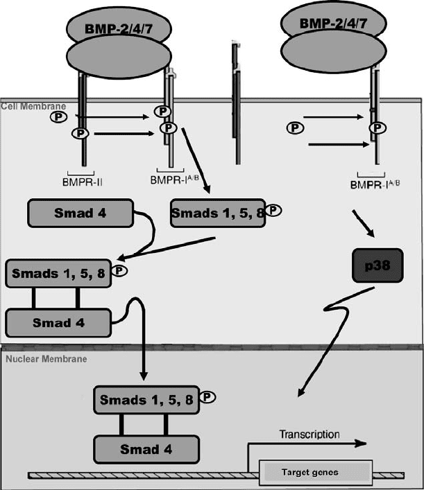
apoptosis. At the cellular level BMPs bind to their transmembrane receptors and initiate a cascade of
phosphorylation events which transduces the signal to upregulate downstream genes. BMPs elicit their
signals through phosphorylation of Smad molecules, which translocate to the nucleus when activated
and regulate the transcription of specific target genes. Recently Hassel et al. [73] have demonstrated that
a Smad independent pathway is activated if the BMP-2 signaling pathway is initiated via BMP-2 induced
signaling receptor complexes instead of preformed receptor complexes (see Figure 21.3). Several studies on
animals have demonstrated the osteoinductive (inducing bone formation) properties of BMPs in healing
nonunions and enhancing spinal fusion. Recombinant human BMP-2 protein delivered to diaphyseal
defects in dogs was able to achieve union (heal a fracture) [74]. A similar study in dogs also showed
promising results using BMP-7 [75]. BMP-7 has been shown to enhance spinal fusion in rabbits [76] and
sheep [77]. Recombinant human BMP-2 successfully healed critical-sized defects in sheep [78], rabbit
[79], and rat [80].
Since their discovery by Dr. Marshall Urist, BMPs have been used in a number of human clinical
trials as well. BMP-7 was effective in healing critical sized defects in the fibula [81] whereas BMP-2
promoted lumbar interbody fusion [82]. Transgenic BMP-2 produced by human mesenchymal stem
cells was effective in bone regeneration [83]. Currently, only BMP-2 is available for clinical use. BMP-7
(rhOP-1) may be approved to treat long bone nonunions secondary to trauma. BMP-2 is approved for
tibial nonunions and in a spinal fusion construct which consists of a spinal fusion cage and rhBMP-2 on
a type I collagen scaffold [84].
21.2.1.6 Platelet Derived Growth Factors
Platelet derived growth factor (PDGF) is composed of two polypeptide chains that may exist as a
homodimer (PDGF-AA, PDGF-BB) or heterodimer (PDGF-AB) [85]. Several reports have indicated
that PDGF-AA and PDGF-BB can enhance wound repair [86], support angiogenesis [87–90], and stim-
ulate cell proliferation in the fetal rat calvarial system and in cultures of osteoblast-like cells derived from
adult human bone explants [91,92]. The PDGF A and B genes act as regulators of cell growth and have been
shown to be chemotactic [88,89,91]. PDGF-BB has been reported to be the most potent PDGF isoform
in skeletal and nonskeletal cells [93]. As a consequence of its role in cell growth, PDGF may exert its effect
by increasing the number of collagen synthesizing cells, although it does not increase collagen synthesis
on a cellular basis [93]. The role of PDGF in osteoblast differentiation may be to increase the number of
cells that can progress into osteoblastic lineage and express the osteoblast phenotype [93]. PDGF expres-
sion at fracture sites in addition to its mitogenic effects indicates a role for PDGF in wound healing and
fracture repair. Systemic administration of PDGF in an osteoporotic animal model demonstrated that
it could stimulate bone formation and improve mechanical strength in long bones and vertebrae [94].
Howes and colleagues showed that subcutaneously implanted demineralized bone matrix augmented
with PDGF could enhance bone healing in a rat model [95]. Locally administered recombinant human
PDGF-BB (rhPDGF-BB) delivered with an injectable collagen gel to rabbit tibial osteotomies enhanced
fracture repair and stimulated osteogenesis [96]. Currently, PDGF-BB is approved by FDA for soft tissue

mikos: “9026_c021” — 2007/4/9 — 15:52 — page7—#7
Tissue Engineering Applications — Bone 21-7
FIGURE 21.3 Smad dependent and independent BMP signaling pathways. (Reprinted from Schmitt J.M., Hwang K.,
Winn S.R., and Hollinger J.O. 1999. J. Orthop. Res. 17: 269–278. With permission from the Orthopedic Research
Society.)
healing and remains as a compelling agent for tissue engineering applications especially in the treatment
of osteoporotic fractures.
Despite the indispensable roles of these factors in osteoblast differentiation and bone formation, there
are a number of concerns about their use in human patients. For instance, one concern with the injection
of a growth factor like IGF-I is hypoglycemia, as reported in a study [97]. Another concern has been the
use of superphysiological doses of these factors in order to trigger a response from the host. In the case of
BMP-2, milligram doses (1.7 to 3.4 mg/dl) have to be used in patients due to diffusion from the wound site
and instability in vivo [98,99]. Another reason for the rapid degradation of BMPs may be the presence of
its natural inhibitors such as noggin and chordin at the fracture site [100,101]. Excessive bone formation
remains a concern due to the risk of bony overgrowth leading to inadvertent fusion of adjacent levels or
compression of the neural elements [102]. The potential side effects also need to be studied in longer time
course experiments and trials for a fair assessment of the outcome.
21.2.2 Stem Cells and Gene Therapy
Mesenchymal stem cells (MSCs) are a population of self-renewing, undifferentiated cells. They can pro-
gress into a number of different cell fates, for example, adipocytic, osteogenic, chondrogenic, fibroblastic.

mikos: “9026_c021” — 2007/4/9 — 15:52 — page8—#8
21-8 Tissue Engineering
Stem cells can be harvested from fat, muscle, and bone marrow and can be genetically engineered to
express bone signaling molecules [103–105]. Bruder et al. [106] implanted human bone marrow MSCs
seeded on a ceramic carrier into the critical-sized defects of the femora of adult athymic rats and observed
evidence for bone formation within 8 weeks. Parietal bone defects in adult sheep were repaired with MSCs
added to a calcium alginate composite [107].
Gene therapy is the process by which genetic material is transferred into a cell’s genome. A gene of
interest can be delivered with the use of nonviral or viral carriers into targeted cell lines. Nonviral delivery
methods include the use of naked plasmid DNA, liposomes, or gene gun. Examples of viral vectors include
adenovirus, adeno-associated virus, lentivirus, herpes simplex virus, Moloney murine leukemia virus and
retrovirus [108]. A method known as ex vivo gene therapy allows the viral infection of the cells to take place
outside the body, thus increasing control over the system. A number of ex vivo studies have been performed
to treat various bone defects in mouse, rabbit, and rat [109]. Human MSCs genetically engineered
with an adenoviral construct expressing BMP-2 were able to form bone and cartilage and regenerate
nonunion fractures in a mouse radius model [53]. An adenoviral construct carrying the BMP-2 gene
was able to achieve spinal fusion in athymic rats [110]. However, the same research group also reported
an immune response with adenoviral BMP-2 delivery into immunocompetent rats rather than athymic
rats [111,112]. Furthermore, FDA has placed a hold on certain gene therapy applications due to safety
concerns.
One method used to improve the efficacy of the delivery of signaling molecules is to use matrix scaffolds.
In Section 21.3 we will overview the importance of biomaterials in tissue engineering and some current
developments in this field.
21.3 The Ideal Scaffold
Autograft bone remains as the gold standard treatment of bone defects in the clinic due to its capability
of providing cells, differentiative factors, and a reliable matrix required for fusion. Autograft bone is
usually isolated from the iliac crest of the patient and can lead to a number of complications including
chronic pain, infection, and fracture. One other option is to use allograft bone in the clinic; however,
in this case immune response and disease transmission remain major concerns. The inadequacies of the
current treatment methods have generated a need for alternative therapeutics for the treatment of bone
defects.
Delivery of an osteoblast-specific gene or a signaling molecule remains a desirable option, but the
therapeutic molecule requires an osteoconductive scaffold for its delivery. The ideal scaffold should
encourage cell attachment, promote and support vascularization, and resist soft tissue forces [113]. The
scaffold needs to be osteoinductive, osteoconductive, and biodegradable; this means that it needs to have
the ability to induce bone formation at a nonbony site, the ability to provide a scaffold for new bone
formation at the delivered site, and the ability to decompose without any toxic components to the cells
and tissues, respectively.
Biomaterials for scaffold design can be classified under two major groups: acellular and cellular systems.
Absorbable filter materials that can promote bone formation without a cellular component are called
acellular systems. Cellular systems have cells embedded in the matrix to guide bone development [112].
Naturally derived matrices are derived from primary components of bone matrix and have natural
affinity to growth factors as well as other osteoinductive factors; however they are difficult to sterilize
and can trigger immune response from the host. Examples include hyaluronic acid, chitosan, and col-
lagen matrices [69,113,114–117]. Inorganic materials include hydroxyapatite, porous coralline, calcium
phosphate cements, and calcium sulfate. Their major advantage is the resemblance to bone structure,
they can be resorbable or nonresorbable, but they are difficult to mold and are brittle [115,118–121].
Synthetic polymers are easy to manufacture and sterilize and they can be designed with controlled release
parameters; however, they may degrade into toxic components and may be difficult to get recognized
by the cells. Examples of synthetic polymers include poly (α-hydroxy acids), polypropylene fumarate,

mikos: “9026_c021” — 2007/4/9 — 15:52 — page9—#9
Tissue Engineering Applications — Bone 21-9
polyanhidrides, polyphosphazenes, polyethylene glycol, and poloxamers [115,122–128]. Ceramics such
as tri-calcium phosphate and hydroxyapatite are biocompatible and display osteoconductivity [129–132];
however, resorption and porosity remain as concerns in these types of matrices. Oxidized cellulose and
oxidized cellulose esters are also biocompatible polymers and have application in surgically implantable
materials [133,134].
New biomaterial composites are being created to overcome the limitations of the different types of
scaffolds mentioned above. Tsuchiya et al. [135] reported that they achieved osteogenesis with the
design of a web-like structured biodegradable hybrid sheet composed of PLGA sheets containing col-
lagen microsponges in their openings when seeded with bone marrow stem cells. These biodegradable
hybrid sheets could be laminated or rolled into any shape. Photoencapsulation of hydrogels in different
layers may help to mimic zonal organization of tissues which may be crucial in tissue engineering of
the cartilage [136]. Recent fabrication techniques allow the synthesis of biomaterials that contain signal
recognition ligands such as RGD domains to enable molecular and cellular responses.
Biocompatible and biodegradable polyurethane scaffolds have also been prepared for bone tissue engin-
eering applications [137–144]. Polyurethanes are prepared by reacting diisocyanates with diols, whereas
polyureas are the reaction product of diisocyanates with diamines (Figure 21.4). To be useful as resorb-
able scaffolds, conventional diisocyanates, such as methylene bis diphenylisocyanate (MDI) and toluene
diisocyanate (TDI), which degrade to carcinogenic and mutagenic compounds [145], cannot be used. To
avoid the toxicity problems associated with aromatic diisocyanates, aliphatic diisocyanates, such as lysine
ethyl ester diisocyanate (LDI), and 1, 4-diisocyanatobutane (BDI), have been reacted with polyether and
polyester polyols to synthesize resorbable polyurethanes [139,146–148].
Diisocyanates react with water to form a disubstituted urea and carbon dioxide gas, which acts as
blowing agent [149]. The water reaction is exploited commercially to manufacture flexible and rigid
polyurethane foams. Zhang et al. [139,140] have prepared porous scaffolds by adding water to an
isocyanate-terminated prepolymer (i.e., the low molecular weight reaction product of a diisocyanate
with a polyol). By varying the concentration of water added, the pore size distribution was con-
trolled to support the growth and proliferation of rabbit bone marrow stromal cells. Porous scaffolds
for the knee-joint meniscus have also been prepared by the solvent casting/salt leaching technique
[150,151].
Bioactive molecules can be incorporated into polyurethanes through the reaction of diisocyanate with
primary amine and hydroxyl groups. Following this approach, Zhang et al. [141] have recently synthesized
a bioactive polyurethane scaffold from lysine ethyl ester diisocyanate (LDI), glycerol, polyethylene glycol
(PEG), water, and ascorbic acid [142]. As the polyurethane degraded, ascorbic acid was released to the
extracellular matrix and stimulated both cell proliferation and type I col and Alp synthesis in vitro. Other
degradation products included lysine and PEG, which are biocompatible. Polyurethanes are potentially
useful biomaterials for preparing bioactive porous scaffolds from both high (e.g., proteins) and low
(e.g., signal recognition ligands) molecular weight bioactive molecules.
In the previous sections, we overviewed some of the required components for bone tissue regeneration:
signaling molecules, cells, and scaffolds. In Section 21.4, we will summarize applications for reconstructive
medicine in a clinical setting. We will review some of the current techniques applied to augment bone
deficits.
21.4 Clinical Reconstruction of Bone Defects
Physical deformities caused by missing or defective tissues affect people of all ages. Most often they are
due to cancer, trauma, or congenital abnormalities. Each year over 500,000 reconstructive procedures
to correct these deformities are performed by plastic surgeons in the United States [152]. There are
two fundamental types of deformities. One is when all tissue elements are present but not according to
normal anatomy. An example is a fracture that heals with bone segments in improper orientations (i.e.,
fracture malunion). The second type is when the tissues are significantly impaired or absent altogether.

mikos: “9026_c021” — 2007/4/9 — 15:52 — page 10 — #10
21-10 Tissue Engineering
N
H
O
R1
O
R2
NC
O
2R1
NH
2
R3
NC
O
R1
NC
O
R1
OHR2
OHH
N
H
O
R1
N
H
R3
N
H
O
R1
N
H
R1
(a)
(b)
(c)
+
+
+
FIGURE 21.4 Reactions of isocyanates with (a) alcohols to form urethane groups, (b) amines to form urea groups,
and (c) water to form disubstituted ureas.
An example is damage caused by cancer radiotherapy (i.e., osteoradionecrosis) or massive trauma (e.g., a
shotgun blast). When a deformity contains all essential elements, repair is possible by rearranging or
augmenting the local tissues. On the other hand, when useful tissues are absent then new tissue must
be supplied. Ideally, tissue replacements should be readily available, easily implanted, reliably incor-
porated into the surrounding normal tissues. This is the goal of tissue engineering for reconstructive
surgery.
It is important to understand current clinical techniques of reconstructive surgery in order to develop
practical new methods. Repairing every deformity involves similar steps of planning, tissue manipulation,
and patient care. During the planning phase, the clinician will assess the defect to determine the exact
form and amount of the deficient tissue. Conventional radiographs and computed tomography (CT)
scanning are the most useful diagnostic tools for the osseous component. Magnetic resonance imaging
(MRI) best demonstrates the soft tissue component. It is important to always consider both elements.
A bone defect due to trauma may have the same anatomic appearance as one caused by cancer, but
the best reconstructive method may be different in each case because of the health and stability of the
surrounding soft tissues. After characterizing the defect, the next step is to select a source of replacement
tissue. The clinician must balance the tissue requirements with the potential morbidity related to harvest.
After considering all of these issues, the clinician and patient discuss them and agree upon a plan for
surgery.
Repairing deformities by tissue replacement is a two-step process that first involves tissue transfer
followed by tissue modification [153]. In the transfer step, tissue is harvested from an uninjured location
(i.e., tissue donor site) and moved into the defect (i.e., recipient site). It is important to understand the
principles that govern this manipulation in natural tissues because they also apply to engineered tissues.
Living tissue may be transferred either as a surgical graft or a surgical flap. Grafts derive a blood supply
from the tissues that surround them in the new location. Except for cartilage and thin pieces of skin, only
small amounts of tissue can be transferred as grafts because survival of the cellular elements by simple
diffusion of oxygen and nutrients is limited to volumes of less than 0.3 cm
3
[154,155]. Success depends
on the potential for angiogenesis and specialized tissue formation that exists in the tissues surrounding
the graft in the new location. During the initial 48 h after transfer, the graft must survive by diffusion
alone [156]. Afterward, blood vessels arising from the tissues surrounding graft begin to align with
and make connections to remnants of blood vessels found in the graft [157]. It takes up to 5 days for
revascularization to occur, depending on the grafted tissue and condition of the tissue bed into which it
is placed. This far exceeds the time during which most whole tissues can survive without a blood supply.
Skeletal muscle tissue, for example, undergoes degeneration within 4 h after being deprived of blood
supply. As new vessels penetrate the graft there is cell-mediated destruction of the degenerating muscle
fibers. The basal laminae and some of the satellite cells appear to be the only elements of the muscle
tissue persists [158]. Bone grafts are unique, however. They essentially are porous, calcified, degradable
mikos: “9026_c021” — 2007/4/9 — 15:52 — page 11 — #11
Tissue Engineering Applications — Bone 21-11
scaffolds that guide tissue formation (i.e., osteoconduction) and deliver a set of bioactive molecules to
induce new bone formation (i.e., osteoinduction). Only a limited number of cellular elements survive and
contribute to healing because of diffusion limitations [159,160]. The surviving cells are mostly located
on the surfaces of the calcified matrix and appear to consist mainly of endosteal lining cells and marrow
stromal cells [161,162]. Even though most cellular components do not survive transfer, bone grafts still
contribute to bone healing because they supply the other essential components of tissue repair. Bone
grafts must be completely replaced over time in order to achieve healing. This can require up to 2
years. They have limited utility for defects greater than 6 cm in length and are avoided when there is
significant local tissue impairment due to significant bacterial contamination, poor vascularity, unstable
soft tissues, or radiation injury in the area of the defect [163]. Tissue engineered bone created without a
capillary system ex vivo in a bioreactor can be expected to perform clinically in a way analogous to a bone
graft.
In contrast to grafts, surgical flaps are tissues transferred with a blood supply independent of the tissues
surrounding the defect. They are called flaps because originally they were actual flaps of skin elevated with
an attachment at the base and rotated into an adjacent area. Over time the definition broadened to include
any unit of skin, muscle, fat, bone, or viscera (e.g., small intestine) that has a discreet vascular source
permitting surgical isolation from the donor site and transfer to a distant recipient site. The most advanced
method of transfer is by detaching the flap completely and reestablishing the blood supply by suturing
the blood vessels of the flap to vessels adjacent to the defect. This is called microvascular surgery because
it involves operating on blood vessels less than 5 mm in diameter using an operating microscope. Bone
transferred in this way allows survival of all tissue elements and yields the most reliable healing. Bone flaps
incorporate more rapidly than grafts and do not require resorption and replacement before achieving
full strength [161]. They are the treatment of choice in circumstances with large defects, significant
soft tissue deficits, or impaired local tissues due to severe trauma, infection, or exposure to radiation
[163–166]. The ultimate goal of bone tissue engineering for reconstructive surgery is to fabricate surgical
bone flaps.
After transfer, the tissue must then be modified to simulate the missing parts. Bones have a complex
three-dimensional shape and must tolerate powerful deforming forces. The relative importance of shape
and load bearing differs based on the anatomic site. The long bones of the extremities function primarily
as load bearing members. Minor shape discrepancies are tolerable as long as there is no significant loss
of strength. On the other hand, craniofacial bones have a limited load bearing function. Their shape
is critical to support and protect the complex and delicate soft tissue structures of the head and neck.
They also determine human facial appearance and play a major role in psychosocial health [168,169].
Therefore, bone replacements in the craniofacial skeleton must maintain a durable shape. In addition,
craniofacial bones have thin soft tissue coverage, and they are located in close proximity to heavily bacteria-
contaminated surfaces of oral and nasal cavities. The oral cavity is one of the most heavily contaminated
areas of the body with numerous bacterial species present including aerobes, anaerobes, fungi, viruses, and
protozoa [170]. The paranasal sinuses are normally not sterile, although the bacterial load appears much
less [171]. It is impossible to perform tissue implantation surgery in this area without a high probability of
bacterial contamination. Therefore, the tissue replacement must be compatible with the thin soft tissues
to avoid erosion and have an intrinsic resistance to infection. These features of the craniofacial skeleton
make fabricating replacements by tissue engineering particularly challenging.
After the surgery is complete, the patient requires special care to recover. The reconstructed areas
must be protected from disruption and infection. Grafted tissues must be stably fixed to prevent shearing
motion at the interface with the tissue bed, which slows the process of revascularization. Flaps must be
closely monitored to rapidly detect thrombosis and occlusion of the blood vessels that supply the tissues.
After complete healing and tissue incorporation, the final step is often a period of rehabilitation to ensure
maximum restoration of function. Finally, additional surgery may be required to make revisions and
improve minor deficiencies that are not able to be avoided during the primary reconstruction or which
may have appeared later due to scar contracture, for example. The entire process can require many months
to completely restore the patient (Figure 21.5a,b and Figure 21.6a–d).
