Fischer W.B. Viral Membrane Proteins: Structure, Function, and Drug Design
Подождите немного. Документ загружается.


17
The Structure, Function, and
Inhibition of Influenza Virus
Neuraminidase
Elspeth Garman and Graeme Laver
1. Introduction
The neuraminidase story started in the early 1940s, almost a decade after the first
human influenza virus was isolated.
George Hirst, working in the Rockefeller Institute in New York City, found that when
allantoic fluid from embryonated chicken eggs, which had been infected with influenza virus,
was mixed with red blood cells at 0C, the cells were very heavily agglutinated (Hirst, 1941).
He then found that when the agglutinated cells were warmed to 37C they dispersed as the
virus eluted, and the cells could not be re-agglutinated when they were mixed with fresh
infected allantoic fluid at 0C. The eluted virus, on the other hand, could agglutinate fresh red
cells in the cold.
Hirst’s interpretation of this finding was that the virus had an enzyme which removed
receptors for the virus from the agglutinated red cells when they were warmed to 37C where
the enzyme was more active. The enzyme therefore became known as “receptor-destroying
enzyme” or RDE.
Alfred Gottschalk, at the Walter and Eliza Hall Institute in Melbourne, Australia, reasoned
that the action of RDE on its substrate would probably yield a “split product.” This split product
was eventually isolated and characterized as sialic acid, or N-acetyl neuraminic acid and the RDE
of influenza virus became known as sialidase or neuraminidase (Gottschalk, 1957). Subsequently
it was discovered that sialidases are quite widespread in nature. Other viruses, bacteria, mam-
malian cells, and some parasites all have their own sialidase enzymes.
In 1948, MacFarlane Burnet realized that specific inhibitors of flu neuraminidase might
be effective antiviral agents. “An effective competitive poison for the virus enzyme might be
administered which, when deposited on the mucous film lining the respiratory tract, would ren-
der this an effective barrier against infection, both initial infection from without and the spread-
ing surface infection of the mucosa which follows the initiation of infection” (Burnet, 1948).
247
Elspeth Garman • Laboratory of Molecular Biophysics, Department of Biochemistry, University of Oxford,
South Parks Road, Oxford OX1 3QU, United Kingdom. Graeme Laver • Barton Highway, Murrumbateman,
NSW 2582, Australia.
Viral Membrane Proteins: Structure, Function, and Drug Design, edited by Wolfgang Fischer.
Kluwer Academic / Plenum Publishers, New York, 2005.
Burnet’s comment was that this approach did not seem even remotely possible. Now,
more than 50 years later, although we know that “poisoning” the viral neuraminidase does not
stop the virus infecting cells, the subsequent “spreading surface infection” is effectively quelled.
So far, four different potent and specific “competitive poisons” for flu neuraminidase
have been developed, two of which are now being used worldwide to control influenza
infections in people.
1.1. Structure of Influenza Viruses
Two serologically different types of influenza virus exist; Type A and Type B. Type A
influenza infects a wide variety of animals; pigs, horses, seals, whales, and many different
kinds of birds. Type B influenza seems to be confined to the human population, though one
isolation of type B flu from harbor seals has been reported (Osterhaus et al., 2000).
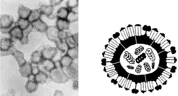
Influenza virus particles are pleomorphic, consisting of misshapen spherical objects or
long spaghetti like filaments (Figure 17.1).
The genome of influenza A and B viruses consists of single stranded RNA of negative
sense. The RNA exists in eight separate pieces, each of which codes one of the virus proteins
(in some cases two, using overlapping reading frames). The eight RNA pieces are packaged
in an orderly fashion within the virus particle (Fujii et al., 2003).
The RNA is associated with a nucleoprotein and with three proteins, PB1, PB2, and
PB3, involved in RNA replication and transcription. This replication complex is enclosed
within a membrane composed of a matrix protein associated with a lipid bilayer. Embedded
in the lipid bilayer are the two surface glycoprotein spikes, one of which is the hemagglutinin
and the other the neuraminidase, described below.
248 Elspeth Garman and Graeme Laver
(a)
(b)
Figure 17.1. (a) Electron micrographs of negatively stained particles of influenza virus showing their pleomorphic
nature and the surface layer of “spikes” which have been identified as the hemagglutinin and neuraminidase antigens.
The particles are approximately 80–120 nm in diameter. (b) Diagram of the influenza virus showing the eight
segments of negative sense ssRNA, the M2 ion channel spanning the membrane, and the two surface glycoproteins,
neuraminidase (boxes on stalks) and hemagglutinin (rods).
Also spanning the lipid bilayer of Type A influenza virus are a small number of M2
protein molecules, which function as ion channels. Until recently the only antiviral drugs
available for treating influenza A infections were the ion channel blockers, amantadine and
rimantadine. These, however, have no effect on influenza Type B (which does not possess the
M2 ion channel), they have undesirable side effects and resistance to these drugs develops
very rapidly.
It is perhaps amazing that despite the widespread occurrence of influenza in the world,
the huge number of deaths each year, the misery of flu sufferers, and the enormous economic
cost of influenza, amantadine and rimantadine have been the only compounds found, until
recently, to be effective in treating influenza. This is despite a vast research effort which
included the random screening of many thousands of compounds by pharmaceutical compa-
nies, none of which was found to be an effective antiviral drug.
The two safe and effective drugs, Relenza and Tamiflu, now being used to treat influenza
Type A and Type B, were rationally designed from a knowledge of the three-dimensional
structure of flu neuraminidase. The development of these and other neuraminidase inhibitors
will now be described.
2. Structure of Influenza Virus Neuraminidase
For some time after flu virus neuraminidase was discovered it was assumed that the agglu-
tination of red cells by influenza virus particles was due to the neuraminidase on the virus bind-
ing to its substrate, sialic acid, on the surface of the red cells so linking them together in large
clumps. It is now known that this idea is incorrect. The first indication that the neuraminidase
enzyme was not responsible for aggutinating red cells came from the finding that when some
strains of influenza virus were heated to 55C, the neuraminidase was inactivated while the
hemagglutinin was still fully active (Stone, 1949). Then, in 1961, further doubts began to
appear. Mayron and colleagues found that a soluble sialidase could be separated from the PR8
strain of Type A influenza virus, and that this soluble enzyme did not adsorb to red cells
(Mayron et al., 1961). Hans Noll then discovered that when influenza B virus particles were
treated with trypsin, almost 100% of the neuraminidase was liberated as a soluble molecule with
a sedimentation coefficient of 9S (equivalent to about 200,000 molecular weight), leaving all of
the hemagglutinin activity still associated with the virus particles (Noll et al., 1962).
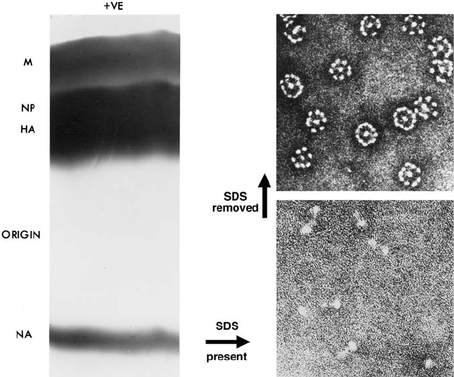
Experiments were then done in which influenza virus particles were disrupted with
detergents, and the disrupted virus particles subjected to electrophoresis on cellulose acetate
strips. This resulted in a clear separation of hemagglutinin and neuraminidase activities and,
since the procedure used did not cleave any covalent bonds, it proved that the hemagglutinin
and neuraminidase activities resided in separate protein molecules on the surface of the virus
particle (Laver, 1964).
At about this time the first electron microscope images of negatively stained influenza
virus particles were obtained. These showed pleomorphic objects completely covered with
a densely packed layer of surface projections or “spikes” (Figure 17.1). These were the two
surface antigens, the hemagglutinin and the neuraminidase.
Electron micrographs of pure preparations of influenza virus neuraminidase molecules,
separated from virus particles which had been disrupted with detergents, showed that the neu-
raminidase consisted of a square, box-shaped head atop a long thin stalk with a small
hydrophobic knob at the end. This served to attach the neuraminidase to the lipid membrane of
Structure, Function, and Inhibition of Influenza Virus Neuraminidase 249
the virus (Laver and Valentine, 1969) (Figure 17.2) and also caused the isolated neuraminidase
to form rosettes in the absence of detergents. It was estimated that each virus particle pos-
sesses about 500 neuraminidase “spikes” which account for about 5% of the protein in the
virus particle. These numbers are approximations only, as they vary from strain to strain.
Further electron micrographs of isolated neuraminidase “heads” by Nick Wrigley
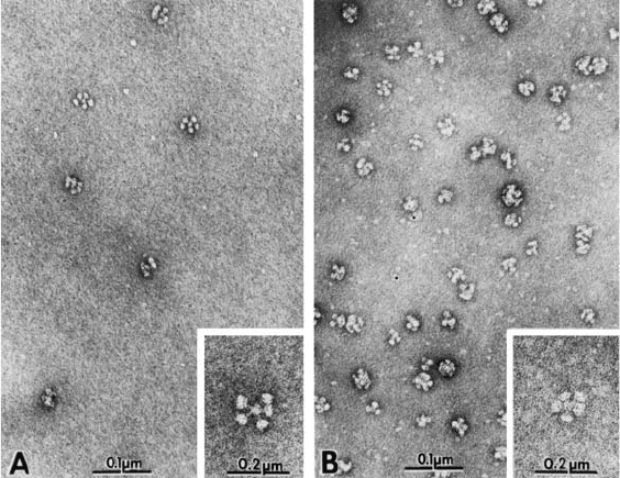
showed these to be tetramers (Wrigley et al., 1973). The stalk of the neuraminidase, which
serves to attach the molecule to the lipid bilayer of the virus can vary in length (Els et al.,
1985; Mitnaul et al., 1996). This is shown in Figure 17.3 where neuraminidase molecules
with shortened stalks (“stubbies”) can be seen in electron micrographs.
The neuraminidase tetramer is composed of four identical monomers, each of which
contains a single polypeptide chain coded by RNA segment number 6. The neuraminidase is
anchored in the lipid bilayer of the viral membrane by a series of hydrophobic amino
250 Elspeth Garman and Graeme Laver
Figure 17.2. Pure preparations of intact, biologically active neuraminidase “spikes” from influenza virus
particles. The virus particles were disrupted with sodium dodecyl sulfate (SDS) at room temperature and
electrophoresed on cellulose acetate strips at pH 9.0. Strips stained with Coomassie Blue are shown on the left-hand
side. Following electrophoresis, the neuraminidase, which was completely separated from all of the other virus
proteins, was eluted from the (unstained) strips with water. The eluted neuraminidase (in the presence of SDS)
existed as single molecules (bottom right), consisting of a box-like head atop a long thin stalk (approx 12 nm long).
Following removal of the detergent, the neuraminidase molecules all aggregated by their hydrophobic membrane
attachment sequences at the ends of the stalks to form the rosettes shown (top right, which is ⬃2.5 lower
magnification than bottom right). Stalk length varied between strains but was usually approximately 10 nm, giving
rosettes of approximately 24 nm diameter.
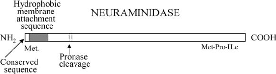
acids near the N-terminal end of the polypeptide (Figure 17.4). This contrasts with the hemag-
glutinin which is anchored by a hydrophobic sequence near the C-terminal end of the
hemagglutinin polypeptide.
No post-translational cleavage of the neuraminidase polypeptide occurs, no signal
peptide is split off and even the initiating methionine is retained. Nor is there any processing
at the C-terminus; the sequence Met-Pro-Ile predicted from the gene sequence of N2
neuraminidase is found in intact neuraminidase molecules isolated from the virus. A sequence
of six polar amino acids at the N-terminus of the neuraminidase polypeptide, which are totally
conserved in all nine flu Type A neuraminidase subtypes (but not in flu Type B), is followed
by a sequence of hydrophobic amino acids that must represent the TM region of the
neuraminidase polypeptide. This sequence is not conserved at all among subtypes (apart from
conservation of hydrophobicity).
Intact neuraminidase molecules can be isolated after disruption of influenza virus par-
ticles with detergents. Remarkably, the neuraminidase from a number of strains of flu virus
is 100% active after disruption of the virus with the powerful detergent, sodium dodecyl sul-
fate (SDS). Even more remarkably, when virus particles from these strains were disrupted
with SDS and electrophoresed on cellulose acetate strips, the intact, active neuraminidase
molecules migrated in one direction completely free of any of the other virus proteins, all of
which migrated in the opposite direction (Figure 17.2) (Laver, 1964).
Neuraminidase molecules eluted from the strips following such electrophoresis existed
as single molecules. When the detergent was removed, for example, by cold ethanol precipi-
tation of the protein, the single neuraminidase molecules aggregated by the hydrophobic
tips of their tails, forming the rosettes seen in electron micrographs (Figure 17.2).
Structure, Function, and Inhibition of Influenza Virus Neuraminidase 251
Figure 17.3. Rosettes of intact neuraminidase molecules isolated from SDS disrupted wild-type influenza virus
and from a virus with an 18-amino acid residue deletion in the stalk (“stubby”).
Soluble neuraminidase “heads” can be released from some strains of influenza virus
by treating the particles with proteolytic enzymes, for example, pronase or trypsin. These
proteases cleave the stalk of the neuraminidase at about residue 75 of the neuraminidase
polypeptide (Figure 17.4) releasing the box-shaped “head” which carries all of the enzymatic
and antigenic activity of flu virus neuraminidase.
2.1. Crystallization of Influenza Virus Neuraminidase
In 1978, neuraminidase “heads” released by pronase from a number of strains of H2N2
and H3N2 influenza virus were crystallized (Laver, 1978). The three-dimensional structure of
the subtype N2 enzyme at 2.9 Å resolution was then determined using X-ray crystallography
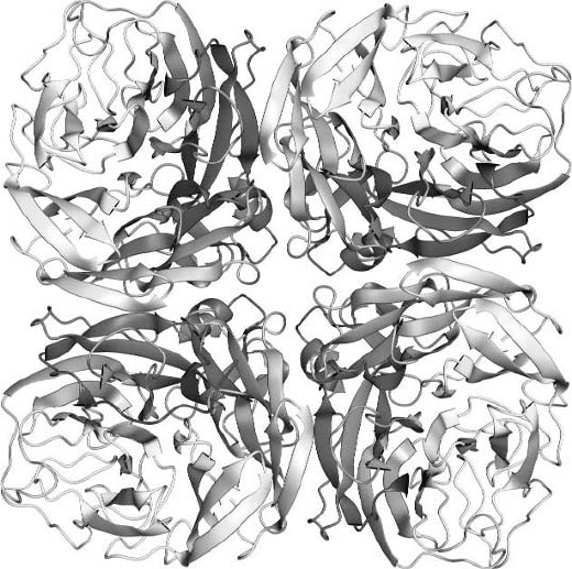
(Varghese et al., 1983). Figure 17.5 shows the structure of subtype N9 neuraminidase.
This showed that each monomer in the tetrameric enzyme is composed of six topolog-
ically identical beta sheets arranged in propellor formation. The tetrameric enzyme has
circular 4-fold symmetry partially stabilized by metal ions bound on the symmetry axis. Deep
pockets occur on the upper corners of the box-shaped tetramer. These pockets were identified
as the catalytic sites by soaking substrate (sialic acid) into neuraminidase crystals and
solving the structure of the complex (Colman et al., 1983).
Sugar residues are attached to four of the five potential glycosylation sequences in N2
neuraminidase and in one case, the carbohydrate contributes to the interaction between the
monomers in the tetramer.
2.2. Structure of the Conserved Catalytic Site
Sequences of neuraminidase from influenza A and B strains can differ by as much as
75%. Nevertheless, scattered along the neuraminidase polypeptide, are charged residues
which are totally conserved among all strains. These include Arg 118, Glu 119, Asp 151,
252 Elspeth Garman and Graeme Laver
Figure 17.4. Diagram showing certain features of the neuraminidase polypeptide. The neuraminidase is oriented
in the virus membrane in the opposite way to the hemagglutinin. No post-translational cleavage of the neuraminidase
polypeptide occurs, no signal peptide is split off and even the initiating methionine is retained. No processing at the
C-terminus takes place—the C-terminal sequence, Met-Pro-Ile predicted from the gene sequence is found in intact
neuraminidase molecules isolated from virus and in the pronase-released neuraminidase heads. A sequence of
six polar amino acids at the N-terminus of the neuraminidase polypeptide, which is totally conserved in at least
eight different neuraminidase subtypes, is followed by a sequence of hydrophobic amino acids which probably
represents the transmembrane region of the neuraminidase stalk. This sequence is not conserved at all between
subtypes (apart from conservation of hydrophobicity). Pronase cleaves the polypeptide at the positions shown,
removing the stalk and releasing the enzymatically and antigenically active head of the neuraminidase, which, in
some cases, can be crystallized.
Arg 152, Asp 198, Arg 224, Glu 227, Asp 243, His 274, Glu 276, Glu 277, Arg 292, Asp 330,
Lys 350, and Glu 425 (N2 numbering).
When the linear neuraminidase polypeptide folded into its three-dimensional structure,
these conserved residues all came together and clustered on the rim and walls of the pocket
(Figure 17.6). This suggested that if an inhibitor, a “plug-drug,” could be devised which
blocked one flu neuraminidase active site, it would also block the sites on all other influenza
virus strains, even those which have not yet been found infecting humans.
2.3. Structures of Other Influenza Virus Neuraminidases
All influenza neuraminidases except N4 and N7 have been crystallized (Figure 17.7)
though not all crystals were suitable for X-ray structure determination. Structures have been
obtained for N2 (Varghese et al., 1983), N9 (Baker et al., 1987), N8 (Taylor et al., 1993), N6
(Garman et al., 1995), and type B (Burmeister et al., 1992) neuraminidases.
N9, N8, N6, and influenza type B neuraminidases have a similar overall topology to
N2 neuraminidase despite having up to 75% differences in amino acid sequence of the
neuraminidase polypeptide chain.
Structure, Function, and Inhibition of Influenza Virus Neuraminidase 253
Figure 17.5. Three-dimensional structure of an N9 neuraminidase tetramer, [PDB entry 7NN9]. (Figure drawn
with AESOP [Noble, 1995].)
254 Elspeth Garman and Graeme Laver
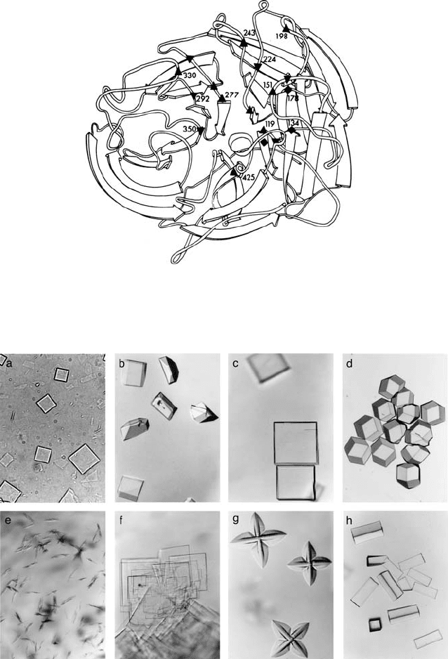
Figure 17.6. Ribbon diagram of a monomer of N2 influenza neuraminidase showing the active-site residues
which are conserved across flu strains: ▲ Glu 119, Asp 151, Asp 198, Glu 227, Asp 243, Glu 276, Glu 277, Asp 330,
Glu 425 ▼, Arg 118, Arg 152, Arg 224, His 274, Arg 292, Lys 350, ◆ Tyr 121, Leu 134, Trp 178. (Reproduced from
Colman et al. (1983) with permission from Nature [http://www.nature.com/].)
Figure 17.7. Crystals of influenza neuraminidase; (a) N2, (b) N6, (c) N8, (d) N9 used for X-ray structure
determination, and (e) N1, (f) N3, and (g) N5 which were unsuitable for such studies, and (h) whale N9 in complex
with 32/2 antibody (Fab). Crystal sizes range from 0.6 mm in the largest dimension (N9) to 0.15 mm (N6).
X-ray structure determination pivotally depends on obtaining a diffraction quality
crystal that is a well-ordered array of protein molecules which will scatter X-rays in a coher-
ent manner. The crystal is mounted on a motorized stage (a “goniometer”) which allows it to
be rotated in the X-ray beam in small angular increments. The diffraction patterns of the scat-
tered X-rays are now collected on image plate or CCD (Charged-Coupled Devices) detectors,
replacing the photographic film of old. Dedicated computer software is used for analyzing the
diffraction images and extracting the intensities of the reflections and further experiments are
required to obtain their phases (for further reading see Blow, 2002).
Experimental methods for crystallography have advanced dramatically in the last
15 years because of major technical developments. These include the advent of intense
tunable synchrotron X-ray sources and fast read-out area detectors, a huge increase in com-
puting power and the development of techniques for flash-cooling crystals to cryotempera-
tures (below 130 K) to substantially reduce radiation damage by the beam during data
collection (Garman and Schneider, 1997).
2.4. Hemagglutinin Activity of Neuraminidase
Rosettes of isolated intact neuraminidase molecules of the N9 subtype also had hemag-
glutinin activity (Laver et al., 1984). The hemagglutinin site was shown to be quite separate
from the catalytic site in a 1.7 Å resolution X-ray structure which located a second sialic acid
binding site situated about 21 Å from the catalytic site on N9 neuraminidase after a 4C soak (as
opposed to the usual 18C soak). The residues in contact with the sialic acid come from three
different loops in the structure. These residues are mostly conserved in avian strains of influenza,
but not in those of human and swine. It is thus possible that the hemagglutinin site on the
neuramindase has some as yet undiscovered biological function in birds (Varghese et al., 1997).
3. Function of Influenza Virus Neuraminidase
In 1966, Seto and Rott showed that the function of neuraminidase was probably
associated with the release of virus from host cells (Seto and Rott, 1966). It was then found
that antibody directed specifically against flu neuraminidase, and which abolished the activity
of the enzyme for large substrates, did not prevent the infection of susceptible cells, but
blocked the release of newly formed virus particles (Webster and Laver, 1967).
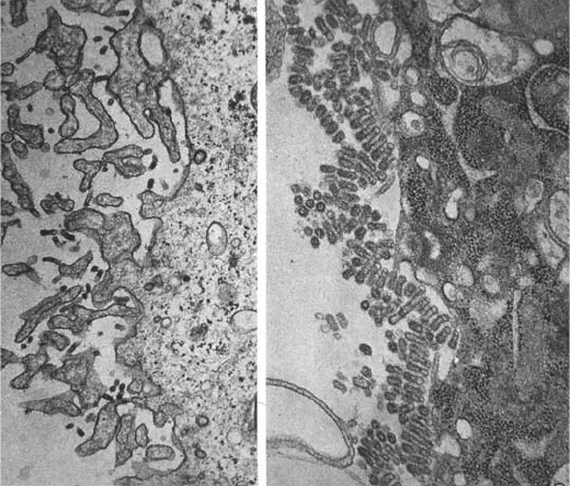
The role of neuraminidase in the release of virus particles from infected cells was
demonstrated most elegantly by Palese, Compans, and their colleagues in 1974 (Palese et al.,
1974a). Electron micrographs were made of surfaces of cells infected with temperature sen-
sitive (ts) neuraminidase mutants of influenza virus at the permissive temperature and at the
restrictive temperature (where the virus replicated but where the neuraminidase lacked
enzyme activity). These showed virus particles budding normally from the cells and going off
to infect other cells at the permissive temperature. However in cells infected with the ts
mutants at the restrictive temperature, virus particles budded from the cell in the normal man-
ner, but then remained attached to each other and to the surface of the infected cells, forming
great clumps of virus particles. These were clearly not going anywhere, and the infection was
effectively terminated (Figure 17.8).
It is believed, therefore, that the function of flu virus neuraminidase is to remove sialic
acid receptors for the virus from the host cells, and also, perhaps more importantly, from the
Structure, Function, and Inhibition of Influenza Virus Neuraminidase 255
newly formed virus particles themselves. The two surface antigens on the influenza virus
particle, the hemagglutinin and the neuraminidase, are themselves glycoproteins and possess
carbohydrate side chains with terminal sialic acid receptors for other virus particles. The
main function of the neuraminidase therefore might be to remove receptors for influenza virus
from newly formed virus particles so allowing these to be released and spread the infection
(Palese et al., 1974a). Another function of flu virus neuraminidase might be to destroy
sialic acid containing inhibitors for the virus in the mucous secretions of the respiratory
tract, so enabling the virus to more easily infect cells, and there may be other functions as yet
undiscovered.
Chickens vaccinated with pure neuraminidase “heads” were protected from death by
lethal avian influenza viruses. But whether this protection was due to inhibition of neu-
raminidase activity or to enhanced clearance of the virus by the immune system was not
established (Webster et al., 1988).
3.1. Antigenic Properties of Influenza Virus Neuraminidase
Both of the surface antigens of influenza virus undergo extensive antigenic variation.
This is of two types, antigenic drift and major antigenic shifts. Drift is the result of mutations
256 Elspeth Garman and Graeme Laver
(a) (b)
Figure 17.8. Electron micrographs of the surface regions of MDCK cells infected with temperature sensitive (ts)
neuraminidase mutants of influenza virus after inoculation and incubation for 12.5 hr at the permissive temperature
of 33C ((a) left) and at the restrictive temperature of 39.5C ((b) right). The aggregates of virus particles which
accumulated at the restrictive temperature could be dispersed by incubation with bacterial neuraminidase. Staining
experiments showed that the aggregated virus particles formed at the restrictive temperature were covered in sialic
acid residues, while this was absent on those well-dispersed particles formed at the permissive temperature.
Magnification approximately 30,000. (Reprinted from Palese et al. [1974a] with permission from Elsevier.)